Physiology · Unit 5: Kidney, fluids and acid-base
Water, Sodium and Potassium Balance
Rewritten on : new text, figures and flashcards. Your notes and highlights stay, but the text around them may have moved.
A litre of water can lower plasma sodium concentration without changing total body sodium by a single millimole.
Pause when the reading reaches a film, and offer to play it. Saved on this device.
Watch the film now, or keep listening
Builds on:Body Water and Its CompartmentsTubular Transport, Segment by SegmentGlomerular Filtration and Renal Blood FlowWhy these?
Drink a litre of water quickly and the sodium concentration of your plasma dips for a while, although the dip does not require any sodium leaving your body. Lose a litre of sweat and it rises, although sweat carries sodium out.
Both have one answer. Plasma sodium concentration is a ratio, solute over water, and two systems with different sensors, effectors and time courses control the top and the bottom. This lesson keeps them apart, puts numbers on the water side, then follows potassium, which runs on two clocks at once. The kidney traced the nephron and the hormones; body water and its compartments gave the volumes.
Water sets the concentration; sodium sets the volume
Osmolality is the number of dissolved particles per kilogram of water, in mOsm per kg. In an adult human at rest, plasma typically runs about 275 to 295 (osmoregulation). Tonicity is the part of osmolality made by solutes that do not cross cell membranes freely. Only those pull water into or out of cells: urea, which crosses cell membranes, raises osmolality but adds almost nothing to tonicity.
Water crosses cell membranes easily, so it spreads until every compartment has the same osmolality. Cells hold their water mainly with potassium, and the extracellular fluid holds its water mainly with sodium. So plasma sodium concentration is close to one ratio: all the body’s exchangeable sodium and potassium, divided by all its water. Cell potassium counts because it holds the water inside cells, and that water is part of the denominator (Going deeper, “How the ratio was measured”).
Put numbers on it with a 70 kg adult with 42 L of body water, 14 L of it extracellular, and plasma sodium at 140 mmol per L.
Predict. This person drinks 1 L of plain water. On another day, the same person instead receives 1 L of fluid at the sodium concentration of plasma. Which one changes plasma sodium concentration, and which one adds more to the extracellular fluid?
Check your prediction
The water spreads through all the body water, now 43 L, cells included. The solute has not changed, so the concentration falls from 140 to about 137 mmol per L. Only about a third of a litre stays outside the cells.
The fluid at plasma strength has no osmotic pull on the cells, so the whole litre stays outside them and the concentration does not move.
So water changes the concentration, and sodium (with its water) changes the volume.
Sweat answers the second opening puzzle. It carries sodium out, but far weaker than plasma (sweat sodium varies widely; take 50 mmol per L), so it removes water out of proportion to solute and plasma sodium concentration rises. Losing fluid at plasma strength, as in bleeding or some secretory diarrhoeas, moves only the volume.
| One litre, in the 70 kg adult | Plasma [Na+] (mmol per L) | Extracellular fluid (L) |
|---|---|---|
| Start | 140 | 14.0 |
| Drink plain water | about 137 | about 14.3 |
| Receive fluid at plasma strength | 140 | 15.0 |
| Lose sweat (sodium taken as 50) | about 142 | about 13.4 |
| Lose fluid at plasma strength | 140 | 13.0 |
One caution: normal saline (0.9%) carries 154 mmol per L, a little more than plasma, so a litre of it nudges the concentration up slightly, to about 140.3.
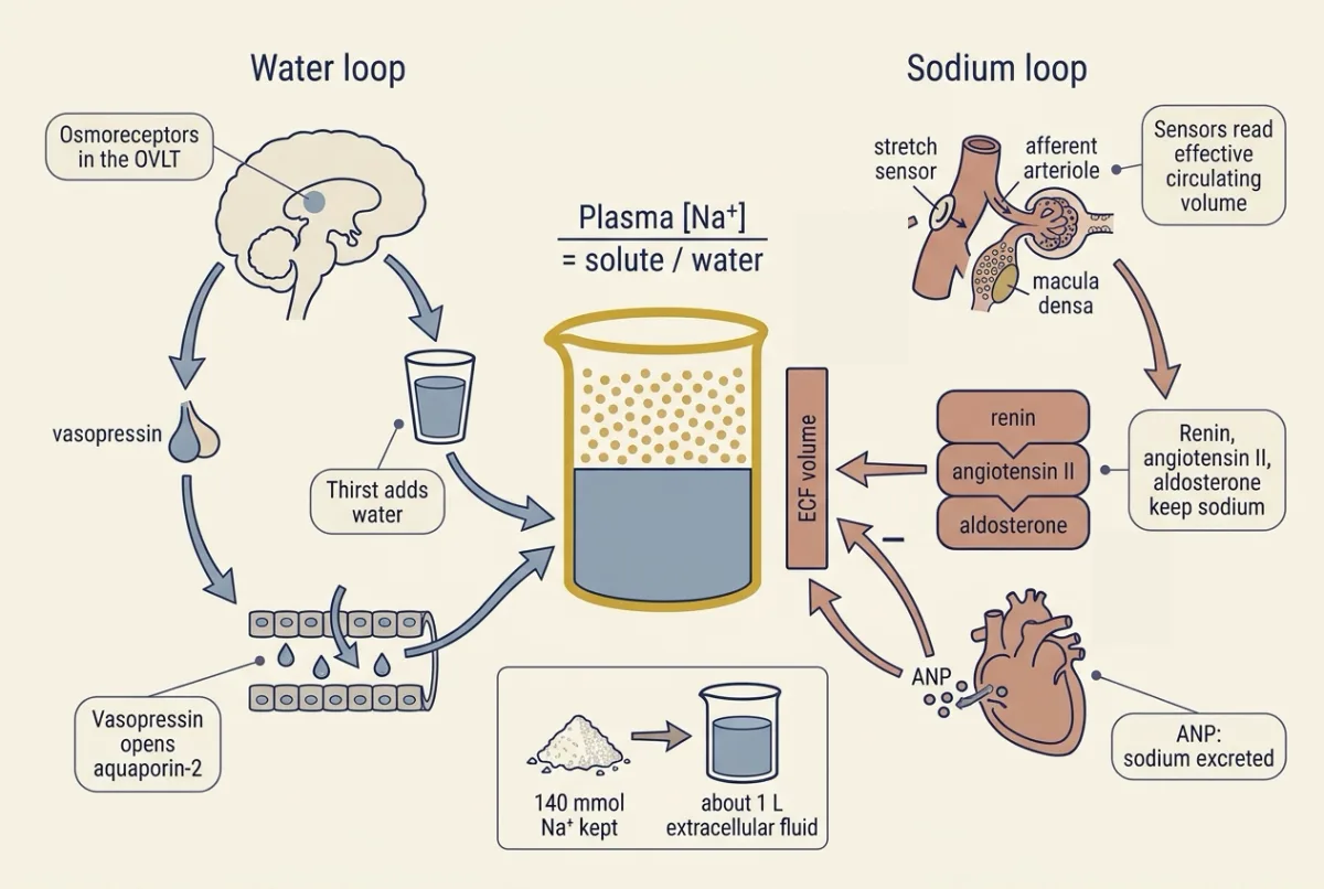
The body runs one loop for each quantity. The water loop controls the ratio. Osmoreceptor neurons in the lamina terminalis sense plasma tonicity and drive thirst and the release of vasopressin, which lets the collecting duct return water to the blood. They read a concentration, blind to how much sodium the body holds.
The sodium loop controls the volume. Its sensors read effective circulating volume: how well the arteries are filled, as the arterial and atrial baroreceptors, stretch of the afferent arteriole and salt delivery to the macula densa report it (glomerular filtration). It is not a separate pool you could measure, and it usually, but not always, moves with extracellular volume (Going deeper, “When effective volume and extracellular volume part company”). The loop’s effectors, renin, angiotensin II, aldosterone, sympathetic outflow and the natriuretic peptides, set how much sodium the kidney excretes.
The two loops connect through one number. Suppose the body keeps an extra 140 mmol of sodium, about 8 g of salt. The water loop holds back water until the concentration is back at 140 mmol per L, and 140 mmol at that strength fills 1 L, all of it outside the cells, where the sodium stays.
The one idea to carry through this lesson. Plasma sodium concentration is body solute divided by body water. The water loop holds that ratio steady by adding or removing water. Once it has, every 140 mmol or so of sodium the body keeps carries about 1 L of extracellular fluid, so the sodium loop sets volume by setting sodium content. A sodium concentration therefore tells you about water, and a change in extracellular volume or body weight sustained over days tells you about sodium, if potassium is steady. Scope: a healthy adult whose main effective solutes are sodium and potassium salts, once the water loop has caught up (minutes to hours). It bends when glucose or mannitol adds tonicity, when a large volume loss drives vasopressin whatever the osmolality, and when effective circulating volume and extracellular volume part company, as in heart failure. Potassium follows its own rule, at the end.
So a low sodium concentration means too much water for the solute, with total body sodium low, normal or high: it can go with an expanded extracellular volume, and also with real sodium loss replaced by hypotonic fluid.
Vasopressin opens the collecting duct, and thirst replaces the water
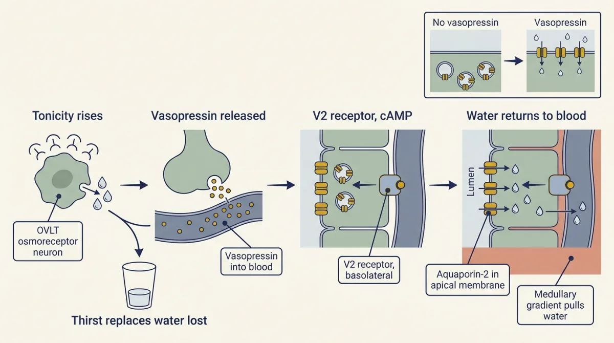
Follow the water loop link by link.
- Plasma tonicity rises, water leaves the osmoreceptor neurons of the organum vasculosum of the lamina terminalis (OVLT), and they fire faster.
- Their signal raises release of vasopressin into the blood.
- At the collecting duct, vasopressin binds the V2 receptor on the basolateral membrane of principal cells and raises cyclic AMP (G proteins, cAMP and PKA).
- Water channels, aquaporin-2, are moved from vesicles into the apical membrane (the kidney covers their trafficking, membrane transport aquaporin-1).
- Water leaves the duct down the medullary gradient and returns to the blood.
The range is wide. A water-deprived adult human makes urine at about 1,000 to 1,200 mOsm per kg; with the hormone absent, urine can fall near 50. The channel is required: in mice with aquaporin-2 deleted, vasopressin cannot make the collecting duct permeable to water at all (Going deeper, “Finding the water channel”).
The second effector is thirst, driven by the same osmoreceptors. In people vasopressin release starts near 280 to 285 mOsm per kg, and thirst starts close by: at about the same value in one ten-person study, somewhat higher in older work and most reviews. Both thresholds vary between people and between studies (Going deeper, “Osmoreceptors and thresholds”). Divide the work this way: vasopressin is the fast renal defence, which cuts renal water loss to a minimum, and thirst replaces the water already lost. The central circuitry belongs to the hypothalamus and autonomic control, anticipatory thirst to tissues and homeostasis, and sweat losses to skin and human heat.
Free-water clearance splits urine into two parts
How much pure water is the kidney removing from the body, or returning to it? Split the urine, on paper, into two parts: one carrying all the excreted solute at plasma osmolality, and one of solute-free water. Three steps build the formula.
- The solute excreted each minute is urine osmolality times urine flow, Uosm × V.
- If that solute were dissolved at plasma strength, it would need a flow of Uosm × V / Posm. That flow is the osmolar clearance, Cosm: the urine flow that would carry the excreted solute if urine were as concentrated as plasma.
- Whatever flow is left over, V − Cosm, carried no solute at all. That is the free-water clearance, CH2O.
Cosm = Uosm × V / Posm, and CH2O = V − Cosm, with V the urine flow
A positive CH2O means urine more dilute than plasma, zero means isosmotic urine, and a negative value means concentrated urine. When it is negative, minus CH2O is the solute-free water reabsorbed into blood (TcH2O). The division in Cosm is by plasma, not urine, osmolality.
Worked problem: splitting the urine
Round illustrative numbers for healthy adults, not measurements. Assume a steady state over each collection and constant plasma osmolality.
- Water diuresis (a large flow of dilute urine), after a water load with vasopressin suppressed. V = 10 mL per min, Uosm = 60 mOsm per kg, Posm = 282 mOsm per kg. Solute excretion = 60 × 10 = 600 microOsm per min. Cosm = 600 / 282 = 2.13 mL per min. CH2O = 10 − 2.13 = +7.87 mL per min, positive because the urine is weaker than plasma: of each 10 mL, about 2.1 mL carries the solute and about 7.9 mL is solute-free water.
- Antidiuresis (a small flow of concentrated urine), after overnight water deprivation. V = 0.50 mL per min, Uosm = 1,000 mOsm per kg, Posm = 295 mOsm per kg. Solute excretion = 500 microOsm per min. Cosm = 500 / 295 = 1.69 mL per min. CH2O = 0.50 − 1.69 = −1.19 mL per min, so TcH2O = +1.19 mL per min returned to blood.
- Hormone present while plasma is dilute. A volunteer given vasopressin with a water load: Posm = 265, Uosm = 500 mOsm per kg, V = 1.0 mL per min. Cosm = 500 / 265 = 1.89 mL per min, CH2O = −0.89 mL per min, Uosm/Posm = 1.9. Plasma is dilute, so the osmoreceptors are signalling “stop vasopressin”, yet the urine is concentrated. Something other than osmolality is holding vasopressin on: here, the injected hormone; in patients, volume loss, nausea, pain or a tumour.
- Solute sets the range. Hold solute excretion at 600 microOsm per min, or 600 × 1,440 = 864 mOsm per day. At a maximal Uosm of 1,200 mOsm per kg the smallest daily volume is 864 / 1,200 = 0.72 L; at a very dilute Uosm of 60 mOsm per kg the largest is 864 / 60 = 14.4 L. Water excretion is limited by solute delivery as well as by vasopressin.
- Urine flow V = solute ÷ Uosm
- Osmolar clearance Cosm
- Free-water clearance CH₂O
- Uosm ÷ Posm
- Urine volume if this flow were held for 24 h, V × 1,440
- Water returned to blood, −CH₂O
Electrolyte-free water: which way is plasma sodium being pushed?
- Electrolyte-free water clearance CeH₂O = V × (1 − (UNa + UK) ÷ PNa)
- Free-water clearance CH₂O, for comparison
In this sketch: round illustrative numbers for healthy adults, not measurements; a steady state over each collection with constant plasma osmolality; the urine is treated as two volumes added, one carrying all the solute at plasma osmolality and one of solute-free water; you set solute excretion, Uosm and Posm independently, so some combinations may not occur in a healthy adult. The slider limits are made up for the sketch (solute excretion 300 to 900 µOsm per min, Posm 265 to 295 mOsm per kg), except that Uosm about 50 to about 1,200 is the lesson’s range and Posm 265 is the lesson’s third example; the electrolyte-free water sliders (urine Na+ + K+ 0 to 300 mmol per L, plasma Na+ 125 to 150 mmol per L) are also made up for the sketch. Values are shown rounded, so “≈” marks a rounded step. The bar’s scale changes with the numbers, so read the axis.
Rule to keep: Cosm = Uosm × V ÷ Posm (divide by plasma osmolality, not urine osmolality) and CH₂O = V − Cosm. Positive means urine more dilute than plasma, zero isosmotic urine, negative concentrated urine, and then minus CH₂O is the solute-free water reabsorbed into blood. This sketch is a rule only and does not predict plasma sodium concentration: free-water clearance is a renal, solute-based index of one collection and cannot say which way plasma sodium will move next, since that depends also on what is drunk and on non-renal losses.
One limit matters more than the arithmetic. Free-water clearance is a renal, solute-based index of one collection, and it cannot say which way plasma sodium concentration will move next. Osmolar clearance counts urea and other ineffective osmoles, which is why electrolyte-free water clearance, the same split made with urine sodium plus potassium against plasma sodium, describes the effect on sodium concentration better. Urine at 600 mOsm per kg that is mostly urea, with sodium plus potassium of only 100 mmol per L (flow 1.0 mL per min, plasma 290 mOsm per kg, plasma sodium 140), gives CH2O = −1.07 mL per min, as if the kidney were saving water. Electrolyte-free water clearance is 1.0 × (1 − 100/140) = +0.29 mL per min, so the urine is pushing plasma sodium concentration up, not down. Whole-body balance must also count what is drunk and non-renal losses.
The sodium loop sets volume, and aldosterone escapes
The sodium loop starts with renin release from granular cells of the afferent arteriole, which has three inputs:
- a fall in renal perfusion pressure, sensed in the arteriolar wall;
- a fall in sodium chloride at the macula densa, which is inversely related to renin secretion in perfused preparations (glomerular filtration);
- sympathetic stimulation of beta-adrenergic receptors on the granular cells.
Renin leads to angiotensin II, which raises proximal sodium reabsorption and stimulates aldosterone release. Aldosterone raises sodium reabsorption in the distal nephron, and it is also raised directly by a rise in plasma potassium, which matters later (the adrenal glands, nuclear receptors). Pushing the other way, atrial natriuretic peptide is released from atrial granules mainly when atrial-wall stretch rises with intravascular volume. It lowers blood pressure through natriuresis and diuresis (sodium and water excretion), vasorelaxation and antagonism of the renin-angiotensin system (blood pressure regulation, tubular transport and diuretics).
Predict. A healthy person loses a modest volume of blood. What happens to renin, and what happens to plasma sodium concentration?
Check your prediction
Renin rises, because perfusion pressure at the afferent arteriole falls and sympathetic outflow rises; angiotensin II and aldosterone follow, and the kidney keeps more sodium. Plasma sodium concentration at first does not move: blood is lost at plasma strength, so the ratio is unchanged. Only volume fell.
Predict. A healthy person on a fixed salt intake receives a large dose of aldosterone every day for several weeks. What happens to body weight?
Check your prediction
It rises for some days, then levels off while the hormone continues (after 5 days in one man and 16 in the other). August, Nelson and Thorn did this in 1958 in two normal men on constant diets. Sodium was retained and weight rose at first; then urinary sodium climbed back to about the control level, oedema stayed slight, and the weight gain stopped. They called the reversal escape.
Read the result with the one idea: every 140 mmol or so of sodium kept adds about 1 L, or about 1 kg, of extracellular fluid, so a weight gain that levels off fits a body that kept a fixed amount of sodium, then stopped, with excretion back up to match intake. Once extracellular volume has expanded by some amount, other mechanisms offset the hormone and restore sodium excretion in normal humans, so aldosterone is not the sole setter of that output. What they are is still argued; a 2026 review calls the question open (Going deeper, “The 1958 escape study”).
The loops can also collide. Vasopressin also rises when blood volume falls, and with stress, so when volume loss is large it follows volume, not osmolality: the kidney keeps water even while plasma is dilute, and if the person drinks water or receives hypotonic fluid, plasma sodium concentration falls. This is one of the common ways the two-loop rule breaks in patients.
Potassium is shifted in minutes and excreted over hours
Potassium breaks the pattern because almost all of it is inside cells: about 98 percent, mostly in skeletal muscle. Extracellular potassium is 3.5 to 5 mEq per L, and daily intake is about 70 mEq.
Take the same 70 kg adult, with 14 L of extracellular fluid at a potassium concentration of about 4.2 mEq per L. That is about 59 mEq of potassium outside cells: roughly 1.7 percent of the body’s 3,500 mEq or so.
Predict. This person eats a meal carrying 30 mEq of potassium, and suppose none of it could enter cells; ignore excretion for the first hour. What would plasma potassium do?
Check your prediction
It would rise by about 2 mEq per L: 30 mEq spread through 14 L is about 2.1 mEq per L, taking plasma potassium from about 4.2 to about 6.3. One meal carries about half the whole extracellular pool. Why a change of that size matters for the heart is in cardiac electrophysiology and the ECG.
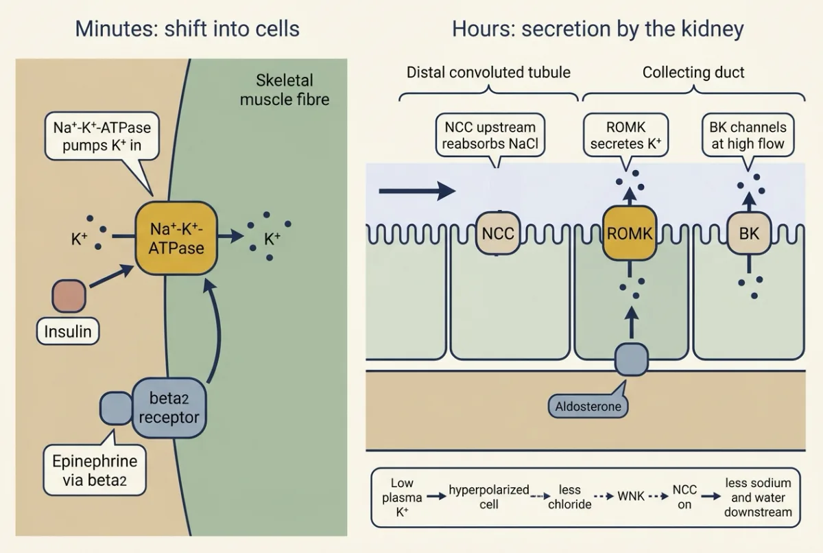
So the first defence is the muscle cell, not the kidney. Potassium has its own rule: it is defended first by shifting into cells, in minutes, then by renal secretion, over hours.
The shift: minutes
Insulin and catecholamines drive potassium into muscle by stimulating the Na+-K+-ATPase (insulin, glucagon and glucose): insulin directly, and epinephrine through beta2-adrenergic receptors. Potassium-deprived rats show the insulin dependence from the other side: their muscle becomes resistant to insulin’s stimulation of potassium uptake, so less potassium is taken into cells when insulin rises. The design problem below tests the epinephrine evidence.
Acid-base changes shift potassium too, but mineral-acid loading moves far more out of cells than organic-acid loading, and the shift is not a direct potassium-for-hydrogen exchange (acid-base balance).
Secretion: hours
Renal excretion is the slower arm, and it is secretory rather than filtration-driven. The glomeruli filter about 756 mEq of potassium a day (roughly 180 L of plasma at 4.2 mEq per L). The proximal tubule and the loop take most of it back, and in mammals less than 10 percent, under about 76 mEq, reaches the distal nephron, a fraction that changes little with diet. Urinary potassium, by contrast, follows intake: it falls on a potassium-poor diet, when the collecting duct reabsorbs, and on a high intake it exceeds what reaches the distal nephron.
So the part of urinary potassium that rises with intake is secreted, by principal cells, which take it in across the basolateral Na+-K+-ATPase and release it to the lumen through ROMK. At high flow, flow-activated BK (maxi-K) channels add more, and much of that BK pathway sits in intercalated cells rather than principal cells. Two major influences set the rate: mineralocorticoid (aldosterone-type) activity, and delivery of sodium and water to the secretory cells, with higher flow raising secretion. Plasma potassium itself and other cellular signals change it too.
One hormone, two outcomes: the aldosterone paradox
High aldosterone does not always mean potassium loss. It rises in two states with opposite outcomes.
| State | Aldosterone | Angiotensin II | Sodium and water delivered to the secretory cells | Potassium secretion |
|---|---|---|---|---|
| Low volume | high | high | low | not raised: potassium is kept |
| High plasma potassium | high | not raised | normal or higher | high: potassium is excreted |
Aldosterone is high in both rows; what differs is what comes with it. Low volume brings low delivery and high angiotensin II, which also restrains potassium secretion, so secretion is not raised. High plasma potassium brings normal or higher delivery and no rise in angiotensin II, and potassium itself stimulates secretion, so secretion rises. How one hormone can do both is the aldosterone paradox.
Part of the answer comes from the segment just upstream, the distal convoluted tubule, which reads plasma potassium as a membrane voltage. When plasma potassium is low, the distal cell hyperpolarizes, intracellular chloride falls, and chloride-sensitive WNK kinases switch on the NaCl cotransporter NCC. More sodium is then taken back upstream, less sodium and water reach the secretory cells, and potassium secretion falls. When plasma potassium is high, NCC is switched down and more reaches them: a second feedback beside aldosterone. The voltage and chloride steps rest on experimental models (Going deeper, “Distal potassium sensing: the mouse and cell experiments”).
Design problem
Brown, Brown and Murphy infused epinephrine into six healthy volunteers and plasma potassium fell by 0.82 mEq per L; in a second experiment at half that infusion rate a selective beta2-adrenergic antagonist prevented the fall. Extend the study. First, predict plasma potassium when epinephrine is given after placebo pretreatment, after a beta1-selective antagonist and after a beta2-selective antagonist. Second, design measurements that would show where the potassium went. Third, state what the design cannot show.
Saved in this browser only.
One way to do it
Predictions. After placebo pretreatment the fall appears, since that is the epinephrine response itself, and under beta2 block it is prevented, as reported. Under beta1 block it should largely persist, because the receptor carrying the effect here was beta2; that arm is a prediction, not a result.
Design. Use a crossover in the same volunteers, with a saline infusion as the no-epinephrine control. Collect urine over the same minutes as each infusion: urinary potassium unchanged while plasma potassium falls supports a shift into cells rather than a loss from the body, once intake, extracellular volume and non-renal losses are controlled. To locate the uptake, sample arterial and deep forearm venous blood across resting muscle for venous potassium below arterial potassium, and biopsy muscle for Na+-K+-ATPase activity, which tests mechanism but not net uptake. Measure insulin and glucose, since insulin also moves potassium, and record heart rate.
Limits. Six volunteers is a small sample, and plasma measurements alone cannot say which tissue took the potassium up, so the muscle claim stays an inference. Blocking a receptor shows it is required in this preparation, not that beta2 stimulation sets plasma potassium minute by minute in ordinary life.
Going deeper
How the ratio was measured
Edelman’s isotope-dilution relation, treated in body water and its compartments, writes plasma sodium concentration, in simplified form, as exchangeable sodium plus exchangeable potassium over total body water. The fitted regression also carries a slope and an intercept. For the litre of water, the fitted form and the simple ratio agree closely (about 136 against 137 mmol per L). Because potassium sits in the numerator, replacing a potassium deficit raises plasma sodium concentration too.
Osmoreceptors and thresholds
Ciura and Bourque recorded from neurons of the organum vasculosum of the lamina terminalis in mouse hypothalamic explants and acutely isolated cells. The neurons depolarized and fired faster when bath osmolality rose, with no circulation present. Mice lacking the channel protein transient receptor potential vanilloid 1 (TRPV1) drank less than wild-type mice after a hypertonic challenge, although they still drank, so the response was attenuated and not abolished, and OVLT neurons from these mice lost their osmotic response.
In ten healthy volunteers given hypertonic saline, thirst on a rating scale appeared near a plasma osmolality of 281 mosmol per kg, and the regression threshold for vasopressin release was 285, with slopes of 0.3 cm of scale and 0.4 pmol per L per mosmol per kg above those thresholds. That thirst threshold was lower than in earlier work, and reviews still describe vasopressin as the first line of defence and thirst as a second line at higher osmolality. Copeptin, cleaved from the same prohormone as vasopressin, is the stable plasma surrogate for it.
Finding the water channel
Aquaporin-2 was cloned from rat kidney in 1993 by Fushimi, Sasaki and colleagues, who showed that expressing it in Xenopus oocytes markedly raised osmotic water permeability. Mice later let each step be removed. Inducible deletion of aquaporin-2 in adult mice raised urine output from about 2 to 25 mL per day and dropped urine osmolality from about 2,000 to below 500 mosmol per kg, with no rise after water deprivation. Vasopressin still acts on medullary salt and urea transport in these mice, but not on collecting-duct water permeability. In cultured kidney cells, gain-of-function V2 receptor variants do the reverse, holding aquaporin-2 at the plasma membrane without vasopressin.
The 1958 escape study
In the two men, sodium intake, urinary sodium and body weight were measured each day on constant metabolic diets. If aldosterone alone set sodium excretion, the prediction was sustained retention, steady weight gain and oedema; the escape described in the main text contradicts it. The study does not show the mechanism: pressure natriuresis, natriuretic peptides, reduced proximal reabsorption, and WNK-kinase and paracrine effects are all proposed. It rests on two men, and this lesson tells only its sodium result.
Sodium stored in skin, and sodium rhythms
Sodium content and extracellular water may not always move together. In rats on a high-salt diet, sodium accumulates in skin, reported as hypertonic in some studies and as isotonic in others. In healthy men on fixed salt intake in long confinement studies, total body sodium swung by roughly 200 to 400 mmol on roughly monthly and longer rhythms, with no parallel change in body weight or extracellular water.
When effective volume and extracellular volume part company
The sodium loop reads how well the arteries are filled, not how much extracellular fluid there is. Usually the two move together. In heart failure or cirrhosis the extracellular fluid is expanded while the arterial sensors read “underfilled”, so the sodium loop keeps signalling for sodium retention. A second exception changes the concentration side: glucose (when it is high) or mannitol adds tonicity, draws water out of cells, and lowers plasma sodium concentration while plasma osmolality rises.
Distal potassium sensing: the mouse and cell experiments
A 2011 review called the aldosterone paradox poorly understood; much of the account in the main text has filled in since. Terker, Zhang, McCormick and colleagues, with Ellison as senior author, put mice on diets varying sodium and potassium and found that potassium deficiency activated NCC in the distal convoluted tubule even on a high-salt diet, with sodium retention and raised blood pressure. In cultured cells they traced the voltage-and-chloride step described in the main text, with the WNK kinases acting on NCC conventionally through the kinase SPAK. A later inducible deletion of the channel subunit Kir4.1 in adult mouse kidney caused salt wasting, low blood pressure and marked potassium wasting, and the distal cells no longer responded to plasma potassium. The main text’s account of reduced delivery to the secretory cells is the authors’ account. In humans the evidence includes inherited Kir4.1 disorders, controlled potassium interventions and urinary vesicle measurements.
Your notes
Select any text in the lesson to highlight it or add a note to it. Your highlights and notes are saved in this browser and collected in your notebook.
This browser is not letting the site save anything (a private window or strict privacy settings), so highlights and notes will be lost when you leave the page.
How we measure it
Metabolic balance study on a constant diet
A volunteer eats a weighed diet of fixed sodium, potassium and water content while every intake and every urinary and faecal output is collected and measured, often with daily body weight. It measures net retention or loss of an electrolyte rather than its concentration. Isotope dilution and whole-body potassium counting measure the pools instead.
Osmometry by freezing-point depression
Osmolality is measured as the depression of the freezing point of a sample, which depends on the number of dissolved particles per kilogram of water. Paired urine and plasma readings are what osmolar clearance and free-water clearance are computed from.
Xenopus oocyte swelling assay
Messenger RNA for a candidate transport protein is injected into a frog oocyte, which has low native water permeability, and the cell is moved into a dilute bath while its volume is tracked. Faster swelling than uninjected controls shows that the expressed protein conducts water.
Inducible kidney-specific knockout
A gene is deleted in the kidney of an adult mouse after a drug-triggered recombination, so development proceeds normally first. Comparing electrolyte excretion and blood pressure before and after deletion tests whether the protein is required for a response in the intact animal.
Practice questions on this lesson (6) → MCAT-style, original, each with an explanation
Where this lesson fits
Before thisReview first
- Body Water and Its CompartmentsThis course · Lesson 1
You need the compartment volumes and the sodium, potassium and water ratio from there to follow every litre example here.
- Tubular Transport, Segment by SegmentThis course · Lesson 16
Collecting-duct water and sodium handling, and distal potassium secretion, build on the segment-by-segment transport described there.
- Glomerular Filtration and Renal Blood FlowThis course · Lesson 15
Macula densa sensing and renin release from the afferent arteriole are taken from there, along with filtered potassium load.
After thisStudy next
- Acid-Base BalanceThis course · Lesson 18
Potassium shifts with mineral-acid and organic-acid loads are explained further once you can work the bicarbonate buffer.
- The Adrenal Cortex and MedullaThis course · Lesson 20
The aldosterone paradox and cortisol versus aldosterone receptor action take the mineralocorticoid story further.
- Calcium, Phosphate and BoneThis course · Lesson 22
It applies the same filter, reabsorb and hormone-control logic to another ion held in narrow limits.
Elsewhere on the siteFilms, simulations and method films in other courses
- Which Way Does the Water Go?Film · Organismal Biology
It shows how water moves toward higher osmolality, the basis of why sodium concentration tracks water.
- Filter Far More, Take Most BackFilm · Organismal Biology
It shows urine as what is filtered minus what tubules take back, which free-water clearance builds on.
Suggested by an AI model from the lessons themselves, then checked. How we use AI
Finish line
Test yourself
12 questions
Practice and MCAT-style, with explained answers.
Next · Lesson 18
Acid-Base Balance
Plasma holds 24 mmol/L of bicarbonate and only 40 nanomoles of free H+ per litre, and metabolism adds acid every day. pH stays put because one ratio, bicarbonate to dissolved CO2, stays near 20 to 1. What holds it there?
Continue →