Physiology · Unit 6: Endocrine systems
The Adrenal Cortex and Medulla
Rewritten on : new text, figures and flashcards. Your notes and highlights stay, but the text around them may have moved.
The kidney's aldosterone receptor binds cortisol just as tightly, and plasma carries far more cortisol. So why does aldosterone run sodium transport?
Pause when the reading reaches a film, and offer to play it. Saved on this device.
Watch the film now, or keep listening
Builds on:Endocrine Axes and How to Test ThemRegulating Arterial PressureWater, Sodium and Potassium BalanceWhy these?
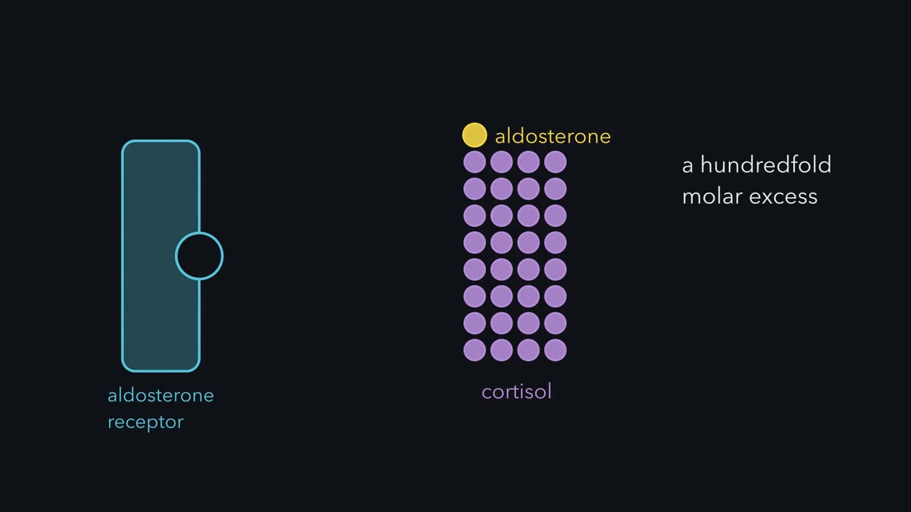 ▶ Watch An Enzyme Guards the Receptor · 2:34
Read the transcript of “An Enzyme Guards the Receptor”
Film transcript: An Enzyme Guards the Receptor
The kidney's aldosterone receptor binds cortisol just as tightly as it binds aldosterone. Adult human plasma carries far more cortisol than aldosterone. A hundredfold molar excess is a commonly cited figure. Yet sodium transport in the kidney follows aldosterone. How? The answer is an enzyme beside the receptor. In 1961, Jean Crabbé used a toad's urinary bladder, which moves sodium from urine side to blood side. He cut each bladder into paired halves and gave aldosterone to one half. After about one hour, its short-circuit current rose above its partner's. Tracer fluxes in eight bladders showed that the extra current was net sodium transport. So in this preparation, aldosterone acts on the epithelium directly.
Now the puzzle. In extracts of kidney and other tissues, mineralocorticoid receptors bound aldosterone and the physiological glucocorticoids with equal affinity. In humans, that glucocorticoid is cortisol, and it circulates at far higher concentrations. On those numbers, cortisol should occupy the receptor nearly all the time. So selectivity is not in the receptor. The answer came in 1988. John Funder and colleagues reported that targets such as kidney, colon and parotid carry an enzyme, 11-beta-hydroxysteroid dehydrogenase. It converts cortisol to inactive cortisone. Aldosterone escapes this conversion. So the enzyme lowers cortisol near the receptor, and the receptor is left to aldosterone. Edwards and colleagues in Edinburgh reached a similar conclusion the same year.
Liquorice fits this idea. Eating large amounts of liquorice was long known to cause sodium retention, low potassium and high blood pressure. Renin and aldosterone are both low. That looks like mineralocorticoid excess without the mineralocorticoid. Glycyrrhetinic acid in liquorice inhibits the enzyme, so cortisol is no longer inactivated in the kidney. It occupies the mineralocorticoid receptor itself. The conclusion has limits. It came from work on rats and corticosterone. The epithelial type 2 form of the enzyme was characterized in the 1990s. Keep this rule. The enzyme beside a receptor is part of what defines a hormone's target tissue.
The kidney’s aldosterone receptor (the mineralocorticoid receptor, which switches on sodium transport) binds cortisol as tightly as it binds aldosterone, and adult human plasma carries far more cortisol (a hundredfold or more, counted in molecules). Yet sodium transport in the kidney follows aldosterone. How?
The short answer: an enzyme in the kidney inactivates cortisol near the receptor (the main part of the answer; Going deeper asks how much it must do). The rest of the lesson earns that sentence, and the same reasoning predicts adrenal disease. The first look at the gland is in the endocrine system.
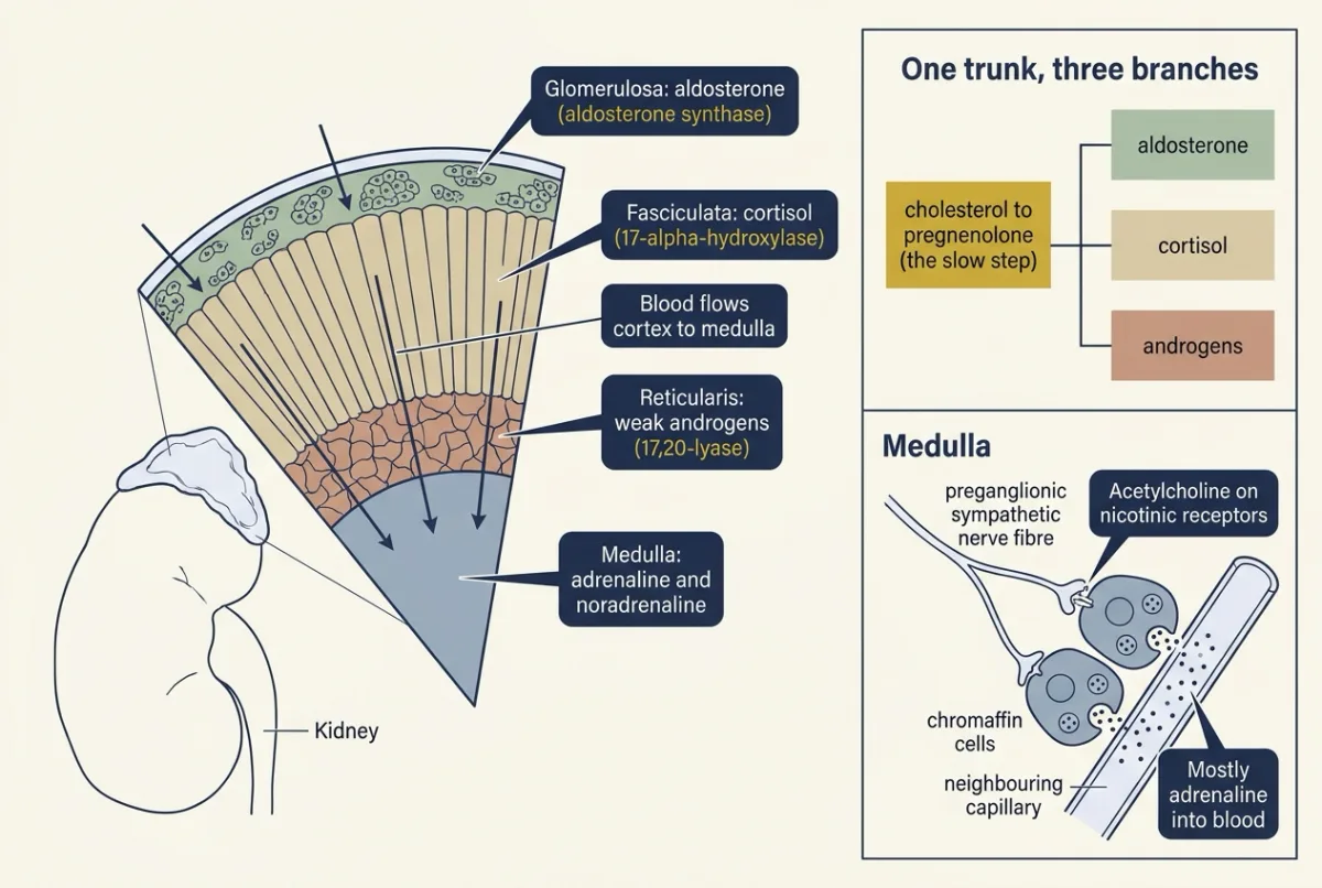
Each adrenal gland is two glands in one capsule. The outer cortex has three zones that make steroids from cholesterol: a salt hormone (aldosterone), a fuel hormone (cortisol) and androgen precursors. The inner medulla is a modified sympathetic ganglion that releases adrenaline and noradrenaline into the blood. Cortisol shifts fuel toward glucose, restrains inflammation and supports blood pressure. Aldosterone makes the kidney keep sodium and lose potassium.
The film at the top of the page covers Crabbé’s experiment and the guard enzyme, both below.
The one idea to carry through this lesson. Read any adrenal state in three steps, in this order. One: the enzymes. A missing enzyme lowers everything after it and backs up the substance just before it. Two: the receptors. Ask what the steroids now present can do at a receptor; some backed-up precursors are hormones themselves. Three: the loops. Only now let the controls respond. Adrenocorticotropic hormone (ACTH) rises when cortisol falls and drives the shared trunk of the pathway (the tree comes next), so extra flow leaves through any branch still open. Renin rises when volume falls and falls when volume expands. Aldosterone follows mainly angiotensin II and plasma potassium, not ACTH.
Scope. The rule predicts the direction of changes in a steady state, compared with a healthy person, not their size, speed or symptoms. Step three must come last; the 11-beta-hydroxylase case below shows why.
One trunk, three branches: the cortical zones
Each adrenal gland sits on the upper pole of a kidney. In the adult human cortex, which enzymes a zone has decides what it makes: step one of the rule, in a healthy gland. Picture the pathway as a tree.
- The trunk, shared by every zone. Cholesterol is cut to pregnenolone by the mitochondrial side-chain cleavage enzyme (CYP11A1). This is the slow step, and adrenocorticotropic hormone (ACTH) speeds it: within minutes by raising cholesterol delivery to the enzyme (through the StAR protein, which moves cholesterol to the inner mitochondrial membrane where the enzyme sits), and over days by inducing the pathway’s enzymes.
- Three branches leave the trunk, one per zone. A few enzymes decide which branch a cell can use. Learn the names in bold; the CYP numbers are for reference.
| Zone (outer to inner) | Main product | The enzyme that decides it |
|---|---|---|
| Glomerulosa | Aldosterone, the main mineralocorticoid (a steroid acting on sodium and potassium balance) | Has aldosterone synthase (CYP11B2), found in this zone only (in adults it is patchy, concentrated in small cell clusters under the capsule; see Going deeper). Lacks 17-alpha-hydroxylase, so it cannot make cortisol. |
| Fasciculata | Cortisol, the main glucocorticoid (named for its effect on blood glucose) | Has 17-alpha-hydroxylase (CYP17A1), 21-hydroxylase and 11-beta-hydroxylase (CYP11B1). Lacks aldosterone synthase. |
| Reticularis | DHEA and androstenedione, mostly weak androgens | Its CYP17A1 acts strongly as a 17,20-lyase, cutting off the steroid’s side chain. |
Two points carry the rest of the lesson. CYP17A1 has two jobs: adding a 17-hydroxyl group (needed for cortisol) and cutting the side chain (needed for androgens). And 21-hydroxylase lies on the routes to cortisol and aldosterone, not to the androgens.
The full routes, step by step
- Glomerulosa: pregnenolone becomes progesterone (by 3-beta-hydroxysteroid dehydrogenase, 3-beta-HSD2); 21-hydroxylase makes 11-deoxycorticosterone (DOC); aldosterone synthase converts DOC through corticosterone to aldosterone.
- Fasciculata: 17-alpha-hydroxylase gives 17-hydroxyprogesterone; 21-hydroxylase gives 11-deoxycortisol; 11-beta-hydroxylase gives cortisol.
- Reticularis: the 17,20-lyase gives DHEA, then DHEA sulfate and androstenedione.
Rats and mice differ from humans here; see Going deeper.
The medulla
The medulla’s chromaffin cells, which arise from the neural crest, release adrenaline (epinephrine) and noradrenaline (norepinephrine) into the blood. Preganglionic sympathetic fibres release acetylcholine onto nicotinic receptors of chromaffin cells, which depolarize and release catecholamines within seconds (hypothalamus and autonomic control). In adult humans most of the output, often given as about four-fifths, is adrenaline. Blood drains from cortex to medulla, and work mainly in rats indicates that this glucocorticoid-rich blood supports phenylethanolamine N-methyltransferase (PNMT), the enzyme that converts noradrenaline to adrenaline.
ACTH drives cortisol; angiotensin II and potassium drive aldosterone
The gland has two separate control systems, and keeping them apart is step three of the rule.
The ACTH loop: cortisol and the androgens
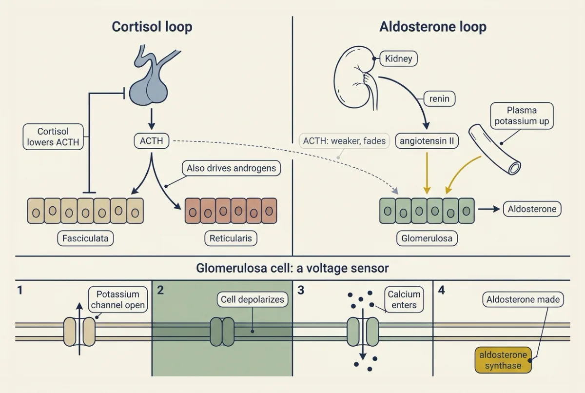
Adrenocorticotropic hormone (ACTH), from the anterior pituitary, binds the MC2 receptor (melanocortin 2 receptor, the ACTH receptor), found mainly on fasciculata and reticularis cells but also in the glomerulosa. It is a G-protein-coupled receptor that signals through Gs and cyclic AMP (cAMP and PKA). Cortisol feeds back to lower ACTH. The loop and its tests belong to endocrine axes and testing and emotion and stress.
The second loop: angiotensin II and potassium drive aldosterone
Aldosterone answers to angiotensin II, the end product of the renin cascade (blood pressure regulation), and to a rise in plasma potassium. ACTH stimulates it too, but less, and with continuous exposure that effect fades.
How does potassium in the blood reach an enzyme inside the cell? The glomerulosa cell is a voltage sensor, and the link is a chain of four steps (membrane transport).
- Potassium channels dominate the cell’s resting conductance (its open ion paths), so it rests near the potassium equilibrium potential, the voltage at which potassium has no net push in or out.
- A rise in plasma potassium moves that equilibrium potential up and depolarizes the cell. Angiotensin II, through its AT1 receptor, closes potassium channels with the same result.
- Depolarization opens voltage-gated calcium channels.
- Calcium drives aldosterone synthase.
Predict. A mutation makes one of the glomerulosa’s potassium channels leak sodium as well. Which way does aldosterone go? Then run step three: what happens to renin and plasma potassium?
Check your prediction
Sodium leaking in depolarizes the cell, as if potassium were high. Calcium channels open and aldosterone is made even when angiotensin II is low. The extra aldosterone retains sodium and raises pressure, which suppresses renin, and it increases potassium secretion, so plasma potassium tends to fall. High aldosterone with low renin, while potassium is normal or low, is a pattern the normal controls do not produce: it points to aldosterone made without regard to renin.
The modern experiment. That mutation is real. Murim Choi, Ute Scholl and colleagues in Richard Lifton’s laboratory (2011) sequenced the coding DNA (the exome) of four aldosterone-producing adenomas, benign tumors, against the same patients’ blood, then sequenced KCNJ5 in 18 more tumor and blood pairs. Recurrent mutations in KCNJ5, a potassium channel gene, appeared in 8 of 22 adenomas and were somatic: in the tumor, not the blood. Expressed with KCNJ3 in cultured 293T cells (a human kidney cell line), the mutant channels conducted sodium and depolarized the cells. Keep apart association, somatic status and function. The study does not show that all primary aldosteronism has this cause (most of these adenomas lacked it), that a cell line behaves like a glomerulosa cell, or why the cells proliferate.
Primary aldosteronism is the disease of this loop, from an adenoma or bilateral hyperplasia. Pressure rises; potassium falls in some patients, but many are normokalemic; bicarbonate may rise (acid-base balance); renin falls. So aldosterone is read against renin: the signature is aldosterone that is high for a suppressed renin (a raised aldosterone-to-renin ratio), even when the aldosterone value itself sits near the reference range.
Predict. Aldosterone retains sodium, and here it is made without limit. Why is a patient with primary aldosteronism not swollen with fluid?
Check your prediction
Sodium reabsorption rises and extracellular volume expands, but only modestly. The higher pressure raises sodium excretion (pressure natriuresis) until output matches intake again. This return to sodium balance despite continuing aldosterone excess is called mineralocorticoid escape, and it involves more than pressure (Going deeper). So edema is not typical: the rise in pressure that limits the sodium retention is itself the clinical cost.
Cortisol shifts fuel, restrains inflammation and supports pressure
Each of cortisol’s three actions has been tested by changing cortisol and watching what follows.
Fuel. In volunteers whose own cortisol was blocked and replaced by infusion, a night-time-like rise in cortisol, compared with a constant basal infusion, increased the liver’s glucose release after a meal, reduced glucose uptake in a way that appeared to reflect insulin resistance, and raised the appearance of the fatty acid palmitate, suggesting lipolysis. Insulin and glucagon changed too, so not every effect is direct. In other volunteers, cortisol increased whole-body proteolysis (protein breakdown). Those amino acids feed gluconeogenesis (integration of metabolism). Cortisol thus raises glucose supply and lowers its use by muscle and fat, opposing insulin (insulin, glucagon and glucose).
Inflammation. Cortisol acts through the glucocorticoid receptor, a ligand-activated transcription factor (nuclear receptors). In mice and cultured cells, glucocorticoids inhibited activation of NF-kappa-B, which switches on many inflammatory genes (TLRs and NF-kappa-B), and inducing its inhibitor, I-kappa-B-alpha, could be a major part of that. Later work found that this induction varies between cell types, and that the receptor also represses inflammatory genes in other ways, for example by binding NF-kappa-B and AP-1 proteins directly.
Pressure. Infusing cortisol into volunteers for five days raised systolic pressure, by a mechanism the authors left unresolved. Part of cortisol’s support of pressure is thought to be permissive: it keeps vascular smooth muscle responsive to noradrenaline and angiotensin II.
Aldosterone acts on the epithelium itself, and an enzyme guards its receptor
By the late 1950s aldosterone was known to make mammals retain sodium. Did it act on sodium-transporting cells directly? Jean Crabbé (1961) used the urinary bladder of a toad, an epithelium that moves sodium from its urine side to its blood side. He cut each bladder into paired halves, mounted each as a sheet between two chambers, and gave aldosterone to one half only. He measured the short-circuit current, the current needed to hold the voltage across the sheet at zero. With the same Ringer solution on both sides and no voltage, nothing drives ions passively, so this current equals net active ion transport when one ion carries it. If the effect required another hormone or blood flow, the isolated sheet should not respond. After about an hour, the treated half’s current rose above its partner’s, and in eight bladders sodium tracer fluxes measured in both directions confirmed that the extra current was net sodium transport.
That ruled in a direct, delayed action on the epithelium and ruled out, in this preparation, a systemic intermediary; pairing removed differences between animals. It did not show which mammalian nephron segment responds (the distal nephron and collecting duct, from later work: tubular transport and diuretics), and the delay suggested, without proving, new protein synthesis, which later toad-bladder work, including from Isidore Edelman’s laboratory, supported.
Worked problem, part 1. These numbers are invented, modelled on Crabbé’s design; he did not report them. Two paired halves each expose 1.00 cm2. Once the response has developed, the aldosterone half carries 100 microamperes and its control 40 microamperes. Assume sodium (charge +1) carries all the net current.
sodium flux (mol per s) = current (C per s) / F, with F = 96,485 C per mol
- Extra current = 100 - 40 = 60 microamperes = 60 x 10−6 C per s.
- Extra flux = (60 x 10−6 C per s) / (96,485 C per mol) = 6.22 x 10⁻¹⁰ mol per s. C per s divided by C per mol gives mol per s.
- Per hour: 6.22 x 10−10 x 3,600 s = 2.24 x 10⁻⁶ mol, so 2.24 micromol per cm2 per h.
- Check: the control alone carries 40 x 10−6 / 96,485 x 3,600 = 1.49 micromol per cm2 per h and the treated half 3.73; the difference is 2.24, a 2.5-fold rise.
If another ion carried part of the current, step 2 would overstate sodium transport; hence the tracers. (Going deeper compares his concentration with plasma levels.)
The guard enzyme: why cortisol does not take the receptor
Now the opening puzzle. In adult human plasma about 90 percent of cortisol is protein-bound, mostly to corticosteroid-binding globulin (CBG, also called transcortin). On the free hormone hypothesis, only the unbound fraction acts. Aldosterone binds CBG weakly, so a larger share of it circulates free. Counting only free hormone, the part that can reach a receptor, cortisol still exceeds aldosterone by roughly a hundredfold (Going deeper derives the range); in total hormone the excess is several times larger.
Even a hundredfold excess should win an equal-affinity receptor nearly every time. Funder, Pearce, Smith and Smith (1988) reported that mineralocorticoid receptors bind aldosterone and the physiological glucocorticoids with equal affinity, and that 11-beta-hydroxysteroid dehydrogenase (11-beta-HSD) in targets such as kidney, colon and parotid protects them by converting cortisol to inactive cortisone. Aldosterone escapes this conversion. The work was done in rats, where the enzyme converts corticosterone to 11-dehydrocorticosterone. Edwards and colleagues reached a similar conclusion that year; the epithelial type 2 isoform was characterized in the 1990s. What the receptor then does is taught in water, sodium and potassium.
The film says the receptor is then “left to aldosterone”. That is the historical conclusion, and Going deeper tests it. Liquorice in large amounts, which causes sodium retention, low potassium and high blood pressure with renin and aldosterone both low, inhibits the enzyme, so cortisol is no longer inactivated in the kidney and occupies the receptor itself.
Predict. In severe Cushing syndrome, cortisol is so high that it overwhelms the kidney’s guard enzyme. Apart from cortisol’s other effects, what happens to sodium, plasma potassium and renin?
Check your prediction
The excess cortisol gets past the enzyme and acts on the mineralocorticoid receptor itself, retaining sodium and lowering potassium. The retained sodium expands volume, which suppresses renin. This is cortisol’s second route to raising pressure.
The enzyme beside the receptor (step one) decides which steroid reaches it (step two): the lesson’s rule, applied at the target instead of in the gland. The film ends on the same point: the enzyme beside a receptor is part of what defines a hormone’s target tissue.
Reading adrenal disease in three steps
You now have what you need to predict an adrenal disease you have never seen. The reminder: enzymes first, then what reaches a receptor, then the loops.
One case all the way through: 21-hydroxylase deficiency
In congenital adrenal hyperplasia (CAH), a child is born missing one enzyme of the cortisol pathway. Take classic salt-wasting 21-hydroxylase deficiency, where enzyme activity is near zero. (In the simple-virilizing form enough enzyme remains to keep aldosterone near normal, although renin is often raised; the androgen story is the same.)
Step one, the enzymes. Cortisol and aldosterone fall, and 17-hydroxyprogesterone, the substrate just before the block, piles up; it is the screening marker.
Step two, the receptors. Nothing piling up is a mineralocorticoid: the block comes before DOC (11-deoxycorticosterone, the aldosterone precursor), so no precursor stands in for the missing aldosterone. Worse, the backed-up progesterone and 17-hydroxyprogesterone act as weak antagonists at the mineralocorticoid receptor and can add to the salt loss.
Step three, the loops. Low cortisol releases the brake on adrenocorticotropic hormone (ACTH). ACTH drives the trunk of the pathway, and the extra flow spills into the androgen branch, which does not need 21-hydroxylase. Androgens rise and virilize affected children (sexual differentiation), and long ACTH drive enlarges the gland: the “hyperplasia”. Meanwhile low aldosterone means sodium is lost, so volume and pressure fall, renin rises and potassium is retained; in the salt-wasting form this becomes a crisis.
Read that off as a row: cortisol down, ACTH up, aldosterone down, renin up, potassium up, androgens up, pressure down.
In this sketch: arrows compare each item with a healthy adult and carry no numbers; one enzyme is blocked at a time; a block lowers its product and everything after it and piles up only its own substrate; the steps add effects in order (step 1 the block itself, step 2 mineralocorticoid activity, step 3 the ACTH and renin loops, including the androgen spill), and readouts not reached yet say “not yet”; ACTH raises or lowers the boxes from pregnenolone to the androgens but not aldosterone; with 11-beta-hydroxylase blocked, ACTH also drives the DOC that piles up in the fasciculata; with 11-beta-hydroxylase blocked, aldosterone synthase in the glomerulosa still works, so corticosterone and aldosterone there follow renin; with the feedback switch off, ACTH ignores cortisol (a switch in the sketch, not a disease or lesion); supplied cortisol with feedback intact lowers ACTH; and blood pressure is shown through the mineralocorticoid route only (cortisol itself also supports pressure, which is left out). These rules are made up to sketch the lesson’s logic. They are not measurements, and cases vary.
A block lowers what comes after it and piles up what comes before it. If cortisol falls with feedback intact, ACTH rises and drives the cortisol and androgen branches harder, and mineralocorticoid activity (aldosterone plus DOC) sets the direction of renin, potassium and pressure.
Predict. 11-beta-hydroxylase makes the last step to cortisol (11-deoxycortisol to cortisol) and, in the cortisol-making zone, the step from DOC to corticosterone. It is not the enzyme the glomerulosa uses to finish aldosterone; that is aldosterone synthase, a different gene. Work the three steps for a child missing 11-beta-hydroxylase. Predict cortisol, ACTH, DOC, androgens, renin, aldosterone and blood pressure. Set the block in the simulation above to check yourself.
Check your prediction
Step one. Cortisol falls; 11-deoxycortisol and DOC, the substrates just before the block, pile up.
Step two. Here the two diseases differ. DOC is itself a mineralocorticoid. It does the job of the missing aldosterone: sodium is retained, potassium secreted, volume expands and pressure tends to rise.
Step three. Low cortisol raises ACTH, which drives more flow into the androgens. The expanded volume suppresses renin, and because aldosterone follows angiotensin II, aldosterone falls too, in a patient with too much mineralocorticoid activity. Potassium is normal or low.
If you predicted high aldosterone, you read the salt retention as aldosterone’s work. Step two says DOC is doing it, and only then do the loops follow: volume expands, renin falls and aldosterone falls with it. The renin loop measures volume and pressure, not which hormone caused them, which is also why aldosterone is always read against renin.
The rest of the table
The same three steps give the other rows.
- Primary insufficiency (Addison disease): the whole cortex is lost. Every product falls and ACTH rises; high ACTH and related peptides from its precursor (POMC) darken skin and mucosa.
- Central insufficiency: only pituitary ACTH is lost. The two inner zones go quiet, but angiotensin II and potassium still drive the glomerulosa, so neither hyperkalemia nor hyperpigmentation appears. Pressure can still fall without cortisol’s support.
- Cushing syndrome from an adrenal adenoma: cortisol is made autonomously, so ACTH is suppressed and the androgen-making zone goes quiet with it.
- Cushing syndrome from too much ACTH, from the pituitary or an ectopic tumor: androgens rise with cortisol. Cushing disease is only the pituitary form of Cushing syndrome. In either form, cortisol high enough to swamp the guard enzyme can lower renin and potassium.
Cases vary.
| Disorder | Cortisol | ACTH | Aldosterone | Renin | Plasma K+ | Adrenal androgens | Blood pressure |
|---|---|---|---|---|---|---|---|
| Primary insufficiency (Addison) | Down | Up | Down | Up | Up | Down | Down |
| Central insufficiency | Down | Low or normal | Near normal | Normal | Normal | Down | Normal or down |
| Cushing, adrenal adenoma | Up | Down | Normal | Normal or down | Normal or down | Down | Up |
| Cushing, pituitary or ectopic ACTH | Up | Up or normal | Normal or down | Normal or down | Normal or down | Up or high-normal | Up |
| Primary aldosteronism | Normal | Normal | Up (or inappropriately normal) | Down | Normal or down | Normal | Up |
| CAH, 21-hydroxylase, salt-wasting | Down | Up | Down | Up | Up | Up (androstenedione; DHEA and DHEAS can be low) | Down |
| CAH, 11-beta-hydroxylase | Down | Up | Down | Down | Normal or down | Up | Up in many |
Design problem
Mineralocorticoid receptors extracted from rat kidney bind aldosterone and corticosterone with about equal affinity, and free corticosterone in rat plasma far exceeds free aldosterone. Yet aldosterone controls sodium transport in the kidney. Design experiments to find out how the kidney’s receptors are kept for aldosterone, and say what each result would mean.
Saved in this browser only.
One way to do it
-
Confirm equal binding in vitro, with labelled steroids and receptors from targets (kidney, colon, parotid) and from hippocampus, a non-epithelial comparison. Equal affinity means selectivity is not in the receptor.
-
Ask what binds in vivo. Give labelled corticosterone to adrenalectomized rats; measure what is bound to kidney receptors and what the steroid has become. Most label as 11-dehydrocorticosterone points to local inactivation.
-
Block the enzyme (11-beta-HSD) with carbenoxolone or glycyrrhetinic acid from liquorice. Predict that corticosterone now occupies kidney receptors and, in the intact animal, that sodium is retained and potassium excreted.
-
Controls: vehicle; the inhibitor alone without glucocorticoid, to exclude a direct action on the receptor; and a glucocorticoid the enzyme cannot inactivate.
Funder and colleagues (1988) concluded that specificity is enzyme-mediated, not receptor-mediated. Limits: rat and corticosterone, the type 2 isoform still to come, and enzyme saturation by very high cortisol shown only later.
Going deeper
Rodents differ: corticosterone and a glomerulosa rhythm
Rats and mice express little adrenal 17-alpha-hydroxylase, so their main glucocorticoid is corticosterone, not cortisol, and they make little adrenal DHEA. That is why the guard-enzyme work in rats speaks of corticosterone.
In mouse adrenal slices, Hu and colleagues (2012) reported spontaneous voltage oscillations in 73 of 85 glomerulosa cells, at a mean of 0.44 Hz at 22 degrees C: an electrical rhythm in mouse cells, not a human hormone pulse rate.
Crabbé’s method, and his concentration
Crabbé used the toad Bufo marinus. Each half-bladder was mounted between two chambers of aerated frog Ringer solution at room temperature, and one half received d-aldosterone at 2.8 x 10⁻⁷ mol per L on the serosal (blood) side; its partner was the control.
Compare the concentration with plasma. 2.8 x 10⁻⁷ mol per L = 280 nmol per L. Take an illustrative adult plasma aldosterone of 15 ng per dL (made up; real ranges differ by laboratory, posture and salt intake), which is 150 ng per L. With a molar mass of 360.4 g per mol, 150 x 10−9 / 360.4 = 4.16 x 10⁻¹⁰ mol per L, or 0.416 nmol per L. The ratio 280 / 0.416 is about 670. The classic shows that the epithelium can respond, not that it responds at physiological concentrations.
How much does the guard enzyme have to do?
First, which excess. Total cortisol of 10 to 20 micrograms per dL (about 280 to 550 nmol per L) against the illustrative aldosterone of 0.416 nmol per L (above) is about 660 to 1,330-fold. If roughly 4 to 10 percent of cortisol and about 37 to 40 percent of aldosterone circulate free (most aldosterone is bound, mainly to albumin), the free ratio is roughly 70 to 360-fold. Total exceeds free by 4 to 10 times.
With equal affinity, each steroid’s share of the occupied receptors equals its share of the two free concentrations. Take round illustrative values: free cortisol 20 and free aldosterone 0.15 nmol per L.
- Unprotected, cortisol holds 20 / 20.15 of the occupied receptors, more than 99 percent.
- For aldosterone to hold half, local cortisol must fall to 0.15 nmol per L: about a 130-fold cut.
- For aldosterone to hold 90 percent, local cortisol must fall to about 0.017 nmol per L: a cut of about 1,200-fold.
That is a lot to ask of one enzyme. Later work proposes, and it is still debated, that in epithelia a receptor occupied by cortisol is also activated much less effectively than one occupied by aldosterone, partly through the NAD+-consuming chemistry of the type 2 enzyme itself. So concentration alone does not quite deliver “left to aldosterone”.
Where the androgens in CAH come from
Human CYP17A1 cuts the side chain of 17-hydroxyprogesterone inefficiently, so in a healthy adrenal most androgen comes through 17-hydroxypregnenolone and DHEA. In 21-hydroxylase deficiency, though, 17-hydroxyprogesterone accumulates so massively that its direct conversion to androstenedione becomes substantial, which helps explain why androstenedione, not DHEA, is the androgen that tracks control. A 5-alpha-reduced “backdoor” route contributes in fetal life and infancy. DHEA and androstenedione are weak at the androgen receptor, but the adrenal also makes 11-beta-hydroxyandrostenedione, which is converted outside the gland to 11-ketotestosterone, a full agonist. These 11-oxygenated steroids carry much of the androgen excess measured in congenital adrenal hyperplasia (CAH).
Two CAH numbers: pressure and the ACTH drive
Raised pressure in 11-beta-hydroxylase deficiency is common but not universal: in one international cohort, 38 of 70 patients who had blood-pressure data were hypertensive (Khattab and colleagues, 2017).
The excess adrenocorticotropic hormone (ACTH) drive in 21-hydroxylase deficiency has been tested from above with crinecerfont, an antagonist at the CRF1 receptor for corticotropin-releasing hormone (CRH) (the hypothalamic hormone that releases ACTH). In 182 randomized adults with classic 21-hydroxylase deficiency, it allowed a 27.3 percent least-squares mean glucocorticoid dose reduction at 24 weeks with androstenedione controlled, against 10.3 percent with placebo (Auchus and colleagues, 2024), consistent with excess CRH-ACTH drive. Replacement was still required.
Primary aldosteronism: escape and how common it is
Mineralocorticoid escape is related to pressure natriuresis but is not just another name for it; it also involves a fall in the thiazide-sensitive NaCl cotransporter of the distal convoluted tubule and a rise in natriuretic peptides, while epithelial sodium channel (ENaC) stays up.
In a recruited cross-sectional cohort, Brown and colleagues (2020) found renin-independent aldosterone production forming a continuum across blood-pressure categories, and a 2025 Endocrine Society guideline suggests screening everyone with hypertension. Where that production comes from, including small aldosterone-producing cell clusters under the capsule that also express aldosterone synthase, remains under study.
The cortisol studies
- Fuel. Dinneen and colleagues (1993) blocked cortisol synthesis in normal volunteers with metyrapone, an 11-beta-hydroxylase inhibitor, and replaced cortisol by infusion either at a constant basal rate or on a schedule mimicking the night-time rise. The effects in the main text are those of the variable schedule after a mixed meal. Simmons and colleagues (1984) found the rise in proteolysis with an eight-hour hydrocortisone infusion in seven normal subjects.
- Inflammation. The NF-kappa-B findings are from Auphan and colleagues (1995); Scheinman and colleagues reported a similar mechanism the same year.
- Pressure. Whitworth and colleagues (1984) infused cortisol into seven subjects for five days at rates meant to resemble ACTH stimulation, and systolic pressure rose from 108 to 129 mmHg.
- Production. By stable-isotope dilution, Esteban and colleagues (1991) measured a mean cortisol production of 5.7 mg per square metre of body surface per day in 12 normal volunteers, lower than earlier estimates by the authors’ account.
Your notes
Select any text in the lesson to highlight it or add a note to it. Your highlights and notes are saved in this browser and collected in your notebook.
This browser is not letting the site save anything (a private window or strict privacy settings), so highlights and notes will be lost when you leave the page.
How we measure it
Short-circuit current with tracer fluxes (Ussing-type chamber)
Mount an epithelial sheet between two chambers and pass whatever current holds the voltage across it at zero. That current equals net active ion transport if one ion carries it, and radioactive tracer fluxes measured in both directions check which ion that is.
Matched tumor and blood sequencing
Sequence the coding DNA of a tumor and of the same patient's blood. A variant found in the tumor but not the blood is somatic, acquired in the tumor's cell lineage, rather than inherited.
Stable-isotope dilution with mass spectrometry
Infuse a hormone labelled with a heavy isotope at a known rate and measure how far the body's own unlabelled hormone dilutes it. At steady state the dilution gives the body's production rate.
Block-and-replace infusion with metabolic tracers
Block the body's own cortisol synthesis with an enzyme inhibitor such as metyrapone, replace cortisol by infusion on a chosen schedule, and follow glucose, amino acid or fatty acid tracers to measure production and uptake.
Practice questions on this lesson (4) → MCAT-style, original, each with an explanation
Where this lesson fits
Before thisReview first
- Endocrine Axes and How to Test ThemThis course · Lesson 2
You need the ACTH-cortisol feedback loop and how it is tested there to read the adrenal disease table here.
- Regulating Arterial PressureThis course · Lesson 10
The renin-angiotensin cascade there is the loop that drives aldosterone and is read against renin here.
- Water, Sodium and Potassium BalanceThis course · Lesson 17
Aldosterone's action on sodium and potassium in the kidney is used here without being re-taught.
After thisStudy next
- Insulin, Glucagon and Blood GlucoseThis course · Lesson 21
Cortisol's opposition to insulin and its raising of glucose supply connects directly to how blood glucose is controlled.
- Sexual Differentiation and the TestisThis course · Lesson 27
Androgen excess from the adrenal in CAH virilizes affected children, which this lesson builds on.
- Exercise: The Whole Body at WorkThis course · Lesson 30
Adrenaline and cortisol release during exercise uses the adrenal medulla and cortex outputs learned here.
Elsewhere on the siteFilms, simulations and method films in other courses
- Half-life and plasma levelSimulation · Organismal Biology
This simulation shows how a hormone's half-life and plasma level behave, as with cortisol bound to its carrier.
- RadioimmunoassayMethod film
Measuring cortisol, ACTH and aldosterone levels to read disease relies on competitive hormone assays.
- The Receptor Is the Gene SwitchFilm · Cell Signaling
Cortisol and aldosterone act through receptors that are themselves gene switches.
Suggested by an AI model from the lessons themselves, then checked. How we use AI
Finish line
Test yourself
9 questions
Practice and MCAT-style, with explained answers.
Next · Lesson 21
Insulin, Glucagon and Blood Glucose
A 75 g glucose drink is six to seven and a half times all the glucose in your blood and tissue fluid. Why does blood glucose rise so little?
Continue →← Lesson 19 · Thyroid Hormone: Tissues Decide What T4 Becomes