Cell Signaling · Unit 3: G-protein-coupled receptors and second messengers
cAMP and Protein Kinase A
Rewritten on : new text, flashcards and film. Your notes and highlights stay, but the text around them may have moved.
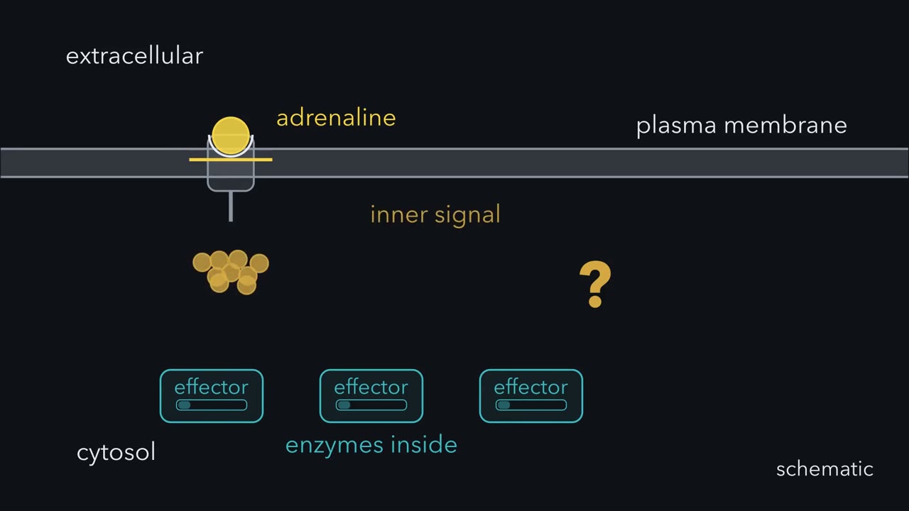
Adrenaline stays outside a liver cell, so how can enzymes inside respond?
Pause when the reading reaches a film, and offer to play it. Saved on this device.
Watch the film now, or keep listening
Builds on:GPCRs and Trimeric G ProteinsPhosphorylation: Kinases and PhosphatasesAdaptors, Docking Proteins and ScaffoldsWhy these?
 ▶ Watch The Messenger Level: Made and Erased · 3:37
Read the transcript of “The Messenger Level: Made and Erased”
Film transcript: The Messenger Level: Made and Erased
Adrenaline stays outside a liver cell. How can enzymes inside respond? Watch the outer signal stop at the membrane while an inner signal begins moving. The answer must connect both spaces without carrying adrenaline across.
The receptor changes shape and activates Gs on the inner face. Gs reaches adenylyl cyclase, in the membrane. The enzyme turns cytosolic ATP into cAMP. The membrane holds the first step beside the inner messenger is produced. cAMP is the second messenger here. Its level changes inside after the first messenger binds outside. Many copies can carry one receptor input toward intracellular effectors. Unlike adrenaline, these copies never need to cross the membrane from outside. The membrane step keeps making messenger while the receptor pathway remains active. Every gold token carries the same chemical information, so quantity becomes adjustable. More tokens raise the level.
Their location and lifetime also matter. A source would let cAMP keep accumulating. Phosphodiesterase provides the opposing process. It opens cAMP's ring and leaves inactive 5′-AMP, erasing the message. That product cannot switch on the kinase that reads cAMP. When removal is proportional to the amount present, it strengthens as cAMP accumulates. A higher level makes encounters with the red enzyme more frequent. The level settles when synthesis and breakdown match. This curve is computed from that first-order model. Now keep the blue source unchanged and strengthen the red removal process. Nothing else in the cell changes during this comparison.
Where will the cAMP level settle: above its old plateau, or below it? Below. At every cAMP level, stronger phosphodiesterase removes more. A lower concentration is enough for removal to match the unchanged source, so the computed plateau falls. The balance returns, but at a different level. Gs raises synthesis, while Gi lowers some forms of cyclase.
Phosphodiesterases set removal. These controls change the messenger before any downstream target is chosen. When adrenaline leaves, the receptor stops activating new Gs. Active Gs lingers until it splits its GTP, so cyclase drive fades. Synthesis falls back to its resting rate.
Phosphodiesterase then returns cAMP toward its resting level. Faster removal makes that return briefer. Phosphodiesterase then returns cyclic amp toward its resting level. The message therefore has an adjustable height and lifetime. Its amount is not the response itself. Several possible effectors wait in the same cytosol. The next question is how a cAMP reader turns that level into a specific action.
From the film
- cAMP levels reflect the balance of synthesis and breakdown.
- Adenylyl cyclase makes cAMP from ATP, while phosphodiesterase converts it to 5′-AMP.
- Gs stimulates adenylyl cyclase, while Gi inhibits some forms of the enzyme.
- When breakdown is first order, the level settles where synthesis and breakdown match; faster breakdown gives a lower plateau and a briefer signal.
Adrenaline binds outside a liver cell, yet enzymes in the cytosol change their activity. The first film showed the relay through cyclic AMP (cAMP); here is the experiment that proved it, then its main reader, protein kinase A (PKA).
Finding the messenger in broken cells
Earl Sutherland later recalled “a widespread feeling among biologists that hormone action could not be studied meaningfully in the absence of organized cell structure.” His laboratory tested it on liver, where adrenaline switches on phosphorylase to start glycogen breakdown.
The researchers homogenized liver and centrifuged it. A pellet held particles later traced to membrane fragments; the clear supernatant held phosphorylase. Every tube also held ATP and magnesium, which both steps needed.
Predict. Hormone added to the supernatant alone left phosphorylase off. Which of these should switch it on: hormone with supernatant plus a little pellet mixed back in, or liquid from a tube of hormone and pellet, added to supernatant?
Check your prediction
Both did. So the hormone acts on the pellet, which hands something to the soluble fraction, where the phosphorylase is. That liquid still worked after boiling and passed through a dialysis membrane, so the transferred factor was small and not an enzyme. Nothing in the tubes was alive: the relay was chemistry.
The paper, by Theodore Rall, Sutherland and Jacques Berthet, came out in 1957. The factor was an adenine nucleotide with one phosphate bonded to the ribose at both the 3′ and 5′ carbons, closing a ring: adenosine 3′,5′-monophosphate, or cyclic AMP. David Lipkin’s chemistry group had isolated the same compound from ATP treated with barium hydroxide, without hormone, and later established its structure.
Sutherland called the hormone the first messenger and cAMP the second messenger. Here, a second messenger is a small intracellular molecule or ion whose level changes after an extracellular first messenger binds, and that carries the signal to intracellular effectors. cAMP is made for the purpose; calcium, another second messenger, is instead released from internal stores.
How much cAMP accumulates?
The first film showed a source filling the pool and a sink draining it; here are the reaction details and the arithmetic behind that picture.
The source is adenylyl cyclase. Its hormone-sensitive forms, in the plasma membrane, convert ATP to cAMP and release pyrophosphate. Whether Gi lowers its activity depends on which cyclase form the cell makes. The prerequisite lesson on GTPase switches develops the G-protein cycle.
The sink is phosphodiesterase, which hydrolyses the ring’s 3′ bond to leave 5′-AMP.
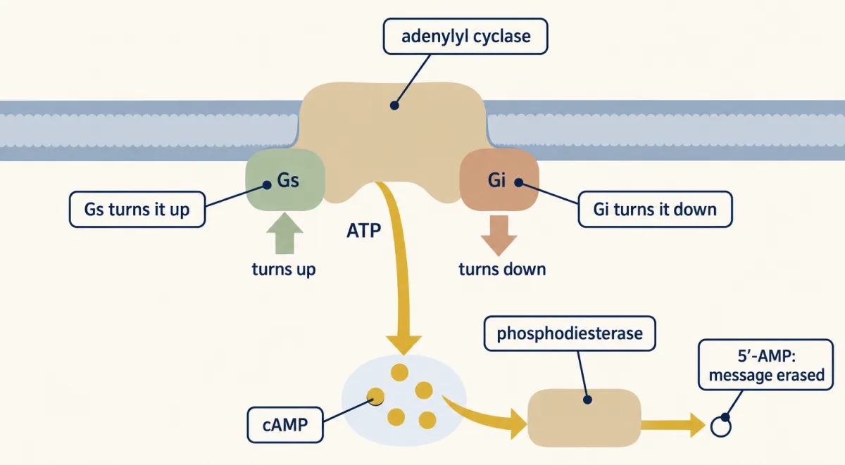
The film’s curve assumed first-order removal, proportional to cAMP concentration (the box above gives its scope). At steady state:
steady-state [cAMP] = synthesis rate ÷ breakdown rate constant
For example, 2 micromolar per second ÷ 4 per second = 0.5 micromolar.
Predict. If both the synthesis rate and the first-order breakdown constant double, what happens to the steady cAMP level?
Check your prediction
Unchanged: the same factor appears above and below the division. Turnover is faster, so the level responds more quickly when either rate changes.
Predict. If cAMP rises high enough to saturate phosphodiesterase, can you still use the simple division rule unchanged?
Check your prediction
No. A saturated phosphodiesterase removes cAMP at a rate no longer proportional to its concentration, so no single breakdown constant describes it.
Raising synthesis and lowering breakdown multiply their effects; the lesson on turning signals off takes up signal duration. Theophylline inhibits phosphodiesterase and strengthens cAMP-linked hormone responses, one of Sutherland’s tests for a cAMP messenger. Caffeine, a close relative, inhibits phosphodiesterase in vitro, although at concentrations reached by drinking coffee it is thought to act mainly at adenosine receptors.
The cAMP level is only half the explanation; the next question is what its kinase can reach.
Read the transcript of “PKA: The Target Sets the Response”
Film transcript: PKA: The Target Sets the Response
Synthesis and breakdown set the cAMP level. Once that messenger activates PKA, why do heart and fat cells respond differently? The messenger is common, but the reachable machinery need not be. That is the puzzle. At low cAMP, PKA rests as paired regulatory and catalytic subunits. Each catalytic subunit is a complete kinase. Each regulatory partner keeps one catalytic site inactive. The regulatory pair acts as an off switch until messenger arrives. A kinase modifies a target protein by transferring phosphate from ATP. The target must enter a groove. A regulatory segment mimics a target and occupies that groove first. Blocking the groove prevents genuine targets from receiving phosphate.
cAMP binds the regulatory subunits and changes their shape. Their blocking segments release the grooves. In the classical model, catalytic subunits come away free. Evidence suggests anchored complexes can act nearby without fully coming apart. Either picture exposes catalytic activity to targets within reach.
Now place the same active PKA across both cells. Both cells receive the same messenger level and display the same kinase. One cell offers green target strips, while the other offers different strips. Must their responses match?
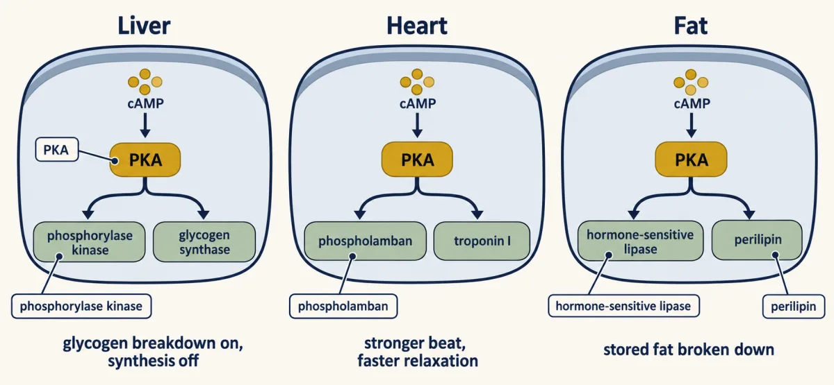
No. An active PKA can modify only compatible targets it reaches. Different cells make different target proteins, so identical messenger and kinase activity can produce different cellular actions. The response follows the proteins present, not a special kind of cAMP. In fat, reachable targets include hormone-sensitive lipase and perilipin, supporting fat breakdown. In heart, other targets alter calcium entry, cycling, contraction, and relaxation. Liver offers another set, including enzymes that control glycogen. The relay is shared; the target set differs. Reach also matters within one cell. An anchoring protein can hold PKA beside a selected substrate.
The address narrows which target can meet the active kinase. Nearby cyclase supplies cAMP, while nearby phosphodiesterase removes it. Experiments reveal local cAMP regions and receptor-specific PKA activity. Local production, local removal, binding, and restricted movement may all contribute. Their importance varies and remains debated. The local patterns are observed; the mechanism is not fully settled. Activation therefore opens the catalytic groove, but access gives the signal meaning. The text now tests that groove with competition and examines the evidence for local signalling. Those experiments separate what is established from what is proposed.
From the film
- PKA responses depend on which targets are nearby.
- Regulatory subunits block PKA catalytic sites until cAMP relieves the inhibition.
- Different cells offer PKA different target proteins, so the same pathway can produce different responses.
- Within one cell, anchoring and local cAMP dynamics help direct PKA toward selected targets.
How cAMP relieves PKA inhibition
The second film showed each of PKA’s two regulatory subunits blocking the groove of one of its two catalytic subunits (each a complete kinase) with a target-mimicking segment. Here is why that segment fits, at the level of sequence.
Many preferred protein kinase A (PKA) targets place a serine or threonine just after basic residues, often a pair of arginines (Arg-Arg-X-Ser/Thr). The regulatory subunit’s blocking stretch copies that pattern. In type I regulatory subunits the phosphate-receiving position holds a residue that cannot accept one: a pseudosubstrate, which fits but cannot be phosphorylated.
Predict. A free target-like peptide, with alanine in place of the serine, is added to purified catalytic subunit. If the experiment supplies far more real substrate, should inhibition become easier or harder to detect?
Check your prediction
Harder. Real substrate competes for the same groove, so enough substrate can displace a free pseudosubstrate peptide. In the whole complex the regulatory subunit grips the catalytic subunit over a much larger surface, so extra substrate does not release it; cAMP does.
Four cAMP, two on each regulatory subunit, relieve the hold. In the classical model, the catalytic subunits come away free. There is evidence that anchored PKA can act on nearby targets without fully coming apart, so take release as the textbook picture, which may not hold everywhere.
Different cells offer different targets
Here is what the film’s targets do:
- Liver: Protein kinase A (PKA) phosphorylates phosphorylase kinase, which activates glycogen phosphorylase (the overview lesson on signal transduction walks through that cascade), and phosphorylates glycogen synthase, inhibiting glycogen synthesis.
- Heart: PKA phosphorylates phospholamban, releasing a brake on the pump that returns calcium to its internal store, and troponin I on contractile filaments. PKA activity also increases calcium entry through L-type calcium channels, producing a stronger beat and faster relaxation.
- Fat: perilipin, the film’s second fat target, is the protein coating the fat droplet.
Catalytic subunits reaching the nucleus can phosphorylate cAMP response element-binding protein (CREB) and change gene expression over a longer time.
Different addresses within one cell
In heart muscle, Hayes, Brunton and colleagues reported that isoproterenol acting at β-adrenergic receptors and prostaglandin E1 both raised cAMP, yet only isoproterenol activated the particulate pool of protein kinase A (PKA) that pellets with cell structures. In 1983 Buxton and Brunton named the result: compartments of cAMP and protein kinase. About twenty years later, Zaccolo and Pozzan imaged local regions of high cAMP in stimulated heart cells from newborn rats.
Predict. If a phosphodiesterase helps confine one local cAMP signal, what should happen when that enzyme is inhibited?
Check your prediction
The signal should spread farther or last longer, so separate responses may begin to mix. That would support a role for local breakdown without proving it is the only barrier.
The film’s local clouds show cAMP compartmentation: cAMP concentrations and effects are not uniform across a cell. Its anchoring proteins are A-kinase anchoring proteins (AKAPs), from the lesson on adaptors and scaffolds; they often hold a phosphodiesterase beside PKA too.
Which candidate contributes most may depend on the preparation. Agarwal, Clancy and Harvey found support for buffering by PKA. Richards and colleagues found similarly slow movement in adult ventricular myocytes but attributed it to tortuosity, the winding paths around cellular obstacles, because saturating binding sites did not speed cAMP. Other studies argue against phosphodiesterase alone explaining gradients. The compartments are observed; their mechanism is not settled.
Design problem
A heart cell and a liver cell both raise cAMP when adrenaline arrives. In liver, glucagon is the main hormone using this route, and adrenaline also acts through calcium. Design how heart contracts harder while liver releases glucose, then explain how one cell could keep two cAMP signals apart.
Saved in this browser only.
One way to do it
Keep receptor, Gs, adenylyl cyclase, cAMP and PKA as the shared relay. In heart, place phospholamban, troponin I and L-type calcium channels with their brake protein Rad within PKA’s reach; PKA phosphorylation of Rad lets more calcium enter. In liver, supply phosphorylase kinase and glycogen phosphorylase, plus glucose 6-phosphatase so glucose can leave.
Within one cell, put each receptor and cyclase near an AKAP carrying PKA, its substrate and a phosphodiesterase, with cAMP-binding proteins or obstacles slowing spread; several mechanisms may contribute.
Going deeper
How the kinase structure established the blocked groove
Walsh, Perkins and Krebs described cAMP-dependent protein kinase in 1968. In 1991 Susan Taylor’s laboratory and Janusz Sowadski’s crystallography group produced the first three-dimensional structure of a protein kinase, PKA’s catalytic subunit. It has two lobes: ATP lies under the small lobe, substrate follows the large lobe, and phosphate transfer occurs in the cleft. Their crystal held a 20-residue pseudosubstrate from a separate inhibitor protein; later structures placed a regulatory subunit’s inhibitor sequence in the same groove. The human genome encodes about 500 protein kinases whose catalytic cores vary around this fold.
Other cAMP readers
Protein kinase A (PKA) is not the only direct reader. Epac is a cAMP-activated exchange factor for the small GTPase Rap. Cyclic-nucleotide-gated channels, including those in olfactory neurons, open when cAMP binds. These effectors sit outside the guiding rule’s PKA-response clause.
Your notes
Select any text in the lesson to highlight it or add a note to it. Your highlights and notes are saved in this browser and collected in your notebook.
This browser is not letting the site save anything (a private window or strict privacy settings), so highlights and notes will be lost when you leave the page.
How we measure it
Cell fractionation by centrifugation
Break cells open, spin the homogenate, and separate the pellet of membrane fragments from the clear supernatant of soluble proteins. Testing each fraction alone and then in combination tells you which part of the cell carries out which step of a response.
Heat-stability test
Heat a sample before asking whether it still works. Most enzymes lose their fold and their activity, while a small molecule usually survives. Sutherland's factor survived, so it was not an enzyme carried over from the membranes.
X-ray crystallography of a kinase bound to an inhibitor
Crystallize the kinase with an inhibitor peptide bound, so that every molecule holds the same shape, then work out the positions of the atoms from the diffraction pattern. The structure shows where the substrate sits and how an inhibitor blocks it; crystals that also hold ATP show where ATP binds.
Fluorescence imaging of cAMP in living cells
Express a sensor protein carrying two fluorescent tags whose energy transfer changes when cAMP binds. The ratio of the two colours, read point by point under a microscope, maps where cAMP is high and where it is low while the cell responds.
Practice questions on this lesson (3) → MCAT-style, original, each with an explanation
Where this lesson fits
Before thisReview first
- GPCRs and Trimeric G ProteinsThis course · Lesson 12
You need to know how a receptor activates Gs and Gi, which set the cyclase rate here.
- Phosphorylation: Kinases and PhosphatasesThis course · Lesson 6
PKA is a kinase, so you need how kinases and phosphatases switch proteins on and off.
- Adaptors, Docking Proteins and ScaffoldsThis course · Lesson 10
The AKAP anchoring that keeps PKA beside its substrate builds on how scaffolds hold signaling proteins.
After thisStudy next
- Turning the Signal Off: Arrestins, RGS Proteins and PhosphodiesterasesThis course · Lesson 16
It takes the phosphodiesterase drain further, showing how cAMP and the receptor are shut off.
- Signaling to the Nucleus: CREB, AP-1 and Immediate Early GenesThis course · Lesson 32
It follows PKA's catalytic subunits into the nucleus to phosphorylate CREB and switch on genes.
- The Shape of a Signal: Size, Duration, Frequency and PlaceThis course · Lesson 35
It develops how duration and place of a cAMP signal give different answers in one cell.
Suggested by an AI model from the lessons themselves, then checked. How we use AI
Finish line
Test yourself
8 questions
Practice and MCAT-style, with explained answers.
Next · Lesson 14
PLC, IP3, DAG and Protein Kinase C
How does a kinase know that a receptor has just cut a lipid, here and now?
Continue →