Physiology · Unit 5: Kidney, fluids and acid-base
Glomerular Filtration and Renal Blood Flow
Rewritten on : new text, figures and flashcards. Your notes and highlights stay, but the text around them may have moved.
Squeeze the arteriole that drains the glomerulus and filtration can rise or fall. Which, and why?
Pause when the reading reaches a film, and offer to play it. Saved on this device.
Watch the film now, or keep listening
Builds on:Capillary Exchange, Lymph and EdemaResistance, Compliance and Local Blood FlowSmooth Muscle and the Myogenic ResponseWhy these?
At rest, an adult human’s two kidneys, well under 1% of body mass, receive about a quarter of the blood the heart pumps, and they extract little of its oxygen: blood leaves them still rich in it.
That is a clue: the flow far exceeds what feeding kidney cells would need. It supplies a filter. In an adult at rest the glomeruli filter about 125 mL of plasma a minute, about 180 L a day, and the tubules return all but 1 to 2 L of it. The kidney traced the nephron and introduced clearance, C = U × V / P. This lesson puts numbers on the filter’s pressures, flows and controls.
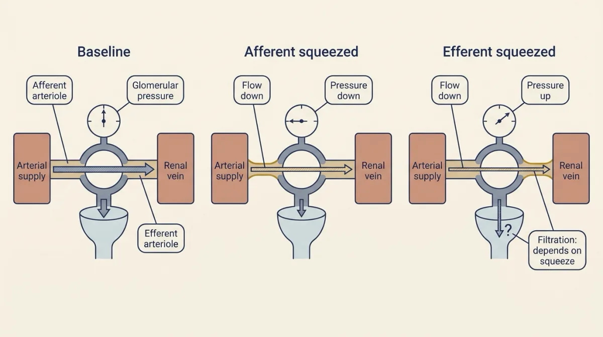
The puzzle: squeeze the efferent arteriole, the vessel that drains the glomerulus, and filtration can rise or fall, depending on how hard you squeeze. One idea explains why.
The one idea to carry through this lesson. Plasma filters wherever the hydrostatic push out of the glomerular capillary (its pressure less the back-pressure in Bowman’s space) still beats the pull of the protein left behind. So glomerular filtration rate (GFR) depends on two things: how high that pressure is, and how much plasma flows through, which slows the concentrating of the protein. The two arterioles set both. Their total resistance sets the flow. The share of that resistance in the efferent arteriole, downstream of the glomerulus, sets the pressure. So the afferent arteriole moves pressure and flow the same way, and the efferent arteriole moves them opposite ways.
The rule assumes a steady state, with arterial and renal venous pressure, the filter itself (Kf, defined below), the pressure in Bowman’s space and plasma protein held constant.
What passes and what stays
Each glomerulus is a tuft of capillaries cupped in Bowman’s capsule. Plasma is pushed into Bowman’s space, the start of the tubule, across a capillary wall of three layers.
- Fenestrated endothelium: a capillary lining perforated by pores, coated with a negatively charged glycocalyx.
- The basement membrane, between the endothelium and the podocytes.
- Podocytes: cells that wrap the capillary in foot processes, with the gaps between them bridged by the slit diaphragm.
Water and small solutes pass freely. Plasma protein mostly stays behind. Human genetics implicates the slit diaphragm: NPHS1 mutations (nephrin) cause congenital nephrotic syndrome of the Finnish type, and NPHS2 mutations (podocin) segregate with steroid-resistant nephrotic syndrome, both with heavy protein loss in urine. Size selectivity comes mostly from the cell layers; how the work of holding protein back is shared with the basement membrane and the glycocalyx is still debated. Basement-membrane charge was long taught as the albumin barrier, but mice with reduced membrane charge did not develop overt proteinuria. Whether charge elsewhere in the barrier, such as the glycocalyx, helps hold back albumin is part of that debate.
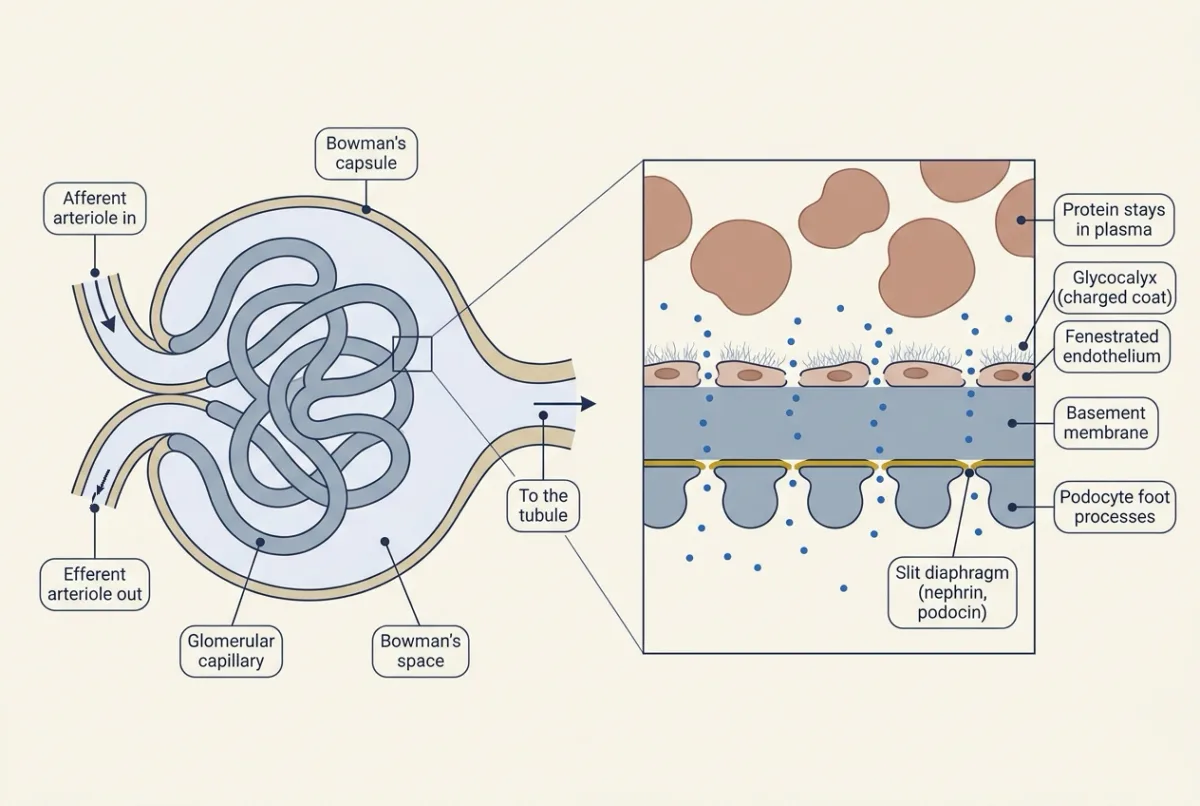
Filtrate leaves its protein behind, so the plasma flowing on carries the same protein in less water: the protein grows more concentrated along the capillary.
Push against pull along one capillary
Unlike most systemic capillaries (capillary exchange and lymph), the glomerular capillary sits between two resistances, the afferent and efferent arterioles, so its hydrostatic pressure barely falls along its length.
Picture two lines along the capillary. The push is the capillary’s hydrostatic pressure, PGC, less a small back-pressure in Bowman’s space, PBS: a nearly flat line. The pull is the oncotic pressure of plasma protein, πGC, its tendency to hold water in the capillary: a line that starts lower and rises as the protein is concentrated. Fluid filters wherever the push beats the pull. The gap between the lines is the net filtration pressure, and it shrinks along the capillary. A fourth force, oncotic pressure in Bowman’s space (πBS), is normally near zero, because almost no protein gets through. These are the four Starling forces: two favour filtration (PGC and πBS) and two oppose it (PBS and πGC).
In words Single-nephron filtration equals the filtration coefficient times the net filtration pressure, averaged along the capillary.
- SNGFR“S N G F R”
- single-nephron GFRnl/min
- Kf“K f”
- ultrafiltration coefficient: filtration per unit net pressure (water permeability times filtering area)nl/min per mmHg
- PGC“P sub G C”
- capillary hydrostatic pressure (push)mmHg
- PBS“P sub B S”
- Bowman's-space hydrostatic pressuremmHg
- πGC“pi sub G C”
- capillary oncotic pressure (pull)mmHg
- πBS“pi sub B S”
- Bowman's-space oncotic pressure, near zerommHg
Sanity check If the pull matches the push all along the capillary, nothing filters, however large Kf is. The equation treats the wall as almost impermeable to protein.
Try one number
Illustrative, human-like numbers: PGC = 55 mmHg throughout, PBS = 15, πGC entering = 25, πBS = 0.
Afferent end: 55 − 15 − 25 = +15 mmHg.
If 18% of the plasma filters out, the remaining 82% holds the same protein, so protein is concentrated 1 / (1 − 0.18) = 1.22-fold by the efferent end.
If oncotic pressure were proportional to concentration, πGC there would be 30.5 mmHg and net pressure +9.5 mmHg. Real oncotic pressure rises faster than linearly, so this underestimates efferent πGC.
Need a refresher? subscripts and Greek letters · rates and “per”
If enough water leaves, the pull can rise to meet the push before the efferent end. Beyond that point nothing filters. This is filtration pressure equilibrium.
Predict. A capillary reaches equilibrium partway along. Now plasma flows through it faster, with the same pressures. Does the protein reach its stopping concentration sooner or later along the capillary, and what happens to filtration?
Check your prediction
Later. Each millilitre of filtrate is drawn from more plasma, so the protein concentrates more slowly. More of the capillary keeps filtering, so SNGFR rises: with equilibrium, SNGFR depends strongly on plasma flow. Without equilibrium flow still matters, only less.
The measured numbers come from Munich-Wistar rats, which have some glomeruli at the kidney surface: Brenner, Troy and Daugharty (1971) measured PGC at about 44 mmHg, roughly half of mean arterial pressure and lower than the illustrative 55 above (learn the directions, not either number), with net filtration pressure averaging about 10 mmHg. In hydropenic rats (mildly water-deprived, so not volume-expanded), oncotic pressure typically rose to equal the hydrostatic difference before the efferent end: equilibrium. Human PGC is inferred rather than measured this way, and whether human glomeruli reach equilibrium is unknown.
Two taps on one hose: the afferent and efferent arterioles
Now the puzzle. Blood enters each glomerulus through the afferent arteriole and leaves through the efferent arteriole. Smooth muscle in both can narrow them (constriction) or widen them (dilation).
Picture a garden hose with a tap near the spigot, a nozzle at the far end and a leaky stretch, the glomerulus, in between. Close the tap a little and less water reaches the leaky stretch, at lower pressure, so less leaks out. Close the nozzle a little and pressure builds in the leaky stretch, so it leaks harder, but less water flows through. The hose has no protein to concentrate, so it misses the part that makes a hard squeeze lower glomerular filtration rate (GFR); that comes next.
The leaky stretch has a pressure, PGC, and a flow, the renal plasma flow (RPF). As in any two resistors in series (resistance, compliance and local flow), the total resistance sets the flow and the efferent share sets the pressure left in the glomerulus.
In words Flow is the pressure drop across the kidney divided by the total resistance. The pressure left in the glomerulus is venous pressure plus the part of the drop that still lies ahead, in the efferent arteriole.
- RBF“R B F”
- renal blood flowmL/min
- Pa“P sub a”
- arterial pressure entering the kidneymmHg
- Pv“P sub v”
- renal venous pressuremmHg
- RA“R sub A”
- afferent (upstream) resistancemmHg per (mL/min)
- RE“R sub E”
- efferent (downstream) resistancemmHg per (mL/min)
- PGC“P sub G C”
- glomerular capillary pressuremmHg
Sanity check If RE were zero, PGC would fall to venous pressure. If RA were zero, PGC would equal arterial pressure. Equal resistances put PGC halfway between the two.
Try one number
Round numbers: Pa = 100 mmHg, Pv = 10 mmHg, and each arteriole has 1 unit of resistance.
Flow: (100 − 10) / (1 + 1) = 45 units.
Pressure: 10 + 90 × 1/2 = 55 mmHg in the glomerulus.
Where it comes from optional
The same blood passes through both arterioles, so flow = total drop / total resistance. The drop across the efferent arteriole is flow × RE, and PGC sits that far above venous pressure. Equivalently, PGC is arterial pressure minus the drop across the afferent arteriole.
The ≈ is there because about a tenth of the blood flow leaves as filtrate before the efferent arteriole, and the capillaries beyond it add resistance. Treat the numbers as round.
Need a refresher? subscripts and Greek letters · rates and “per”
Predict. Start from the example: 1 unit each, flow 45, PGC 55 mmHg. Double the efferent resistance to 2 units. Which way do flow and PGC move? Then put it back and double the afferent resistance instead.
Check your prediction
Efferent doubled: total resistance 3, so flow = 90 / 3 = 30, down by a third. The efferent share is now 2/3, so PGC = 10 + 90 × 2/3 = 70 mmHg, up 15.
Afferent doubled: total resistance 3 again, so flow is again 30. The efferent share is now 1/3, so PGC = 10 + 90 × 1/3 = 40 mmHg, down 15. With the push-and-pull numbers above (PBS 15, entering π 25), 40 mmHg leaves no net pressure at all: a hard afferent squeeze can stop filtration, which shows how steeply GFR depends on PGC.
Same loss of flow, opposite change in pressure.
From pressure and flow to GFR
GFR rises with both PGC and plasma flow. The filtration fraction (FF) is the share of plasma flow that is filtered, GFR / RPF.
- Afferent constriction lowers pressure and flow together: two losses, so GFR falls. The textbook shortcut treats GFR and RPF as falling in proportion, leaving FF unchanged. But protein concentrates as water leaves, so with a lower PGC the pull catches up sooner and FF tends to fall; how much depends on Kf and on how close the capillary runs to equilibrium. Afferent dilation does the reverse.
- Efferent constriction sets the two effects against each other. Higher PGC pushes more fluid out. Lower flow concentrates the protein sooner, so the pull catches up earlier. With a mild squeeze the pressure gain wins and GFR rises slightly. With a hard squeeze the pressure gain levels off, because PGC can never exceed arterial pressure, while flow keeps falling, so GFR falls. Throughout, a larger share of the plasma is filtered, so FF rises. That answers the puzzle: it depends on how hard you squeeze.
- Efferent dilation raises flow and lowers PGC. From the usual resting state the pressure loss wins, so GFR and FF fall.
| Change | Plasma flow | PGC | GFR | Filtration fraction |
|---|---|---|---|---|
| Afferent constriction | down | down | down | unchanged in the shortcut; tends to fall |
| Afferent dilation | up | up | up | unchanged in the shortcut; tends to rise |
| Efferent constriction, mild | down | up | up a little | up |
| Efferent constriction, severe | down a lot | up | down from its peak (below the starting value only at very high resistance) | up |
| Efferent dilation | up | down | down | down |
- GFR
- Renal plasma flow, RPF
- Renal blood flow, RBF
- Filtration fraction, FF
- Glomerular capillary pressure, P_GC
- Resistance: total, and efferent share
- Net pressure, afferent end then efferent end
In this sketch: steady state, adult human at rest, both kidneys. Blood flow Q = (arterial pressure − renal venous pressure) / (afferent resistance + efferent resistance), ignoring the volume filtered in the series resistance, and P_GC = arterial pressure − Q × afferent resistance. The capillary is cut into 100 segments; each filters Kf / 100 × (P_GC − P_BS − pi) and stops filtering when its net pressure is not positive. Pi rises in proportion to protein concentration (a linear rule: the lesson notes real oncotic pressure rises faster than linearly, so this underestimates pi downstream), and there are no feedback dynamics. From the lesson’s worked problem (made-up round numbers): P_BS 15 mmHg, afferent pi 25 mmHg, hematocrit 0.45, P_GC 55 mmHg, GFR 125 mL per min, RPF 694 mL per min, FF 0.18. Renal venous pressure is 10 mmHg, as in the lesson’s series example, so the two arterioles share the baseline resistance equally, as in the lesson. Made up for the sketch: an arterial pressure of 100 mmHg at baseline, Kf (chosen so baseline GFR is 125 mL per min), the slider ranges, the autoregulation rule (afferent resistance rescaled so that P_GC stays at 55 mmHg over 80 to 180 mmHg when the efferent resistance is at baseline, a stand-in for the lesson’s “about 80 to 180 mmHg”) and a 1 mmHg tolerance for calling net pressure zero. In this sketch the size of the FF change with afferent resistance and of the GFR rise and fall with efferent resistance come from these made-up numbers and are much larger than the lesson’s “rises slightly”: only the directions carry over. All numbers are schematic, and arteriole thickness is drawn thinner for higher resistance, not to scale.
Rule to keep: the total resistance of the two arterioles sets plasma flow, and the efferent share sets P_GC, so the afferent arteriole moves pressure and flow the same way and the efferent arteriole moves them opposite ways. As water leaves and protein stays, oncotic pressure rises and net pressure shrinks along the capillary.
In the simulation, raise the efferent resistance step by step and watch GFR climb, level off and start to fall (within the slider’s range it stays above its starting value) while FF keeps rising, and see where oncotic pressure catches up. Its numbers are made up and its oncotic pressure is simply proportional to protein concentration, so only the directions, not the sizes, carry over.
Two drugs as tests of the rule
Predict. Angiotensin II constricts both arterioles. When perfusion pressure falls it is usually described as acting more on efferent AT1 receptors, supporting GFR better than plasma flow; treat that preference as a description, not a law. A patient whose kidneys are poorly perfused depends on it, and starts an ACE inhibitor, which blocks angiotensin II formation. What happens to PGC and GFR?
Check your prediction
Losing angiotensin II relaxes the efferent arteriole most. Its share of the resistance falls, so PGC and GFR fall, most when perfusion pressure is already low. An angiotensin receptor blocker (ARB) does the same, which is one reason clinicians check plasma creatinine after starting either drug.
Prostaglandins dilate the afferent arteriole. NSAIDs block their synthesis, so the afferent arteriole can constrict and GFR can fall, most when angiotensin II and other constrictors are already high.
Why the controls act upstream
Predict. Arterial pressure rises from 100 to 130 mmHg with both arterioles at 1 unit. Which single arteriole could you adjust to bring both flow and PGC back to where they were?
Check your prediction
Unadjusted: flow = 120 / 2 = 60 and PGC = 10 + 120 / 2 = 70 mmHg. Both are too high.
Raise the afferent resistance to 5/3 of a unit. The total is 8/3, so flow = 120 × 3/8 = 45. The efferent share is 3/8, so PGC = 10 + 120 × 3/8 = 55 mmHg. Both are back.
The efferent arteriole cannot do this. Relaxing it to 0.6 of a unit brings PGC back to 55 mmHg, but flow rises to 120 / 1.6 = 75. Only the afferent arteriole can cancel a change in arterial pressure on both at once.
Measuring it in a person: clearance
In a person we measure both kidneys together, by clearance. If a substance is freely filtered and neither reabsorbed nor secreted, everything filtered reaches the urine, so U × V = glomerular filtration rate (GFR) × P, and its clearance, U × V / P, is the filtration rate. Inulin behaves this way, so inulin clearance is GFR.
In 1926 Poul Brandt Rehberg, in August Krogh’s Copenhagen laboratory, tested the idea on himself with creatinine; a 2025 review summarises his result as the 180 L of filtrate a day above, so about 99% of filtered water must be reabsorbed. That made filtration with massive reabsorption quantitatively workable in one human, although creatinine turned out not to be a pure filtration marker.
Plasma flow takes a second marker, para-aminohippurate (PAH). PAH is filtered and also secreted, so one pass through the kidney removes roughly 90% of it from arterial blood; that fraction is the extraction. Because extraction is incomplete, PAH clearance slightly underestimates plasma flow and is called effective renal plasma flow (ERPF).
In words Plasma flow is PAH clearance divided by the share of PAH removed in one pass. Blood flow is plasma flow divided by the plasma share of blood. Filtration fraction is filtration rate over plasma flow.
- CPAH“C sub P A H”
- PAH clearance (ERPF)mL/min
- EPAH“E sub P A H”
- PAH extraction in one pass, about 0.9none
- RPF“R P F”
- renal plasma flowmL/min
- Hct“hematocrit”
- fraction of blood that is red cellsnone
- RBF“R B F”
- renal blood flowmL/min
- GFR“G F R”
- filtration rate (inulin clearance)mL/min
- FF“F F”
- filtration fractionnone
Sanity check If extraction were complete (EPAH = 1), PAH clearance would equal plasma flow. FF cannot exceed 1. Only plasma is filtered, so FF uses plasma flow, not blood flow.
Need a refresher? subscripts and Greek letters · rates and “per”
Worked problem: filtration, flow and a drifting marker
Made-up round numbers, adult human at rest, steady state, both kidneys. Inulin behaves ideally, PAH extraction is 0.90 (assumed), hematocrit 0.45 and cardiac output 5.0 L per min.
- GFR. Uinulin = 25 mg per mL, V = 1.0 mL per min, Pinulin = 0.20 mg per mL. C = 25 × 1.0 / 0.20 = 125 mL per min, or 125 × 1,440 = 180 L per day.
- Flows. UPAH = 12.5 mg per mL, PPAH = 0.020 mg per mL. ERPF = 12.5 × 1.0 / 0.020 = 625 mL per min. RPF = 625 / 0.90 = 694 mL per min. Renal blood flow (RBF) = 694 / 0.55 = about 1,260 mL per min, about 25% of cardiac output.
- Filtration fraction. 125 / 694 = 0.18, or 125 / 625 = 0.20 using ERPF.
- Creatinine. Take production as a constant 1.0 mg per min (illustrative) and creatinine as filtered only. At steady state excretion equals production, so Pcr = 1.0 / 125 = 0.008 mg per mL = 0.8 mg per dL. If GFR halves, excretion falls first and creatinine accumulates. Plasma creatinine climbs over days until excretion again matches production, at 1.6 mg per dL.
But proximal tubule cells also secrete creatinine through transporters, so creatinine clearance exceeds GFR, and a drug that blocks those transporters, such as cobicistat, raises plasma creatinine without changing GFR. Creatinine-based estimates also depend on muscle mass, and scaling GFR to 1.73 m2 of body surface is a convention.
Holding GFR steady: myogenic response and tubuloglomerular feedback
In mammals renal blood flow (RBF) and glomerular filtration rate (GFR) stay nearly constant over renal perfusion pressures of about 80 to 180 mmHg: autoregulation. Two mechanisms do most of it, both acting mainly on the afferent arteriole.
In the myogenic response, stretch of arteriolar smooth muscle depolarizes it, cytosolic calcium rises and it contracts. Bayliss described the reaction in arteries in 1902 (smooth muscle). The stretch sensor is still debated.
Tubuloglomerular feedback reads the tubule instead of the pressure. It runs through the macula densa, a plaque of cells at the end of the thick ascending limb, where each tubule touches its own glomerulus. The direction first: more NaCl arriving constricts the afferent arteriole; less NaCl dilates it, and granular cells, the renin-secreting cells in the arteriole wall, release renin. NaCl enters macula densa cells through sodium-potassium-chloride cotransporter 2 (NKCC2), the cotransporter that loop diuretics block. When NaCl delivery rises, the macula densa cells release ATP. Adenosine formed from it constricts the afferent arteriole through adenosine A1 receptors (A1 is Gi-coupled; G proteins), so PGC and SNGFR fall. In some preparations ATP may also act directly on P2X receptors.
Sun and colleagues in Schnermann’s group (2001) used micropuncture in anesthetized wild-type mice and mice lacking A1 receptors. They perfused the loop of Henle at 0 and then 30 nl per min and recorded proximal stop-flow pressure, the pressure in a proximal tubule blocked downstream, which follows PGC. If A1 receptors carry the signal, it should fall in wild type only. It fell 6.8 mmHg in wild type and 0.4 mmHg in knockouts, and baseline GFR, blood pressure and heart rate did not differ. So A1 receptors are required for the feedback response in these mice, and a mediator bypassing A1 signalling contributes little in this preparation. The experiment does not say which cell needs the receptor or where the adenosine comes from, does not show that feedback sets resting GFR and tests no human; because the receptor is missing for life, development may compensate.
Diabetes gives a second test. High plasma glucose raises filtered glucose, and the proximal tubule reabsorbs more of it, with sodium, through sodium-glucose cotransporter 2 (SGLT2). Less NaCl reaches the macula densa, the afferent arteriole dilates, and PGC and GFR rise: hyperfiltration. Whether blunted feedback or other preglomerular dilation dominates is open.
Predict. An SGLT2 inhibitor is given to a rat with early diabetes. Which way do NaCl at the macula densa, the afferent arteriole, PGC and SNGFR move?
Check your prediction
More NaCl reaches the macula densa, feedback constricts the afferent arteriole, and PGC and SNGFR fall. In rats with early streptozotocin diabetes, dapagliflozin raised early distal chloride by about 70% and sharply lowered SNGFR.
In adults with type 1 diabetes under clamped glycemia, Cherney and colleagues (2014) measured inulin and PAH clearance before and after eight weeks of empagliflozin. In the high-GFR group GFR fell from about 172 to 139 mL per min per 1.73 m2, effective renal plasma flow (ERPF) fell and renal vascular resistance rose. That study had no control arm, and clearances cannot say which arteriole changed. Vessel diameters can: in Zucker diabetic fatty rats, luseogliflozin reduced afferent dilation without changing efferent diameter, and an A1 antagonist blocked the effect, which fits feedback acting on the afferent arteriole through A1 receptors. SGLT2 at the tubule is in tubular transport and diuretics.
Design problem
Three drugs each lower GFR, measured by inulin clearance. Drug A also lowers PAH clearance. Drug B raises it. Drug C leaves it unchanged. For A and B, which arteriole changes fit, and what would the filtration fraction tell you? For C, what could explain it, and what extra measurement would tell the possibilities apart?
Saved in this browser only.
One way to do it
Drug A lowered GFR and PAH clearance together. If PAH extraction is unchanged (check with a renal vein sample: a drug that blocks PAH secretion lowers its clearance without lowering flow), plasma flow fell. Single-arteriole changes that fit: afferent constriction, or hard efferent constriction; constriction of both arterioles also fits. FF tells the two single-arteriole changes apart: it stays the same or falls with afferent constriction, and rises with hard efferent constriction, where plasma flow falls much more than GFR.
Drug B lowered GFR while plasma flow rose. That is what lower PGC with higher flow looks like, so efferent dilation fits, and FF fell.
Drug C fits neither arteriole alone. It could lower Kf (less filtering area or a less permeable wall), raise Bowman’s-space pressure (for example by blocking tubules), or constrict the afferent arteriole while dilating the efferent just enough to keep total resistance, and so flow, the same. PGC separates the paired arteriole change (PGC lower) from the other two (PGC about unchanged). Tubule or Bowman’s-space pressure then separates blockage (raised) from a Kf fall (not raised). Neither can be measured in a person, so this needs an animal preparation with micropuncture.
Going deeper
Measuring pressure in a rat glomerulus
In Brenner’s servo-nulling method, a pump applies whatever pressure keeps the saline interface at the tip of a capillary pipette from moving; that pressure equals the pressure in the vessel. Brenner, Troy and Daugharty (1971) reported in cmH2O: PGC about 60 and net filtration pressure about 14, the 44 and 10 mmHg quoted above.
Design problem: measuring Kf
Filtration pressure equilibrium hides Kf. You study one surface glomerulus in an anesthetized, hydropenic Munich-Wistar rat, as Brenner’s group did. Pipettes give PGC and tubule pressure, arterial and efferent arteriolar blood give oncotic pressures, and a timed proximal collection gives SNGFR. Made-up values: SNGFR 30 nl per min, PGC 45 mmHg, PBS 10 mmHg, afferent oncotic pressure 19 mmHg, efferent 35 mmHg. What can you say about Kf? Design a way to measure it.
One way to do it
SNGFR = 0.50 nl per s. The hydrostatic difference is 45 - 10 = 35 mmHg, so net pressure is 16 mmHg at the afferent end and 0 at the efferent end. Equilibrium was reached at an unknown point. If net pressure falls fastest where it is highest, as the usual curves show, mean net pressure is at most about (16 + 0) / 2 = 8 mmHg. So Kf is at least 0.50 / 8 = 0.0625 nl per s per mmHg: a minimum, not a value.
Move the capillary out of equilibrium. Raise glomerular plasma flow, for example with an infusion of rat plasma, until efferent oncotic pressure stays below 35 mmHg, then integrate net pressure along the capillary using a model of oncotic pressure against protein concentration. Illustrative result: SNGFR 0.80 nl per s, net pressure 19 mmHg falling to 7 mmHg, integrated mean 10 mmHg, so Kf = 0.80 / 10 = 0.08 nl per s per mmHg, the value Deen and colleagues (1973) reported. Repeat at a second plasma flow: Kf should not change, and SNGFR’s rise with flow should come from mean net pressure, as they found.
Limits: compare the same rats before and after; anesthesia and surgery alter hemodynamics; surface glomeruli may not represent deep nephrons; this is a rat value. Do not infer filtration fraction from the oncotic ratio, because oncotic pressure is not proportional to protein concentration.
Where the filtrate goes: kidney, ureter and bladder
Each kidney has an outer cortex, where the glomeruli lie, and an inner medulla of loops and collecting ducts, which drain into the renal pelvis and the ureter. Peristalsis carries urine down the ureter. Each ureter crosses the bladder wall obliquely, so rising bladder pressure presses it shut and limits backflow.
The bladder wall, the detrusor, is smooth muscle with features of both single-unit and multi-unit types (smooth muscle). The internal urethral sphincter is smooth muscle at the bladder neck, not under voluntary control (a distinct ring in males; in females the bladder neck and proximal urethra act as a functional sphincter); the external urethral sphincter is voluntary skeletal muscle.
During storage, sympathetic fibres in the hypogastric nerve relax the detrusor through beta-3 adrenoceptors and contract the bladder neck through alpha-1 adrenoceptors, while the pudendal nerve keeps the external sphincter contracted. Voiding is a brainstem reflex, worked out mainly in cats and supported in humans by imaging: bladder stretch afferents reach the periaqueductal gray, which relays to the pontine micturition centre (Barrington’s nucleus). Its output excites sacral preganglionic neurons, conventionally called parasympathetic (a label now debated), whose pelvic nerve fibres excite neurons in pelvic and bladder-wall ganglia; these postganglionic neurons release acetylcholine, which contracts the detrusor through muscarinic M3 receptors, while sympathetic and pudendal outflow are inhibited and both sphincters relax. Cortical control of the reflex matures in childhood.
How clearance was worked out
The logic of clearance was worked out in a live dispute: does urine begin as a filtrate that the tubules mostly reabsorb (the line from Carl Ludwig), or do kidney cells secrete it (the line of William Bowman and Rudolf Heidenhain)? Arthur Cushny’s 1917 book argued for filtration and reabsorption against Heidenhain’s urea-based objection that about 70 L of filtrate a day would have to be reabsorbed.
Rehberg took a load of creatinine by mouth and measured it in plasma and timed urine hourly. Urea is partly reabsorbed, so its clearance runs low; creatinine is concentrated in urine far more, so it should come closer to the filtration rate. His clearance varied between periods, from about 50 to 200 mL per min. He could not exclude tubular handling, and a 2023 history argues that the high plasma creatinine from his oral load caused tubular secretion that inflated the values.
Homer Smith’s work led to inulin as the glomerular filtration rate (GFR) marker (Shannon and Smith, 1935) and to para-aminohippurate, PAH, for plasma flow (Smith and colleagues, 1945); see osmoregulation.
Barrier, resistance and transporter details
Endothelial pores. The fenestrated endothelium has pores about 60 to 100 nm wide covering, in different estimates, a fifth to a half of its surface.
Resistance to water flow. In hydrodynamic models the endothelium adds little resistance to water flow; the basement membrane and the filtration slits, mainly the slit diaphragm, each supply roughly half.
Creatinine transporters. Proximal tubule cells take creatinine up through OAT2, OCT2 and OCT3 and release it through MATE1 and MATE2-K.
Who found tubuloglomerular feedback. A 2015 review credits Goormaghtigh with the juxtaglomerular idea (1937), Thurau and others with the feedback hypothesis in the early 1960s, and the Navar and Schnermann laboratories with demonstrating its role in autoregulation in the 1970s.
Your notes
Select any text in the lesson to highlight it or add a note to it. Your highlights and notes are saved in this browser and collected in your notebook.
This browser is not letting the site save anything (a private window or strict privacy settings), so highlights and notes will be lost when you leave the page.
How we measure it
Clearance measurement (inulin and PAH)
Bring a marker to a steady plasma level, collect timed urine, and compute U x V / P. Inulin clearance gives GFR; para-aminohippurate (PAH) clearance gives effective renal plasma flow, which becomes true plasma flow only after dividing by an extraction that is measured with a renal vein sample or assumed.
Glomerular micropuncture with servo-nulling pressure measurement
In rats with surface glomeruli, a fine pipette filled with concentrated saline is placed in a capillary or tubule, and a servo pump applies whatever pressure keeps the fluid interface at the tip from moving; that pressure equals the pressure in the vessel. Combined with timed tubular collections and blood sampled before and after the glomerulus, it gives single-nephron GFR, pressures and oncotic pressures.
Loop of Henle perfusion with stop-flow pressure
Perfuse the loop of one nephron from the late proximal tubule at set rates while recording early proximal flow or the pressure in a proximal tubule blocked downstream. The fall in that pressure as loop flow rises measures the strength of tubuloglomerular feedback.
Knockout mice
Delete one gene, here the adenosine A1 receptor, in every cell and compare the mutants with wild-type mice in the same preparation. It tests whether a pathway needs that gene, but cannot say in which cell the gene acts, and development may compensate.
Practice questions on this lesson (8) → MCAT-style, original, each with an explanation
Where this lesson fits
Before thisReview first
- Capillary Exchange, Lymph and EdemaThis course · Lesson 9
The four Starling forces across a capillary are taught there and reused here for the glomerular capillary.
- Resistance, Compliance and Local Blood FlowThis course · Lesson 8
You need two resistors in series, where total resistance sets flow, to treat the two arterioles here.
- Smooth Muscle and the Myogenic ResponseThis course · Lesson 4
The myogenic response in the afferent arteriole is the stretch-triggered contraction taught there.
After thisStudy next
- Tubular Transport, Segment by SegmentThis course · Lesson 16
It follows the filtrate down the nephron and treats SGLT2, which sets the macula densa signal used here.
- Water, Sodium and Potassium BalanceThis course · Lesson 17
Filtered load and its tubular handling decide how the kidney balances sodium and water.
Elsewhere on the siteFilms, simulations and method films in other courses
- Filter Far More, Take Most BackFilm · Organismal Biology
It shows why the kidney filters about 180 litres of plasma a day and takes most of it back.
- Starling forces along a capillarySimulation · Organismal Biology
It shows flow as pressure difference divided by resistance, which governs flow through the two arterioles.
- Radioactive and stable isotope tracersMethod film
Clearance of inulin and PAH works by following a marker substance through the kidney, which this film shows.
Suggested by an AI model from the lessons themselves, then checked. How we use AI
Finish line
Test yourself
13 questions
Practice and MCAT-style, with explained answers.
Next · Lesson 16
Tubular Transport, Segment by Segment
Every stretch of the nephron has its own door for sodium, and most of those doors are drug targets. Block a different one and sodium, potassium, calcium and magnesium move in a different, largely predictable pattern.
Continue →