Physiology · Unit 6: Endocrine systems
Calcium, Phosphate and Bone
Rewritten on : new text, figures and flashcards. Your notes and highlights stay, but the text around them may have moved.
Your kidneys filter about ten grams of calcium a day and you lose about a fifth of a gram in urine. What sets that last fraction?
Pause when the reading reaches a film, and offer to play it. Saved on this device.
Watch the film now, or keep listening
Builds on:Glomerular Filtration and Renal Blood FlowTubular Transport, Segment by SegmentEndocrine Axes and How to Test ThemWhy these?
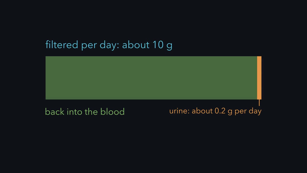 ▶ Watch A Small Step Sets Calcium Loss · 2:33
Read the transcript of “A Small Step Sets Calcium Loss”
Film transcript: A Small Step Sets Calcium Loss
Your kidneys filter about ten grams of calcium a day. You lose about a fifth of a gram in urine. So almost all the filtered calcium goes back into the blood. What sets that last small fraction? Work it out with made-up round numbers, for a reference adult at steady state. In adult plasma, roughly half the calcium is ionized, about 40 percent bound to protein, and about 10 percent complexed. Calcium bound to protein is not filtered. A GFR of 125 mL/min is 180 L a day. Total calcium is 100 mg/L, and 60 percent is filterable. So the filtered load is 10,800 mg per day. Most of that is reabsorbed along the tubule. Roughly 60 to 70 percent of filtered calcium is reabsorbed in the proximal tubule.
20 to 25 percent is reabsorbed in the thick ascending limb, largely between cells. The distal convoluted and connecting tubules take back roughly 5 to 10 percent, through cells. Parathyroid hormone, or PTH, fine-tunes this small step and also acts upstream. Urinary calcium in the example is 200 mg per day. That is 1.85 percent of the filtered load, so 98.15 percent is reabsorbed. Now let reabsorption fall to 97 percent, with the same filtered load, as when PTH falls. Excretion becomes 324 mg per day, 1.62 times the original. A fall of about 1.15 percentage points moves excretion by 62 The parathyroid chief cells carry the calcium-sensing receptor, which reads ionized calcium. A rise in ionized calcium activates the receptor and inhibits PTH secretion. The response is steep: a small change in calcium moves secretion a long way. PTH raises active calcium reabsorption in the distal nephron.
So when PTH falls, distal reabsorption falls, and with the same filtered load, urinary calcium rises. Keep the rule. The kidney filters about 10 g of calcium a day and reabsorbs about 98 The distal step that PTH regulates handles under a tenth of the load. Yet that step fine-tunes urine calcium, along with filtered load.
About 99 percent of the calcium in an adult human body is in bone, yet the parathyroid glands read only a sliver of the remaining 1 percent: the free calcium ions in plasma. That sliver stays in a narrow range while bone is dissolved and rebuilt. How?
The pressure on that sliver is easy to miss. In round numbers, the roughly 14 litres of extracellular fluid hold about 840 mg of filterable calcium (14 L × 60 mg per L; filterable means free or bound to small ions, not to protein), and the kidneys filter about 10,800 mg a day: the whole stock passes the filter roughly once every two hours, and almost all of it has to come back. Holding a pool that small and that fast-moving takes a loop that senses it directly.
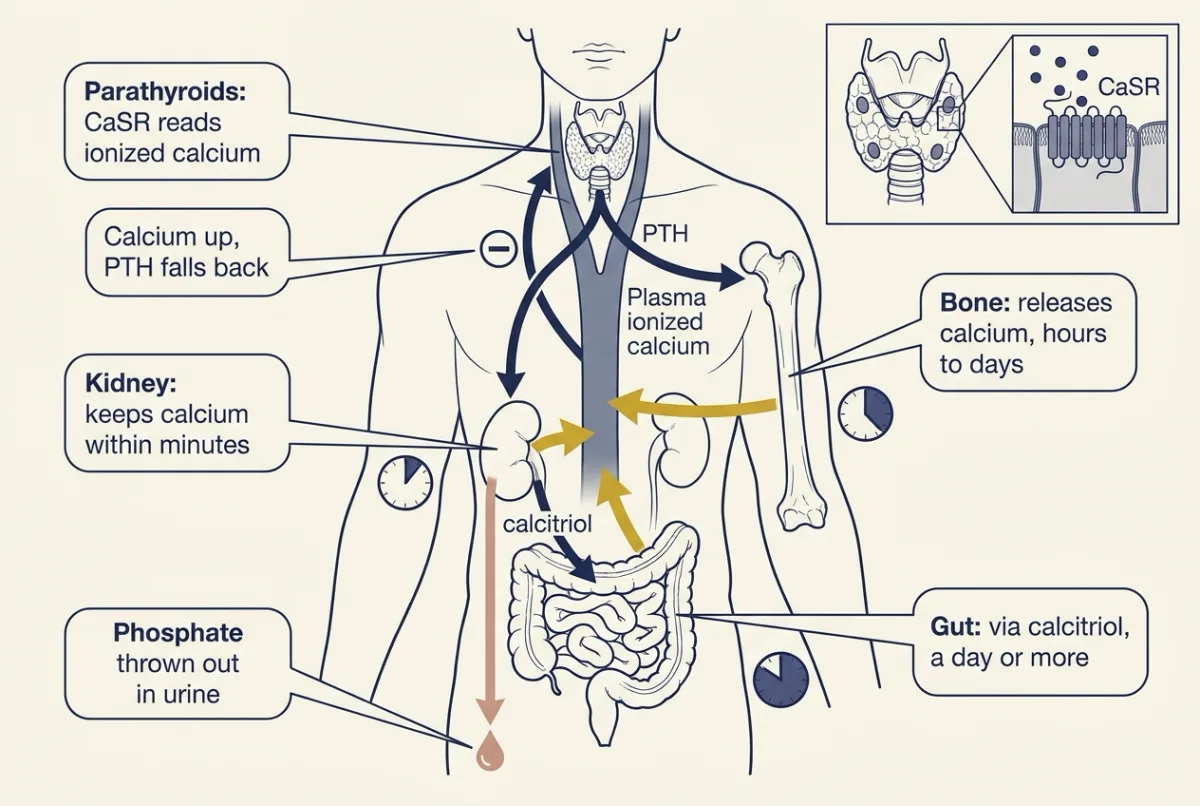
The one idea to carry through this lesson. Plasma ionized calcium is the regulated variable. The parathyroid calcium-sensing receptor reads it and sets parathyroid hormone (PTH) by negative feedback: less calcium, more PTH. PTH opens three doors that raise plasma calcium, each on its own clock: kidney reabsorption (minutes), bone resorption (hours for the first release, days for new osteoclasts), and gut absorption through the calcitriol that PTH has the kidney make (a day or more). At the kidney PTH does the opposite to phosphate and throws it out, so PTH raises calcium and lowers phosphate. Phosphate has its own brake, made by bone: FGF23.
The rule holds for an adult at steady state with working kidneys; chronic kidney disease, the last case here, is where it breaks.
What is regulated: ionized calcium
Bone holds the reserve: that 99 percent of body calcium, and roughly 85 percent of its phosphate. Remodeling, osteoclasts resorbing a patch and osteoblasts refilling it, takes months at each site, a few weeks to resorb and several months to refill, and sets the long-term traffic with blood. The fast exchange with plasma happens at bone surfaces.
Plasma calcium sits in three pockets. Roughly half of the total is free ionized calcium, about 40 percent is bound to protein, mostly albumin, and about 10 percent is complexed with citrate, phosphate and bicarbonate. Only the ionized fraction acts on cells and is sensed and regulated.
Labs report total calcium in mg per dL and ionized calcium, read by an ion-selective electrode, in mmol per L. Calcium’s molar mass is about 40 g, so 1 mmol/L is about 4 mg/dL: an ionized calcium of 1.2 mmol/L is about 4.8 mg/dL, about half of a total of 9.5 mg/dL. Formulas that adjust total calcium for albumin do not reliably reproduce it: in 22,658 Alberta adults with total and ionized calcium measured in the same samples (albumin in 10,659 of them), albumin-adjusted calcium generally did no better than unadjusted total calcium (Desgagnés and colleagues, 2025).
Predict. In alkalosis, fewer hydrogen ions compete for albumin’s binding sites, so albumin binds more calcium. What happens to total calcium, and to ionized calcium?
Check your prediction
Total calcium stays the same and ionized calcium falls: calcium has only moved from the free pocket to the albumin pocket, so a normal total can hide a drop in the regulated variable. Nerves and muscles respond to ionized calcium, and the drop can make them overexcitable, felt as tingling in the fingers.
One sensor, one hormone, three doors
Follow one disturbance through the loop: ionized calcium dips a little, say after a day of meals low in calcium.
The sensor. The parathyroid glands, usually four, sit on the back of the thyroid. Their chief cells carry the calcium-sensing receptor (CaSR), a G protein-coupled receptor (G proteins) that binds ionized calcium outside the cell. High calcium keeps the receptor active and parathyroid hormone (PTH) secretion down; a dip takes the brake off. The response is steep, so a small change in calcium moves PTH a long way, which keeps the range narrow. (Calcium signaling covers calcium inside cells; this lesson is about the calcium outside them.)
The hormone. Parathyroid hormone (PTH) is a peptide. It acts through a G protein-coupled receptor that raises cAMP (cAMP and PKA). It opens three doors, on three clocks.
- Kidney, within minutes. PTH raises active calcium reabsorption in the distal convoluted and connecting tubules, so less calcium leaves in urine.
- Bone, over hours to days. PTH’s receptors sit on osteoblast-lineage cells, including osteocytes, so PTH reaches osteoclasts mainly through them (direct effects on osteoclast-lineage cells, reported in culture, are unproven in the body): they raise RANKL relative to its decoy, osteoprotegerin (OPG), and more osteoclasts form (the bone door, below). Some calcium comes off bone surfaces within hours; new osteoclasts take days.
- Gut, over a day or more. PTH stimulates the kidney’s 1-alpha-hydroxylase, the enzyme that makes active vitamin D, calcitriol. Calcitriol acts through the vitamin D receptor, a transcription factor (nuclear receptors), so its effect waits on new proteins. Then more calcium is absorbed from food.
Calcium rises, CaSR becomes active again, and PTH falls back. That is negative feedback.
Predict. Bone mineral is calcium phosphate, and PTH dissolves it. So when PTH rises, which way should plasma phosphate go?
Check your prediction
Down, which surprises most people. At the kidney PTH does the opposite to phosphate. It pulls the sodium-phosphate cotransporter NaPi-IIa out of the proximal tubule’s brush border, so less filtered phosphate is reabsorbed and more is lost in urine.
Compare the sizes. Hydroxyapatite, the bone mineral, carries about 2 mg of calcium per 1 mg of phosphate (as phosphorus, the way labs report it), so releasing an extra 200 mg of calcium from bone each day adds only about 90 mg of phosphate from bone alone. The kidneys filter about 5,300 mg of phosphate a day (180 L × 33 mg per L × 90 percent filterable), and raising the fraction excreted from 15 to 20 percent throws out an extra 270 mg or so. The kidney wins: the PTH signature is calcium up, phosphate down. It holds because PTH’s effect on excretion is large, and only while the kidneys can excrete (chronic kidney disease, at the end, is the test).
Calcitriol has two jobs. Vitamin D is made in skin (skin and human heat), 25-hydroxylated in the liver (absorption and the liver) and 1-hydroxylated in the kidney, giving calcitriol, 1,25-dihydroxyvitamin D. First, it is PTH’s slow arm: it raises calcium absorption in the gut, and active phosphate absorption too. Second, it suppresses PTH gene transcription, so a hormone PTH makes turns PTH down.
Calcitonin, from the thyroid’s parafollicular C cells (thyroid hormone), stops the crawling of isolated osteoclasts on glass or plastic within minutes (Chambers and Magnus, 1982), yet its role in normal adult human calcium control appears modest.
The kidney door, in numbers
The film at the top of the page is this section’s worked example: why a small, regulated step sets the day’s calcium loss.
Glomerular filtration rate (GFR) is in glomerular filtration and clearance in the kidney. Protein-bound calcium is not filtered; the ionized and complexed fractions, roughly 55 to 60 percent of total, are. Of the filtered calcium, roughly 60 to 70 percent is reabsorbed in the proximal tubule and 20 to 25 percent in the thick ascending limb, largely between cells. The distal convoluted and connecting tubules take back roughly 5 to 10 percent through cells, the step where parathyroid hormone (PTH) does most of its fine-tuning. Both PTH and calcium-sensing receptor (CaSR) also act upstream: CaSR in the thick ascending limb lowers calcium reabsorption when calcium is high. Phosphate is reabsorbed mainly in the proximal tubule, the step PTH and FGF23 turn down. Why thiazides lower urinary calcium and loop diuretics raise it is in tubular transport and diuretics.
The distal step handles no more than a tenth of the filtered load (5 to 10 percent), so it sounds minor. But 5 to 10 percent of about 10,800 mg a day is 540 to 1,080 mg, about 2.7 to 5.4 times the day’s urinary calcium of about 200 mg. The regulated step is small next to what is filtered and large next to what is lost.
Worked problem: calcium and phosphate in a day
Made-up round numbers for a reference adult at steady state, with bone formation equal to resorption, no sweat losses and a filterable calcium fraction of 60 percent.
In words The fraction excreted is the day's urinary loss divided by the filtered load, and the filtered load is GFR times the filterable plasma concentration.
- FE“F E”
- fractional excretion: the share of the filtered load that reaches urine. One minus FE is the fraction reabsorbed.no unit
- E“E”
- the amount excreted in urine per daymg/day
- GFR“G F R”
- glomerular filtration rateL/day
- Pf“P sub f”
- the filterable plasma concentration: for calcium, the ionized and complexed part, not the protein-bound partmg/L
Sanity check GFR × Pf, the bottom of the fraction, is the filtered load. If nothing were reabsorbed, FE would be 1. Using total instead of filterable calcium inflates the filtered load and makes FE look too small.
Need a refresher? units and prefixes · rates and “per”
- Filtered load. GFR 125 mL per min = 0.125 L per min x 1,440 min per day = 180 L per day. Total plasma calcium 10 mg per dL = 100 mg per L, 60 percent filterable: 60 mg per L. Filtered load = 180 x 60 = 10,800 mg per day.
- Fractional reabsorption. Urinary calcium is 200 mg per day. Fractional excretion = 200 / 10,800 = 0.0185 (1.85 percent); 98.15 percent is reabsorbed.
- Balance. With bone in balance, net intestinal absorption equals urinary excretion, 200 mg per day. At an intake of 1,000 mg per day, fecal calcium = 800 mg per day.
- Sensitivity. Now let PTH fall, so fractional reabsorption drops from 98.15 to 97.0 percent with the filtered load held the same.
Predict. Roughly how much does the day’s urinary calcium change?
Check your prediction
Excretion = 10,800 × 0.030 = 324 mg per day, 1.62 times the original: a fall of about 1.15 percentage points in reabsorption moves excretion by 62 percent, because the small regulated step acts on a huge flow.
Holding the filtered load fixed is a what-if, as in the film. In life PTH usually falls because calcium rose, and a rise in calcium raises the filtered load, switches off PTH and acts on CaSR in the thick ascending limb. All three raise urine calcium.
- Filtered load = GFR × 1.44 × filterable calcium
- Urinary excretion = filtered load × (1 − fractional reabsorption)
- Net intestinal absorption needed = excretion − net bone release
- Distal step PTH fine-tunes (5–10% of filtered load)
In this sketch: made-up round numbers for a reference adult at steady state, starting from the lesson’s worked problem (GFR 125 mL per min, filterable calcium 60 mg per L, 98.15 percent reabsorbed, bone formation equal to resorption, no sweat losses). The 1.44 is 1,440 minutes per day divided by 1,000 mL per L. The slider ranges, including the 0 to 200 mg per day of net bone release, are made up for the sketch. Each slider is moved on its own and filterable calcium is held at the value you set; no hormones are simulated, so the sketch does not show how the body would set or respond to any of these values. In life a rise in calcium moves several of these together: it raises the filtered load, lowers PTH and acts on CaSR in the thick ascending limb, and all three raise urine calcium. The 5 to 10 percent distal share is the lesson’s round range; the sketch does not model which segment changes when you move the slider. A fall of about 1.15 percentage points in reabsorption, from 98.15 to 97.0 percent, moves excretion by 62 percent in the lesson’s example. At whole-body balance, net intestinal absorption equals urinary loss plus net bone accretion (net bone release is negative accretion).
Rule to keep: filtered load = GFR × filterable calcium, and excretion = filtered load × (1 − fractional reabsorption).
- Phosphate. Suppose plasma phosphate is low. A blood sample and a spot urine taken together give these made-up values: plasma phosphate 2.0 mg per dL, urine phosphate 40 mg per dL, plasma creatinine 1.0 mg per dL, urine creatinine 100 mg per dL. This lesson’s reference adult, with normal phosphate, excretes about 15 percent of filtered phosphate, and a kidney conserving phosphate should excrete less than that. Is this one holding on?
In words The fractional excretion of phosphate is urine phosphate times plasma creatinine, divided by plasma phosphate times urine creatinine.
- UP“U sub P”
- urine phosphatemg/dL
- PP“P sub P”
- plasma phosphatemg/dL
- UCr“U sub C r”
- urine creatininemg/dL
- PCr“P sub C r”
- plasma creatininemg/dL
Sanity check If the kidney excreted every bit of filtered phosphate, FE(P) would be 1, or 100 percent. A kidney conserving phosphate gives a small fraction.
Try one number
FE(P) = (40 × 1.0) / (2.0 × 100) = 40 / 200 = 0.20, or 20 percent; 80 percent is reabsorbed.
Where it comes from optional
Step 1, phosphate clearance = UP × V / PP, where V is urine flow.
Step 2, creatinine clearance = UCr × V / PCr, taken as an estimate of GFR.
Step 3, FE(P) = phosphate clearance / creatinine clearance = (UP × V / PP) / (UCr × V / PCr). V is on top and bottom and cancels, which is why a spot urine works. This assumes creatinine clearance approximates GFR and phosphate is freely filtered, with both samples taken together.
Need a refresher? subscripts and Greek letters · rates and “per”
A fifth is no small fraction, so this kidney is not holding on: that points to renal phosphate wasting, for example from PTH or FGF23. The calculation cannot say which.
The bone door: RANKL and OPG
Three cells matter here (muscle, bone and movement has more). Osteoblasts lay down matrix, osteoclasts resorb it, and osteocytes, former osteoblasts walled into the matrix, stay linked to one another by fine processes. The matrix is collagen stiffened by a calcium phosphate mineral, hydroxyapatite. Osteoblast-lineage cells and osteocytes display RANKL, a cytokine of the tumor necrosis factor family. Its receptor, RANK, sits on osteoclast precursors, and engaging it drives their formation and activation. Osteoprotegerin (OPG) is a soluble decoy: it binds RANKL and keeps it off RANK. Picture a key, a lock and a dummy lock that swallows keys. RANKL relative to OPG is a main control on how many osteoclasts form; precursor supply, M-CSF and other signals also matter.
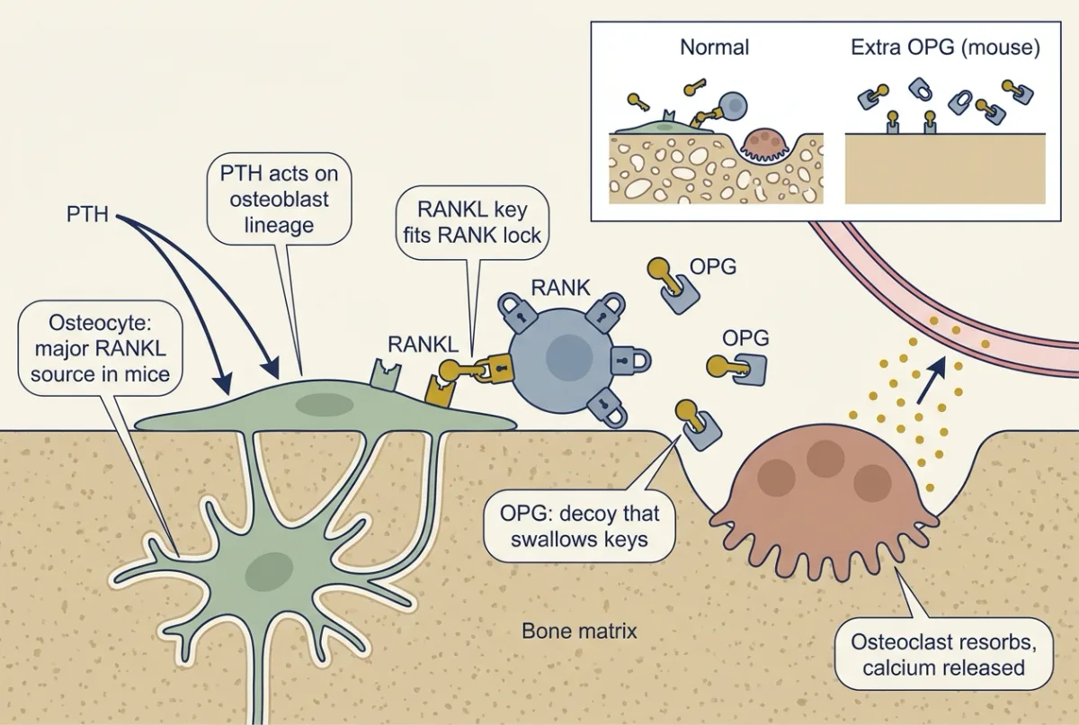
The decoy’s power showed first in mice. Simonet and colleagues (Cell, 1997) made transgenic mice that expressed OPG from the liver; they developed profound but nonlethal osteopetrosis, an excess of dense bone, because with the keys swallowed too little bone was resorbed. Which cell supplies the keys? Two studies that deleted RANKL in mouse osteocytes, each with a different genetic tool, got severe osteopetrosis and fewer osteoclasts. So in mice osteocytes are a major source of the RANKL that remodeling needs (the tools’ limits are in Going deeper).
This is the door parathyroid hormone (PTH) uses (step 2 above). Continuous PTH excess causes net bone loss even though formation also rises; intermittent exposure behaves differently and should not be lumped with it.
Phosphate’s own brake: FGF23
Parathyroid hormone (PTH) throws phosphate out, but PTH is set by calcium, and calcitriol brings phosphate in with the calcium. Phosphate needs its own control, and bone provides it. Osteocytes and osteoblasts make FGF23, which acts on the kidney through a fibroblast growth factor (FGF) receptor paired with the co-receptor Klotho. It does two things there:
- it lowers phosphate reabsorption in the proximal tubule, as PTH does;
- it lowers calcitriol, the opposite of PTH.
The second effect makes sense for a phosphate hormone: calcitriol raises gut phosphate absorption, so lowering calcitriol cuts phosphate intake at the source. Calcitriol, in turn, raises FGF23. The two form a second, smaller loop that runs on phosphate. FGF23 answers mainly to phosphate and calcitriol, not to the calcium-sensing receptor, so it is a phosphate hormone rather than an arm of the calcium response. Calcium still tugs on it: in rodents low calcium holds FGF23 down, which would spare calcitriol and so fits keeping the gut door open.
The modern experiment: mice without FGF23
FGF23 was first tied to rare disorders in which too much FGF23 activity makes the kidney lose phosphate. Is it needed normally? Shimada and colleagues (Journal of Clinical Investigation, 2004) deleted Fgf23 in mice.
Predict. If FGF23 is a normal phosphate brake, what should happen to serum phosphate and calcitriol in mice that lack it?
Check your prediction
Both should rise. Compared with littermates, the null mice had high serum phosphate, increased renal phosphate reabsorption and elevated calcitriol, with abnormal bone mineralization. In the same group’s injection study, recombinant FGF23 lowered serum phosphate even in parathyroidectomized rats, so that effect did not need PTH.
So endogenous FGF23 is necessary for normal phosphate and vitamin D handling in the mouse. A lifelong, whole-body deletion cannot separate direct renal effects from those of high calcitriol or development, and says nothing quantitative about humans.
The three hormones side by side, in an adult with working kidneys:
| Hormone | Made by | Plasma calcium | Plasma phosphate | Calcitriol |
|---|---|---|---|---|
| PTH | Parathyroid chief cells | Up | Down | Up |
| Calcitriol | Kidney (from vitamin D) | Up | Up | (it is calcitriol) |
| FGF23 | Osteocytes, osteoblasts | Little direct effect | Down | Down |
FGF23’s effect on calcium is small rather than absent: by lowering calcitriol it slowly reduces gut calcium absorption, and in mice it also raises calcium reabsorption through TRPV5 in the distal tubule (Andrukhova and colleagues, 2014).
Predicting the disorders from the loop
The rule once more, in short: ionized calcium sets parathyroid hormone (PTH); PTH brings calcium in through kidney, bone and gut and throws phosphate out; FGF23 brakes phosphate. Each setting below (untreated, in adults) is that loop with one part broken. Name the broken part and the arrows follow.
Primary hyperparathyroidism: PTH stuck high, with normal kidney function. A parathyroid source secretes PTH that ionized calcium no longer suppresses normally, so PTH is high or inappropriately normal, that is, normal when high calcium should have suppressed it (endocrine axes and testing). All three doors open: bone resorption rises through RANKL, distal calcium reabsorption rises, and calcitriol rises with gut absorption behind it. Urinary calcium can still be raised, because plasma calcium and so the filtered load rise. NaPi-IIa leaves the brush border, so phosphate is often low-normal or low.
Hypoparathyroidism: PTH missing.
Predict. Calcium, phosphate, PTH and calcitriol: which way does each go?
Check your prediction
Calcium low, phosphate high, PTH low or inappropriately low, calcitriol low or low-normal. Losing PTH removes its boost to distal calcium reabsorption, its phosphaturia and its drive to calcitriol: all three doors close, and the kidney keeps phosphate it would have thrown out.
Vitamin D deficiency: the gut door short of raw material. Why might calcitriol be normal early on, with stores low? The loop compensates: PTH rises, and high PTH stimulates 1-alpha-hydroxylation, so calcitriol can be normal or even high. The same PTH throws out phosphate, so phosphate is often normal and can be low. Later calcitriol may fall, and calcium usually, and phosphate often, fall with it.
Chronic kidney disease (CKD): the kidney door fails. At steady state the kidneys must excrete the phosphate the gut absorbs, say 800 mg a day, and excretion is glomerular filtration rate (GFR) × filterable plasma phosphate (about 90 percent of the total) × FE(P). Turned around, plasma phosphate = intake ÷ (GFR × 0.9 × FE(P)); at 180 L per day and 15 percent, 800 ÷ (180 × 0.9 × 0.15) = 33 mg per L, about 3.3 mg/dL. Three cases in round numbers:
- GFR 180 L per day, FE(P) 15 percent: plasma phosphate of about 3.3 mg/dL does it.
- GFR 60 L per day: at the same FE(P), plasma phosphate must reach about 9.9 mg/dL; if FGF23 and PTH push FE(P) to 50 percent, about 3.0 mg/dL is enough.
- GFR 15 L per day: even at FE(P) 50 percent, plasma phosphate must reach about 11.9 mg/dL.
The amount excreted is 800 mg a day in every case; what has to rise is the fraction. So moderate CKD keeps phosphate normal largely by raising FE(P). A large cross-sectional cohort found FGF23 elevated while phosphate and PTH were still normal (Isakova and colleagues, 2011), which fits FGF23 rising first without proving the order. Later, the kidney retains phosphate, calcitriol falls and PTH rises. In advanced CKD even the largest possible rise cannot keep phosphate normal: FE(P) stops at 100 percent, and at a GFR of 15 L per day excreting every filtered milligram still leaves plasma phosphate near 5.9 mg/dL. Phosphate settles high and climbs from there despite high PTH: PTH’s signature fails where the rule said it would.
The table collects the patterns: directions, not criteria, which stage, treatment and other disease can change.
| Setting | Calcium | Phosphate | PTH | Calcitriol |
|---|---|---|---|---|
| Primary hyperparathyroidism | High | Low-normal or low | High or inappropriately normal | High or high-normal |
| Vitamin D deficiency, early | Often normal | Often normal, can be low (PTH phosphaturia) | High | Normal, or even raised |
| Vitamin D deficiency, later | Usually falls | Often falls | High | May fall |
| Early chronic kidney disease (CKD) | May stay normal | May stay normal | Often rising | May begin to fall |
| Advanced CKD | Normal or tending low | Usually high | High | Low |
| Hypoparathyroidism | Low | High | Low or inappropriately low | Low or low-normal |
Design problem
PTH raises RANKL relative to osteoprotegerin (OPG), and in mice osteocytes are a major source of RANKL. Design an experiment in adult mice to test whether PTH needs osteocyte RANKL to raise plasma calcium, and say what each outcome would mean.
Saved in this browser only.
One way to do it
Delete a floxed RANKL allele with an inducible osteocyte Cre, switched on in adults so bone develops normally, with floxed-only and Cre-only controls on the same inducer. Confirm the deletion in sorted cells. Then give continuous PTH to every group and read plasma ionized calcium over hours and days, osteoclast number on bone surfaces, and urine calcium and calcitriol, since the kidney and gut doors are still open.
If calcium rises in the first hours in both groups but the deleted mice add no osteoclasts and fall behind over days, the slow part of the bone door needs osteocyte RANKL and the early release does not. If calcium rises with no new osteoclasts, the kidney and gut doors may be carrying the load. If everything rises normally, osteocyte RANKL is dispensable here or another source compensates. Any result is about mice in this setting.
Going deeper
The main line above does not depend on these details.
Bone as a tissue, and the joints
Compact bone is built of osteons, cylinders of matrix around a central canal; spongy bone is a lattice of struts called trabeculae. Bone ends at most synovial joints are covered by hyaline articular cartilage, which has no blood vessels and reduces friction. Ligaments join bone to bone and tendons join muscle to bone. By structure, joints are fibrous, cartilaginous or synovial.
How large PTH’s phosphate effect must be
Parathyroid hormone (PTH) lowers phosphate because its effect on excretion is large, not automatically. Take the steady-state relation used in the chronic kidney disease (CKD) case, plasma phosphate = input ÷ (GFR × filterable fraction × FE(P)), with 180 L per day, 90 percent filterable, 800 mg per day of input and FE(P) 15 percent: about 3.3 mg/dL. If PTH adds 200 mg per day of input (bone plus gut) and raises FE(P) to 25 percent, phosphate falls to about 2.5 mg/dL. At 18 percent it would not fall (about 3.4 mg/dL). Break-even here is about 19 percent.
How RANKL and OPG were found
The pieces came from three papers, not one. Simonet and colleagues’ 1997 osteoprotegerin (OPG) mice (above) came with a second result: OPG blocked ovariectomy-associated bone loss in rats. Lacey and colleagues (Cell, 1998) identified the ligand OPG binds as a cytokine that drives osteoclast differentiation. Hsu and colleagues (PNAS, 1999) showed, in mouse bone-marrow progenitors, that it acts through RANK. Other laboratories described the same molecules independently under other names. The ovariectomized-rat result bridges to estrogen and bone in the ovarian cycle.
Which cell’s RANKL: the genetic tools
Nakashima and colleagues (Nature Medicine, 2011) deleted RANKL with a Dmp1-Cre transgene aimed at osteocytes, and the mice developed severe osteopetrosis. That Cre also acts in some osteoblasts and lining cells, so Xiong and colleagues (PLoS ONE, 2015) repeated it with a Sost-Cre that spares those cells, though it is also active in some marrow hematopoietic cells, and saw a threefold fall in osteoclast number in cancellous bone.
Either result carries a limit worth keeping in mind: osteopetrosis after an osteocyte deletion means osteocyte RANKL is necessary in that setting, not that osteocytes are the only source at every age, site or in humans.
The experiments behind the kidney and gut steps
- The calcium-sensing receptor was cloned from bovine parathyroid by Brown, Hebert and colleagues (Nature, 1993).
- PTH’s retrieval of NaPi-IIa from the proximal brush border was shown in rat kidney in vivo and in isolated perfused mouse tubules (Traebert and colleagues, 2000).
- In thyroparathyroidectomized rats on a low-calcium diet, production of active vitamin D appeared to depend on the parathyroid (Garabedian and colleagues, 1972).
- In mice lacking the vitamin D receptor, a high-calcium, high-phosphate rescue diet with lactose, which restored ionized calcium, prevented rickets and the rise in PTH. That points to impaired intestinal calcium absorption as the cause (Li and colleagues, 1998).
- In wild-type mice, two days of calcitriol raised active jejunal phosphate transport while the paracellular route was unchanged (Hernando and colleagues, 2021).
- In rats, calcitriol inhibits PTH gene transcription (Silver and colleagues, 1986).
FGF23 in more detail
Klotho was identified as the co-receptor that lets FGF23 act on the kidney by Urakawa and colleagues (2006). In rodents, injected FGF23 lowered renal mRNA for 1-alpha-hydroxylase and raised it for 24-hydroxylase, the enzyme that inactivates vitamin D metabolites. In normal mice, injected calcitriol raised serum FGF23 within four hours. Calcium matters too: calcium-deficient rats had lower circulating FGF23 (Rodriguez-Ortiz and colleagues, 2012), and in mice low calcium blunted the rise in FGF23 that calcitriol and PTH otherwise cause (David and colleagues, 2013).
The cohort cited for FGF23 rising first is the baseline analysis of 3,879 adults in the Chronic Renal Insufficiency Cohort, nearly all in CKD stages 2 to 4; “still normal” there means mean phosphate and median PTH.
Calcitonin: a hormone that lowers calcium
Calcitonin’s discovery came from dogs. By the early 1960s PTH alone explained calcium control: when calcium rose, PTH secretion fell. Copp, Cameron, Cheney, Davidson and Henze (Endocrinology, 1962) isolated the thyroid-parathyroid tissue of fasted, anesthetized dogs and perfused it through its own circulation. If high calcium only switched PTH off, systemic calcium should fall no faster than existing PTH and its effects waned. Perfusion with blood containing 12 mg of calcium per 100 mL did lower systemic plasma calcium. The group compared it with removing the glands, the most complete PTH withdrawal possible, and calcium fell faster after high-calcium perfusion, so the authors argued the fall could not be PTH withdrawal alone. They inferred a calcium-lowering factor and named it calcitonin.
That ruled in an active, calcium-lowering signal in this dog preparation and ruled out PTH withdrawal as the only response. It did not locate the source: Copp’s group placed it in the parathyroid, and later work in the thyroid’s C cells.
Your notes
Select any text in the lesson to highlight it or add a note to it. Your highlights and notes are saved in this browser and collected in your notebook.
This browser is not letting the site save anything (a private window or strict privacy settings), so highlights and notes will be lost when you leave the page.
How we measure it
Isolated gland perfusion in an intact animal
Separate a gland's blood supply from the rest of the circulation in an anesthetized animal and perfuse it with blood of chosen composition, while sampling systemic blood. Copp's group used it in dogs to change the calcium reaching the thyroid-parathyroid tissue alone and to watch what its venous output did to the rest of the body.
Transgenic and cell-specific knockout mice
Add a gene under a chosen promoter, delete it everywhere, or delete it in one cell type with a Cre line and a floxed allele. Overexpression of osteoprotegerin tested what excess OPG can do; deletion of Fgf23 and osteocyte-targeted deletion of RANKL tested whether that molecule or that cell source is needed for normal bone or mineral handling in mice.
Ion-selective electrode measurement of ionized calcium
A calcium-selective membrane electrode develops a potential that depends on the free calcium activity of the sample, so it measures the ionized fraction directly rather than total calcium. The reading depends on pH and sample handling, because protein binding of calcium changes with pH.
Fractional excretion from paired plasma and urine
Measure a solute and creatinine in the same plasma and urine samples; the ratio of the two clearances gives the fraction of the filtered solute that is excreted, without a timed urine collection. It assumes creatinine clearance approximates GFR and the solute is freely filtered.
Practice questions on this lesson (5) → MCAT-style, original, each with an explanation
Where this lesson fits
Before thisReview first
- Glomerular Filtration and Renal Blood FlowThis course · Lesson 15
You need GFR and filtered load from there to calculate how much calcium and phosphate the kidney filters.
- Tubular Transport, Segment by SegmentThis course · Lesson 16
The distal and proximal tubule steps for calcium and phosphate here build on how each nephron segment handles ions.
- Endocrine Axes and How to Test ThemThis course · Lesson 2
Reading PTH as high or inappropriately normal uses how feedback axes are tested.
After thisStudy next
- Exercise: The Whole Body at WorkThis course · Lesson 30
Whole-body exercise work draws on the muscle and bone calcium handling set up here, though only loosely.
Elsewhere on the siteFilms, simulations and method films in other courses
- Tap, Drain and a SensorFilm · Organismal Biology
Hormone released into blood, carried to cells with a receptor: the basic pattern PTH and calcitriol follow.
- Cells Compute With SwitchesFilm · Biochemistry
PTH acts through a cell-surface receptor and cAMP signalling, so this shows how that kind of relay works.
- Calcium gradientSimulation · Cell Signaling
Calcium kept low inside cells explains why the free calcium fraction is a regulated signal.
Suggested by an AI model from the lessons themselves, then checked. How we use AI
Finish line
Test yourself
10 questions
Practice and MCAT-style, with explained answers.
Next · Lesson 23
Gut Motility, Secretion and Gut Hormones
A glass of water is usually half gone from your stomach in about 10 minutes. A rich, high-calorie drink may take about 2 hours to be half gone. What holds the rich drink back?
Continue →