Cell Signaling · Unit 3: G-protein-coupled receptors and second messengers
GPCRs and Trimeric G Proteins
Rewritten on : new text, flashcards and film. Your notes and highlights stay, but the text around them may have moved.
Adrenaline strengthens the heartbeat but relaxes the airways. How can the same hormone give opposite orders?
Pause when the reading reaches a film, and offer to play it. Saved on this device.
Watch the film now, or keep listening
Builds on:A Field Guide to ReceptorsBinding: Affinity, Occupancy and Dose–ResponseGTPase Switches: The TimerWhy these?
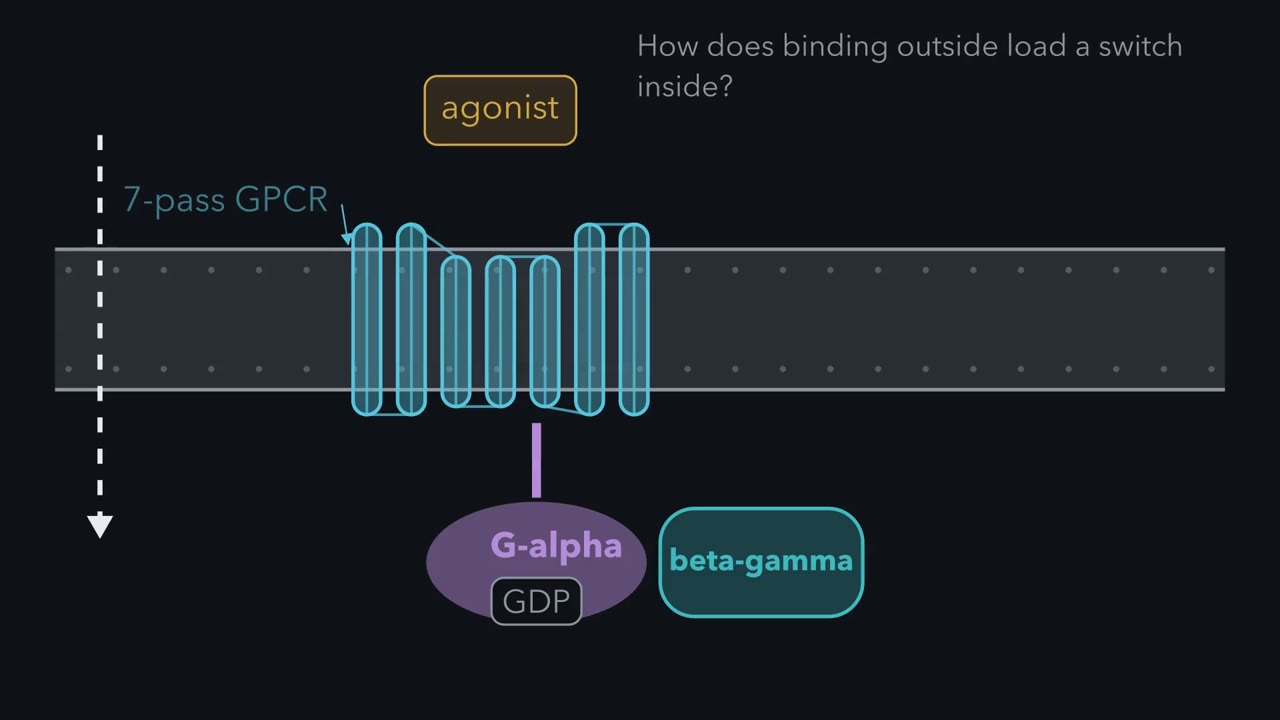 ▶ Watch How a Receptor Loads a Switch · 3:36
Read the transcript of “How a Receptor Loads a Switch”
Film transcript: How a Receptor Loads a Switch
How can binding outside a cell load a molecular switch inside it? Follow one membrane as the message crosses without the ligand crossing. A G-protein-coupled receptor, or GPCR, is one chain woven repeatedly through the membrane. Its outer pocket and inner face are physically connected. An agonist is a ligand that favors receptor activation. It settles into the pocket and favors an active shape. One cytoplasmic helix swings outward, opening a cavity below. A membrane-bound G protein waits below as a trimer. Purple Gα alpha holds gray GDP.
Teal βγ stays pressed against it. In this resting arrangement, the switch is off. The Gα alpha tail rises into the receptor cavity, bringing the whole trimer to the active receptor. Docking changes Gα's grip on GDP. The gray nucleotide leaves first. Green GTP can then occupy the empty site. The receptor has acted as an exchange factor, not as a source of GTP. It remains ready to load another G protein while agonist stays bound. GTP changes the Gα -alpha surface and frees βγ to engage targets. The textbook picture draws them apart.
Activated Gi may instead rearrange without fully separating. Either way, both surfaces can regulate effectors. Hydrolysis later returns Gα -alpha to GDP and favors reassembly. Now the cell chooses what those surfaces meet.
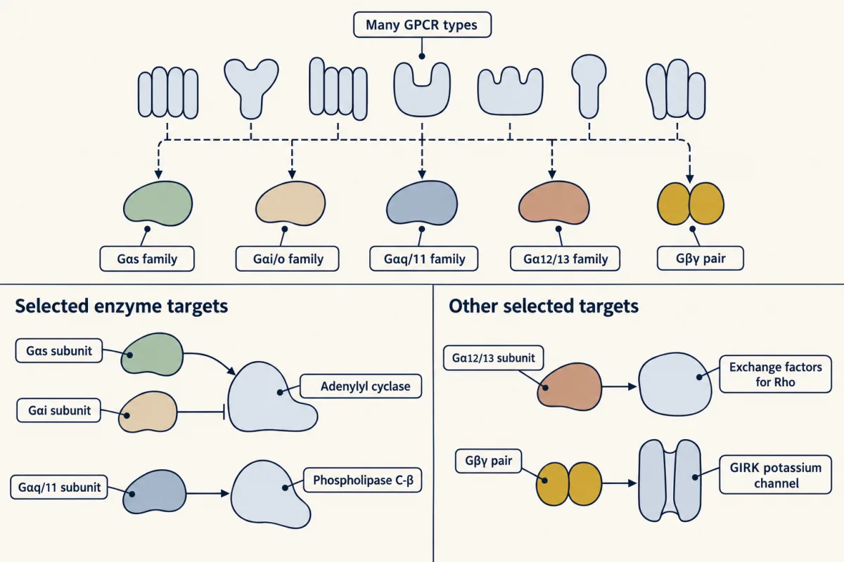
Follow the hourglass. Many receptors narrow to a few Gα -alpha families and βγ, then widen toward enzymes and channels. One branch raises cAMP. Another lowers it. Another raises calcium. βγ can open a potassium channel. Each family points to a main direct target, not a guaranteed exclusive partner. The receptor makes the first selection. The cell then supplies whichever enzymes, channels, and scaffolds are present. So one messenger can carry different meanings in different cells. The same acetylcholine can therefore meet different wiring. In a pacemaker cell, the receptor on screen loads Gi, and freed βγ opens potassium channels. Firing slows.
In airway muscle, a different receptor loads Gq, and phospholipase C-β -beta raises calcium. The muscle contracts. The input matches; the cellular paths differ. So do not predict an answer from the ligand alone. Trace the receptor subtype, the G protein it loads, and the targets available. That path predicts the cell's first response.
Dose and efficacy still change how strongly the receptor is activated. The wiring decides what that activation means.
From the film
- GPCR subtype, G protein, and downstream proteins decide the first response.
- An agonist can open a cytoplasmic receptor cavity where a trimeric G protein docks.
- The active receptor promotes GDP release, allowing GTP to bind Gα.
- Gα-GTP and βγ can regulate different targets.
Adrenaline makes the heart beat harder, relaxes airway muscle and narrows skin blood vessels. The hormone matters, but it cannot specify those opposite outcomes by itself.
The one idea. G-protein-coupled receptor (GPCR) subtype, G protein, and downstream proteins decide the first response.
Scope: the first response through the canonical GPCR–heterotrimeric-G-protein pathway in a particular cell. Ligand dose and efficacy still matter, one receptor may use several G proteins, and prolonged signaling recruits other partners.
From an outside pocket to an inside switch
The first film drew this relay as cartoons; here are the real parts.
A G-protein-coupled receptor (GPCR) is one protein chain that crosses the plasma membrane seven times. Each crossing is an alpha helix of twenty or more mostly water-avoiding amino acids. The amino end lies outside the cell and the carboxyl end inside, the architecture from the receptor field guide.
“Lots of inputs/ligands.” The fold accepts very different inputs: adrenaline, acetylcholine, dopamine, histamine, glucagon and other peptide hormones, odorants, bitter and sweet tastes, and light. Roughly a third of approved drugs act on a GPCR.
An agonist activates its receptor (the lesson on binding and dose-response). In the beta-2-adrenergic receptor, adrenaline binds among the helices toward the outer face. Comparing the receptor’s inactive crystal structure with one caught holding Gs (with a high-affinity agonist bound) shows the largest change inside: the cytoplasmic end of helix 6 moves outward by about 14 angstroms, opening the cavity where the tail of G alpha docks.
The heterotrimeric G protein has three subunits. G alpha holds a guanine nucleotide; G beta and G gamma bind so tightly that they act as one beta-gamma unit. Lipid tails on G alpha and G gamma hold the trio at the membrane’s inner face, as in the lesson on lipid anchors.
The active receptor acts as a guanine-nucleotide exchange factor (GEF): it promotes GDP release from G alpha, and GTP, more plentiful than GDP in the cytosol, fills the empty site. In the textbook picture G-alpha–GTP then leaves beta-gamma; in living cells, activated Gi may instead rearrange without fully separating.
active receptor + Gα(GDP)·βγ + GTP → active receptor + Gα(GTP) + βγ + GDP
The receptor appears on both sides because it is a catalyst, the source of the gain from the lesson on signaling circuits. One activation cycle ends when G alpha hydrolyses GTP and the GDP-bound trimer reforms.
Predict. A mutant G alpha can bind GDP but cannot release it. The receptor still binds agonist. What happens to the first downstream response?
Check your prediction
It fails. The receptor activates, but G alpha cannot release GDP to load GTP, so the G protein stays inactive.
”All the pathways”: four G-alpha families, then many targets
The human genome has 16 G-alpha genes in four sequence families.
| G-alpha family | Main direct target | First consequence | Example receptors |
|---|---|---|---|
| Gs | adenylyl cyclase, activated | cAMP rises | beta-adrenergic, glucagon |
| Gi/o | adenylyl cyclase, inhibited | cAMP falls; beta-gamma acts | M2 muscarinic, alpha-2-adrenergic, CB1 cannabinoid |
| Gq/11 | phospholipase C-beta | PIP2 becomes IP3 and DAG; calcium rises | alpha-1-adrenergic, M3 muscarinic |
| G12/13 | exchange factors for Rho | actin rearranges; cells change shape and move | thrombin, lysophosphatidic acid |
Beta-gamma is a signal too: in heart pacemaker cells, beta-gamma released from Gi opens the GIRK potassium channel, short for G-protein-gated inwardly rectifying potassium channel. Potassium flows out and moves the membrane potential away from the firing threshold. G-alpha-i also lowers cAMP, reducing current through pacemaker channels that cAMP opens directly.
“Lots of downstream effectors.” This is the hourglass the first film drew.
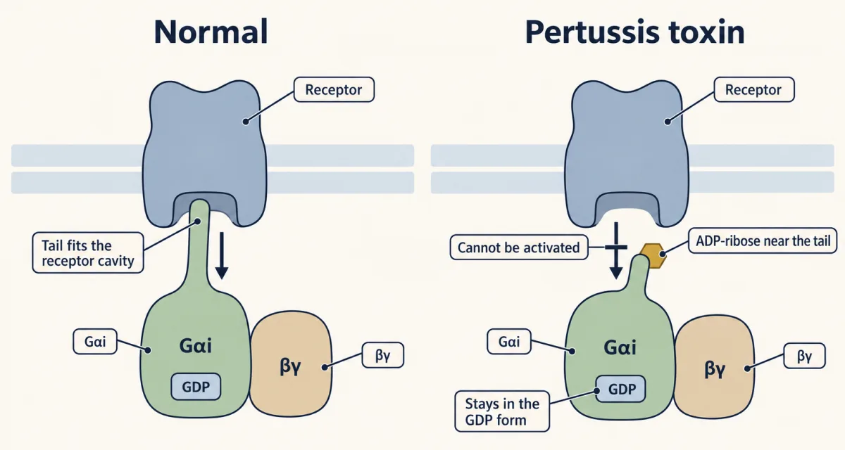
Predict. A receptor agonist lowers cAMP. Pertussis toxin leaves basal cAMP unchanged but prevents that fall. Which family carried the response?
Check your prediction
Gi/o. Pertussis toxin, an enzyme from the whooping-cough bacterium, adds ADP-ribose to a cysteine near the tail of G alpha-i and G alpha-o, so the tail cannot engage the receptor and the protein stays GDP-bound. Cholera toxin, from the lesson on GTPase switches, instead jams Gs on.
If pertussis toxin abolishes a response, that response most likely runs through Gi/o. The reverse test is weaker: Gz, a Gi/o-family member, lacks the toxin site, so toxin resistance does not exclude that family.
One ligand, different cellular answers
The first film traced acetylcholine: through M2 and Gi it slows pacemaker cells, and through M3 and Gq it contracts airway muscle. One transmitter quiets one tissue and excites another. Acetylcholine also marks the rule’s boundary: at skeletal muscle it meets an ion channel from the receptor field guide, not a G-protein-coupled receptor (GPCR).
Adrenaline has a larger cast: nine human adrenergic receptor genes. Three alpha-1 subtypes couple mainly to Gq, three alpha-2 subtypes to Gi, and three beta subtypes to Gs.
Predict. Assign one adrenergic subtype and G-alpha family to make the heart beat harder, relax airway muscle and narrow skin vessels. Then predict the effect of blocking only the heart subtype.
Check your prediction
Skin vessels: alpha-1 on Gq, because raised cytosolic calcium contracts smooth muscle. Airway muscle: beta-2 on Gs, because raised cAMP favors relaxation there. Heart: beta-1 on Gs. Heart and airway cells interpret the same messenger differently because different downstream proteins wait for cAMP (the next lesson follows that messenger).
Blocking only beta-1, the reasoning behind beta-1-selective beta-blockers, lowers heartbeat rate and force during adrenaline signaling and leaves airway and vessel receptors alone. A blocker that also hits beta-2 removes adrenaline’s airway relaxation, a risk for a person with asthma. This is the assignment the body mainly uses.
Predict. The thrombin receptor on platelets activates both Gq and G12/13. If a drug blocked only the Gq branch, which output in the table could remain?
Check your prediction
The G12/13 branch could still activate Rho exchange factors and reshape the cell, even if the Gq-dependent calcium rise were lost.
So far ligands have only switched receptors on. But efficacy, how far a bound ligand moves the response, can also push it below where it started.
Read the transcript of “Above, At, or Below Basal”
Film transcript: Above, At, or Below Basal
The first film followed a receptor as it loaded a G protein. If an empty receptor already signals, where can a ligand move the response? A receptor population can signal even when every binding pocket is empty. Individual receptors sample less-active and more-active shapes. If some active shapes engage G proteins, the population produces basal activity. That starting signal becomes the reference for classifying what a ligand does. An agonist raises signaling above that line. In the simple picture, it favors the more-active receptor population. Gold agonist blocks settle into open receptor pockets. More receptors remain signaling-competent, so the meter rises.
Agonist describes the measured effect, not merely the ability to bind. A neutral antagonist behaves differently. By itself, it leaves basal signaling approximately unchanged. Its job appears when another ligand arrives. Antagonist blocks occupy the pockets, so the incoming agonist finds few free receptors. At this agonist dose, the meter stays near basal instead of rising. Neutral describes the drug's effect on basal activity. An inverse agonist moves the response below basal. In the simple picture, it favors the less-active receptor population. Red-edged ligand blocks settle into those receptors. Fewer receptors engage G proteins, and the meter falls.
The prefix inverse refers to signaling direction, not to opposite binding. Now remove measurable basal activity from the assay. The meter starts at zero. A neutral antagonist cannot lower it. Neither can an inverse agonist reveal a further decrease. Added alone, they look the same. Distinguishing them requires a receptor system with a visible basal signal. Real ligands can produce intermediate changes, even at a saturating concentration.
Carazolol, bound in inactive β2-adrenergic receptor structures, is a partial inverse agonist. In an assay, it lowers basal signaling without producing the full reduction possible there. Efficacy means the response a bound ligand can produce. Partial describes its efficacy in that measured pathway, not incomplete binding in this assay. The active-versus-inactive picture is a useful first model, not a claim of only two rigid structures. Receptors sample ensembles of shapes. Efficacy is measured for one response in one cell. A ligand classed in one assay may class differently in another. Keep the meter and assay beside the receptor whenever you name efficacy.
Use one reference line and three questions. Does the ligand raise signaling above basal? Does it leave basal unchanged while blocking another ligand? Or does it lower signaling below basal? Those observations separate agonism, neutral antagonism, and inverse agonism without pretending the receptor is a simple switch.
From the film
- Basal activity separates agonists, neutral antagonists, and inverse agonists.
- An agonist raises signaling above basal activity.
- A neutral antagonist leaves basal activity unchanged while blocking other ligands.
- An inverse agonist lowers signaling below basal activity.
Ligand efficacy, and why “GPCRs can be unfaithful to trimeric G proteins”
The second film classed ligands by how they move basal activity, a receptor population’s signaling with no ligand: an agonist raises it, a neutral antagonist leaves it while blocking other ligands, an inverse agonist lowers it. Its meter was an operational test, not a claim that a G-protein-coupled receptor (GPCR) has only two rigid structures.
For many receptors only a small fraction is active at any moment, so basal activity can be too low to measure. Many drugs long called antagonists proved to be inverse agonists once tested where it was visible. Carazolol, the beta-blocker bound in the inactive beta-2 receptor structures from 2007, is a partial inverse agonist.
The four-family table also simplified: as the thrombin receptor showed, one receptor can couple to several G-alpha families, and the dominant partner can differ between cell types, so a coupling measured in one cell line is a hypothesis about the next. Efficacy is measured the same way: a ligand classed for one response in one cell may be classed differently for another.
Time adds another partner. Kinases phosphorylate a receptor that has stayed active, then arrestin binds its tail, blocks further G-protein coupling and helps pull the receptor into the cell. It also binds other signaling proteins, but whether arrestin signals on its own without G proteins remains debated, a question the lesson on turning signals off develops.
Predict. Two cell lines have equal amounts of a Gs-coupled receptor known to undergo beta-arrestin-dependent desensitization, such as the beta-2-adrenergic receptor, but one lacks both beta-arrestins. After prolonged agonist exposure, which line should retain more G-protein signaling?
Check your prediction
The line lacking beta-arrestins. Its initial response can be normal, but it loses a major brake on repeated receptor–G-protein coupling, so desensitization is reduced.
Going deeper
How the seven-pass plan was found
Rhodopsin, the light-catching pigment of retinal rod cells, was too greasy to sequence whole. Paul Hargrave’s laboratory in the United States and Yuri Ovchinnikov’s in Moscow cut it at methionine and sequenced the pieces one amino acid at a time. By 1983 both had published the complete cow-rhodopsin sequence, including seven water-avoiding stretches long enough to span a membrane.
In 1986 Robert Lefkowitz’s laboratory at Duke and a Merck group cloned a beta-adrenergic receptor gene. Brian Kobilka, then a cardiology fellow, had worked toward it for almost two years. Its sequence resembled rhodopsin, seven spans included. The only other seven-pass protein then known also used light, and Lefkowitz later recalled speculation that this plan marked light-sensitive molecules. The adrenaline receptor showed instead that it marked receptors that signal through G proteins.
How many GPCR genes
The human genome holds roughly 800 G-protein-coupled receptor (GPCR) genes; Lefkowitz’s Nobel lecture says almost a thousand. About half are odorant receptors, while a mouse has about a thousand odorant-receptor genes alone.
How the pertussis test named CB1
An orphan receptor is a receptor gene with no known ligand. In 1990 Lisa Matsuda and colleagues in Tom Bonner’s group had a rat-brain gene predicting a seven-pass protein. Allyn Howlett’s laboratory had shown that cannabinoids lower cAMP dose-dependently in nerve-cell preparations, that only one mirror-image form works, and that pertussis toxin blocks the effect. Expressed in cells that lacked it, the orphan showed those properties: it was CB1, the cannabinoid receptor, which couples to Gi/o.
Your notes
Select any text in the lesson to highlight it or add a note to it. Your highlights and notes are saved in this browser and collected in your notebook.
This browser is not letting the site save anything (a private window or strict privacy settings), so highlights and notes will be lost when you leave the page.
How we measure it
Cyanogen bromide cleavage and Edman sequencing
Cyanogen bromide cuts a protein chain at methionine, and Edman chemistry then removes and identifies one amino acid at a time from the end of each fragment. Overlapping fragments are pieced into the full sequence. This is how rhodopsin was read before its gene was cloned.
Hydropathy analysis
Score each amino acid in a sequence for how well it tolerates water, then average the scores along the chain. A run of about twenty water-avoiding residues is long enough to cross a membrane as a helix. Seven such runs in rhodopsin and in the β-adrenergic receptor gave the seven-pass model years before a crystal structure of either existed.
Expression of a cloned receptor in cells that lack it
Put the gene for a candidate receptor into a cultured cell line that does not make it, add test ligands, and measure a second messenger such as cAMP. A response that appears only in cells carrying the gene, rises with dose, and is lost after pertussis toxin is strong evidence for both the ligand and the G protein family.
Crystallography of stabilised receptors
GPCRs are scarce and unstable in detergent, so crystals were obtained by holding the receptor in one shape: an inverse agonist in the pocket with T4 lysozyme fused in for the inactive state, an agonist with the G protein and a llama antibody fragment for the active one. Comparing the two structures shows how the active receptor differs from the inactive one.
Practice questions on this lesson (10) → MCAT-style, original, each with an explanation
Where this lesson fits
Before thisReview first
- A Field Guide to ReceptorsThis course · Lesson 4
It introduces the seven-pass receptor architecture and the receptor classes that this lesson builds on.
- Binding: Affinity, Occupancy and Dose–ResponseThis course · Lesson 3
You need agonist, antagonist and occupancy from there to place the inverse agonist as a third kind of drug.
- GTPase Switches: The TimerThis course · Lesson 7
The active receptor acts as the GEF for Gα, so you need the GDP-GTP switch and its timer from there.
After thisStudy next
- cAMP and Protein Kinase AThis course · Lesson 13
It follows Gs and Gi through adenylyl cyclase to cAMP and the kinase that reads it.
- PLC, IP3, DAG and Protein Kinase CThis course · Lesson 14
It takes the Gq/11 branch onward: phospholipase C-β cuts PIP2 into IP3 and DAG.
- Turning the Signal Off: Arrestins, RGS Proteins and PhosphodiesterasesThis course · Lesson 16
It explains how receptor phosphorylation and arrestin block further G-protein coupling, the step this lesson only previews.
Suggested by an AI model from the lessons themselves, then checked. How we use AI
Finish line
Test yourself
15 questions
Practice and MCAT-style, with explained answers.
Next · Lesson 13
cAMP and Protein Kinase A
Adrenaline stays outside a liver cell, so how can enzymes inside respond?
Continue →