Physiology · Unit 5: Kidney, fluids and acid-base
Acid-Base Balance
Rewritten on : new text, figures and flashcards. Your notes and highlights stay, but the text around them may have moved.
Plasma holds 24 mmol/L of bicarbonate and only 40 nanomoles of free H+ per litre, and metabolism adds acid every day. pH stays put because one ratio, bicarbonate to dissolved CO2, stays near 20 to 1. What holds it there?
Pause when the reading reaches a film, and offer to play it. Saved on this device.
Watch the film now, or keep listening
Builds on:Glomerular Filtration and Renal Blood FlowVentilation, Perfusion and Gas ExchangeTubular Transport, Segment by SegmentWhy these?
Adult human arterial plasma at rest holds about 24 millimoles of bicarbonate per litre and about 40 nanomoles of free hydrogen ions per litre.
Metabolism in a US adult on a typical diet adds roughly 50 mEq of non-volatile acid a day, yet pH stays near 7.40, held on three clocks: buffers in seconds, lungs in minutes, kidneys over days. The lungs control carbon dioxide; the kidney controls bicarbonate with two jobs: it reclaims the bicarbonate it filters, and it makes new bicarbonate by excreting acid, mostly as ammonium made from glutamine. How do two organs on such different clocks hold one ratio?
Water, pH and weak bonds gave you pH = pKa + log([A-]/[HA]), and the kidney the nephron; here both get numbers.
The one idea to carry through this lesson. pH follows one ratio: bicarbonate over dissolved CO2, 20 to 1 at pH 7.40. The lungs set the bottom of the ratio, starting within minutes; the kidney sets the top over days. A disturbance pushes one term. Compensation moves the other term in the same direction, which brings the ratio back toward 20: usually partway, sometimes nearly all the way in chronic respiratory disorders (pH back inside the normal range, 7.35 to 7.45), and never past 7.40 to the other side.
Its scope: human plasma at 37 C, one disturbance at a time, at the steady state the compensating organ reaches. Two limits:
- The top is not wholly independent of the bottom. When pCO2 rises, non-bicarbonate buffers, mostly hemoglobin, take up the H+ made from the extra CO2, so bicarbonate rises a little within minutes (numbers below), before the kidney does anything. That is buffering, not compensation. It is why plasma bicarbonate on its own mixes respiratory and metabolic effects.
- The ratio says how acidic plasma is, not which acid. The anion gap, at the end of the lesson, is the tool for that.
pH follows a ratio
Two kinds of acid load the body. A volatile acid leaves as a gas: carbon dioxide, removed by the lungs. A non-volatile acid, such as sulfuric acid from sulfur-containing amino acids or an organic acid such as lactic acid or a ketoacid, cannot be breathed off; it is buffered, and the kidney excretes the net fixed-acid load. Lactate and ketoanions can also be metabolised, which gives bicarbonate back. A disturbance that starts with pCO2 is respiratory; one that starts with non-volatile acid or base is metabolic.
Picture pH as the reading of a fraction. On top sits bicarbonate, the base, which the kidney keeps. On the bottom sits dissolved CO2, the acid, which the lungs keep. Double both and pH does not move; change one alone and it does.
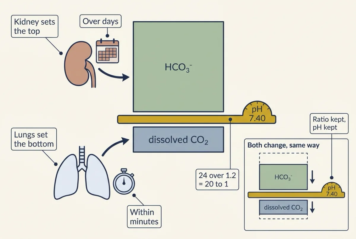
In symbols, for human plasma at 37 C, with pCO2 in mmHg and bicarbonate in mmol/L:
In words pH equals six point one plus the base-ten log of bicarbonate divided by the dissolved carbon dioxide, which is zero point zero three times the carbon dioxide tension.
- pH“p H”
- the pH of the plasmano unit
- 6.1“six point one”
- an apparent pK for the combined reaction CO2 + H2O = HCO3- + H+ in plasma at 37 C, hydration and ionization together
- [HCO3-]“bicarbonate”
- the plasma bicarbonate concentrationmmol/L
- 0.03“zero point zero three”
- the solubility of CO2 in plasma; it converts pCO2 into dissolved CO2. It is not the pK.mmol/L per mmHg
- pCO2“p C O 2”
- the carbon dioxide tension of the plasmammHg
Sanity check At rest, 24 / (0.03 × 40) = 20, and pH = 6.1 + 1.30 = 7.40. pH follows the ratio, not either number alone.
Worked example · try your own numbers
The worked problem below: bicarbonate 12 mmol/L, pCO2 27 mmHg. Dissolved CO2 = 0.03 × 27 = 0.81 mmol/L; 12 / 0.81 = 14.8; pH = 6.1 + 1.17 = 7.27.
Your numbers Change any box; the answer updates.
Need a refresher? log scales
Because the 6.1 folds hydration and ionization together, it is not the pK of carbonic acid, which by itself is a much stronger acid.
Predict. A sample has a perfectly normal bicarbonate of 24 mmol/L, but its pCO2 has risen to 60 mmHg. What is its pH?
Check your prediction
Dissolved CO2 is 0.03 × 60 = 1.8 mmol/L. The ratio is 24 / 1.8 = 13.3, and pH = 6.1 + 1.12 = 7.22. A normal bicarbonate does not mean a normal pH: the bottom of the fraction rose, so the ratio fell.
Why this buffer works: it is open
Now the puzzle. The pair’s pK of 6.1 sits 1.3 units below 7.40, so at pH 7.40 only about one molecule in 21 of the pair is CO2. In a sealed bottle, every proton you add turns a bicarbonate into CO2, so the small bottom of the ratio grows fast and pH crashes. Closed, this would be a poor buffer. In the body the lungs remove that CO2, so the bottom of the ratio stays put and only the top shrinks.
Predict. Add 10 mmol/L of strong acid to a made-up plasma with no other buffers (bicarbonate 24, pCO2 40), once sealed in a syringe and once with pCO2 held at 40 mmHg. Where does pH end up in each?
Check your prediction
In both, the acid turns 10 mmol/L of bicarbonate into CO2, so bicarbonate falls to 14. Sealed, the new CO2 stays dissolved: 1.2 + 10 = 11.2 mmol/L, and pH falls to about 6.20. Open, dissolved CO2 stays at 1.2 and pH falls only to about 7.17.
As buffering power (the acid needed per unit fall in pH near 7.40), the open system manages about 55 mmol/L per pH unit and the sealed one about 2.6: roughly twentyfold. A closed pair of the same concentration with its pK at 7.4 would manage about 14.5, so in a bottle the CO2/bicarbonate pair is several times worse than a well-matched buffer. Being open is what makes it the dominant buffer of extracellular fluid. Alveolar ventilation holds the bottom term (ventilation and perfusion).
- pH
- [H⁺], nmol/L
- HCO₃⁻ to dissolved CO₂ ratio
- Bicarbonate, mmol/L
- Dissolved CO₂, mmol/L
- pCO₂ (dissolved CO₂ ÷ 0.03), mmHg
- Same load, the other way
- pCO₂ that would restore 20:1
In this sketch: one made-up compartment of human plasma at 37 C with no hemoglobin, protein, phosphate, cell or bone buffering and no renal response, so it shows direction and rough size and no more. pH = 6.1 + log10(HCO₃⁻ / (0.03 × pCO₂)) with the lesson’s pK of 6.1 and CO₂ solubility of 0.03 mmol/L per mmHg, and [H⁺] = 10^(9 − pH) nmol/L. The acid load takes bicarbonate away mmol for mmol. Open: the gas phase holds pCO₂, so dissolved CO₂ stays where it was. Closed: total CO₂ in the sample (bicarbonate plus dissolved CO₂) stays constant, so dissolved CO₂ rises by the amount of bicarbonate lost. Compensated: pCO₂ is set to Winter’s fit for metabolic acidosis, 1.5 × bicarbonate + 8 mmHg, never above the starting pCO₂; a fit with real scatter, not a law. Made up for the sketch: the slider ranges, the starting values you choose, and a floor of 1 mmol/L of bicarbonate (the logarithm fails at zero), so a load is capped at 1 mmol/L below the starting bicarbonate. The equation is applied across the whole slider range without checking that plasma could sit there, and the sketch gives no interpretation, classification or reference range.
Rule to keep: pH follows the ratio of bicarbonate to dissolved CO₂, not either number alone; added acid lowers the numerator either way, but only a system open to CO₂ keeps the denominator from climbing, and lungs that lower it move the ratio back toward 20, partway.
Uncatalysed interconversion of CO2 and bicarbonate is slow, with a half-time of seconds to tens of seconds, while blood spends about one second in a pulmonary capillary; carbonic anhydrase closes that gap.
Being open also gives the lungs a second role. In a metabolic acidosis bicarbonate falls; within minutes breathing begins to deepen and pCO2 falls too, reaching its full, steady-state extent over about 12 to 24 hours. Both terms fall, so the ratio moves back toward 20. That is compensation, and the last section measures how far it goes.
Buffering is not removal: from seconds to days
None of the fast steps removes acid. Follow a load of non-volatile acid.
Seconds. Outside cells, bicarbonate takes up the protons and becomes CO2 for the lungs, with plasma proteins and phosphate taking a smaller share. A different load, the H+ made from CO2 itself inside red cells, is buffered mainly by hemoglobin (carrying oxygen and carbon dioxide). Inside cells, buffering only limits how far and how fast intracellular pH moves; returning it to its set point takes acid-base transporters (membrane transport).
Minutes. The lungs breathe off the CO2 the buffering made, then breathe harder (the compensation above).
Hours. Cells and bone take up a further share, possibly a large one. In cultured neonatal mouse calvariae (skull bones), acute metabolic acidosis led bone to take up hydrogen ions into its mineral phase, with surface carbonate depleted and sodium and potassium leaving.
Days. Every proton a buffer holds is still in the body, and bicarbonate spent on a fixed-acid load is gone until the kidney makes more, or until a retained lactate or ketoanion is metabolised. Ketoacids add to the daily load (insulin, glucagon and glucose). Whether lactate formation itself releases protons in working muscle is contested, and exercise physiology takes up that debate.
Laboratories also report base excess for the metabolic part of a sample; this lesson reads bicarbonate with pCO2.
The kidney’s two jobs
Only the kidney can finish the job. Reclaiming filtered bicarbonate stops a loss but makes no new base; making new bicarbonate by excreting acid is the only way to repay what the day’s acid consumed.
Why the first job matters: human kidneys make about 160 to 170 L of ultrafiltrate a day (glomerular filtration). At 24 mmol/L, 165 L carries about 4,000 mmol of bicarbonate, roughly eighty times the day’s non-volatile acid. Losing just 3 percent, about 119 mmol, would cost more base than that acid consumes.
Job 1: reclaim filtered bicarbonate
The proximal tubule takes back 60 to 70 percent of filtered water and NaCl and a greater proportion of the NaHCO3. Reclaiming is indirect. Apical NHE3 exchanges Na+ in for H+ out, and a V-type H+-ATPase secretes more H+. Carbonic anhydrase IV, anchored on the luminal membrane, turns H+ and filtered HCO3- into CO2 and water, fast enough to keep up with the fluid moving past the cells. CO2 enters the cell, cytosolic carbonic anhydrase II rehydrates it, the H+ is secreted again, and the HCO3- leaves across the basolateral membrane on the Na+-dependent cotransporter NBCe1. One bicarbonate vanishes from the lumen and one appears in the blood; no new base is made.
NHE3 knockout mice and human carbonic anhydrase II deficiency support this route (Going deeper). The segment table and carbonic anhydrase inhibition as a probe are in tubular transport and diuretics.
Predict. A carbonic anhydrase inhibitor such as acetazolamide blocks the enzyme in the proximal tubule. What happens to bicarbonate in the urine, to plasma bicarbonate, and to the kidney’s output of new base?
Check your prediction
The indirect route needs carbonic anhydrase, so less filtered bicarbonate is reclaimed. Bicarbonate appears in the urine and plasma bicarbonate falls. The primary failure is reclaiming: base the body already had is lost in the urine. On the balance sheet, net acid excretion falls because its bicarbonate term grows, and the alkaline urine also carries less titratable acid and ammonium. The glutamine-to-ammonium machinery that makes new base is not the drug’s main target.
Job 2: make new bicarbonate
Why not simply pour H+ into the urine? Urine pH bottoms out near 4.5, about 800 times plasma’s free H+, yet a made-up day’s urine of 1.5 L at that pH carries only about 0.05 mmol of free H+, against a daily acid load near 50 mEq. Free protons cannot be the cargo. Buffers have to carry the acid out, and the kidney’s balance sheet counts them:
net acid excretion = titratable acid + NH4+ - HCO3-
New bicarbonate is made in proportion to that net acid excretion. It has two carriers.
Titratable acid is protons carried on urinary buffers, mainly phosphate. It is normally about one-third to one-half of the total, and it is limited by how much phosphate reaches the urine.
Ammonium is the other half to two-thirds. Proximal cells metabolise each glutamine to two ammonium ions and two bicarbonate ions, and the bicarbonate goes to the blood. If the ammonium leaves in the urine, the body keeps that bicarbonate free and clear: it is as if the kidney had excreted an H+ and returned a bicarbonate. If the ammonium goes back to the blood instead, the liver disposes of it in a step that uses up the same bicarbonate, and the gain is cancelled (protein turnover). So only excreted ammonium counts. People on a typical Western diet excrete 30 to 40 mmol of ammonium a day.
Under a sustained acid load, ammonium carries most of the increase. In acidosis the proximal tubule takes up more glutamine and citrate and recovers less phosphate, raising both terms, but titratable acid is capped by urinary phosphate, so most of the extra acid leaves as ammonium. In acute acidosis distal H+ secretion rises at once while ammonium production lags; in chronic acidosis ammonium excretion is high and urine pH can rise to about 6, so urine pH is a poor index of ammonium excretion.
In the collecting duct, type-A intercalated cells pump H+ into the lumen through a V-type H+-ATPase and return the new HCO3- made inside the cell to the blood on the basolateral Cl-/HCO3- exchanger AE1. A second type, type-B, runs the mirror arrangement and secretes bicarbonate. In mice it also does much of the collecting duct’s chloride reabsorption, and so affects salt balance and blood pressure; in people its role is known mostly from rare cases (Going deeper). No master renal pH sensor that sets this output has been identified.
How does ammonia reach the collecting-duct lumen? The textbook account was diffusion and trapping: NH3 crosses membranes freely and is trapped as NH4+ in acid fluid, so deleting an ammonia transporter should matter little. Sophie Biver and colleagues (2008) acid-loaded Rhcg knockout mice beside wild-type controls and microperfused their collecting ducts. The knockouts could not acidify urine normally because ammonium excretion was impaired, and their ducts showed reduced apical NH3 permeability and impaired transepithelial NH3 transport. Protein-mediated transport is required in the mouse collecting duct. The result does not show whether Rhcg carries NH3 or NH4+, says nothing about humans, and, tested under acid loading, does not show what sets baseline excretion.
Human genetics tests the type-A cell directly. Karet and colleagues found the gene for the proton pump’s B1 subunit mutated in families with distal renal tubular acidosis and hearing loss.
Predict. Follow a loss-of-function B1 mutation through the system. What happens to urine pH, net acid excretion, plasma bicarbonate and pCO2?
Check your prediction
Apical H+ pumping falls, so the duct builds a smaller gradient and urine pH stays higher. Less phosphate is titrated and less ammonium is held in the lumen, so net acid excretion falls. Less new bicarbonate is made, so plasma bicarbonate and pH fall. The acid that is not excreted is buffered elsewhere, bone included. Ventilation rises, so pCO2 falls: the lungs compensate, as the rule predicts. The last section adds what happens to plasma chloride.
Reading a blood gas
Compensation: the other organ pushes the same way
- Metabolic acidosis. Bicarbonate falls first. Breathing deepens and pCO2 falls, as above.
- Metabolic alkalosis. Bicarbonate rises. Breathing slows a little and pCO2 rises.
- Respiratory acidosis. pCO2 rises first. Bicarbonate rises about 1 mmol/L per 10 mmHg at once, by buffering, and about 5 per 10 mmHg over days as the kidney makes new bicarbonate (one prospective study of steady-state chronic hypercapnia, Gonzalez and colleagues, 2018).
- Respiratory alkalosis. pCO2 falls. Bicarbonate falls a little at once, and more over days as the kidney excretes less acid; altitude’s falling bicarbonate is told in control of breathing.
How far would the other term have to move to put pH all the way back at 7.40? Far enough to restore 20 to 1. In a metabolic acidosis with bicarbonate at 12 mmol/L, that means pCO2 = 12 / (0.03 × 20) = 20 mmHg. In a respiratory disorder it means bicarbonate changing 6 mmol/L for every 10 mmHg of pCO2, more than the 1 or about 5 observed. Real people fall short of both lines: compensation usually blunts the pH change rather than erasing it.
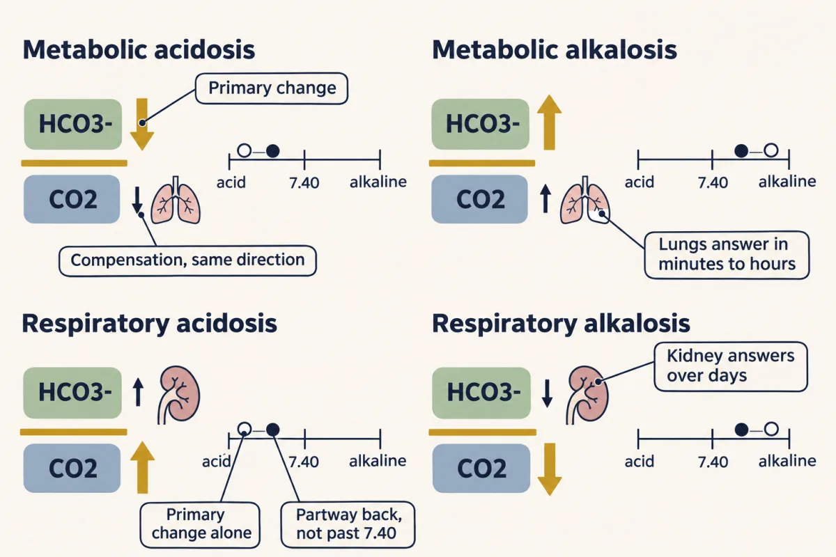
So two numbers that move in opposite directions, or a compensation far bigger or smaller than expected, mean two things are going on at once.
A recipe in four steps
- Is pH low or high? That names the problem: acidemia or alkalemia.
- Which term moved in the direction that explains it? A low pH comes from a fall in bicarbonate (metabolic) or a rise in pCO2 (respiratory). That term is the primary one; the other is usually the compensation.
- Is the compensation the expected size? Compare the other term with an empirical fit for that disorder.
- For a metabolic acidosis, compute the anion gap, corrected for albumin, to ask what kind of acid it is.
Worked problem
A made-up sample of human plasma at 37 C, at steady state, against a normal of bicarbonate 24 mmol/L, pCO2 40 mmHg, pH 7.40 and albumin 4.0 g/dL. The sample: pCO2 27 mmHg, bicarbonate 12, Na+ 140 and Cl- 104 mmol/L, albumin 2.0 g/dL.
Step 1: pH. Dissolved CO2 is 0.03 × 27 = 0.81 mmol/L. The ratio is 12 / 0.81 = 14.8, well below 20, and pH = 6.1 + 1.17 = 7.27: an acidemia. In concentration terms, [H+] = 10−7.27 mol/L, which is 109 − 7.27 = about 54 nmol/L, against 40 at pH 7.40.
Step 2: which term moved? Bicarbonate fell, which lowers pH; a primary rise in pCO2 would have raised bicarbonate. So this is a metabolic acidosis: acid gained or base lost. pCO2 fell too, but a falling pCO2 raises pH: it is the lungs answering, not the cause.
Step 3: is the compensation the right size? For metabolic acidosis the usual fit is Winter’s formula: expected pCO2 = 1.5 × bicarbonate + 8, plus or minus 2 mmHg. Here that is 1.5 × 12 + 8 = 26, so anything from 24 to 28 fits, and the measured 27 does, short of the 20 that full compensation needs. Without any compensation, at a pCO2 of 40, pH would have been 7.10; the lungs bought back about 0.17 of a pH unit.
Predict. Suppose the same patient’s pCO2 were 36 mmHg instead of 27. What would pH be, and what would that tell you?
Check your prediction
Dissolved CO2 is 0.03 × 36 = 1.08 mmol/L, the ratio is 12 / 1.08 = 11.1, and pH = 6.1 + 1.05 = 7.15. pCO2 is well above the 24 to 28 the fit expects, so ventilation is below the fit. That points to a second, respiratory acidosis on top of the metabolic one: the patient may be tiring, sedated or unable to breathe harder. A pCO2 well below 24 would mean the opposite, an added respiratory alkalosis.
Step 4: what kind of acid? The anion gap compares the main measured cation with the main measured anions: gap = [Na+] − [Cl-] − [HCO3-] = 140 − 104 − 12 = 24 mmol/L, against 12 in this lesson’s normal sample (140 − 104 − 24). The gap stands for anions the panel does not measure, less the cations it does not measure; albumin is the largest part. Two kinds of metabolic acidosis behave differently:
- Acid added with an unmeasured anion, such as lactic acid or a ketoacid. Its H+ uses up bicarbonate and its anion stays behind, so the gap rises.
- Bicarbonate lost directly, or not replaced, as from the gut or the kidney. Chloride takes its place, so plasma chloride rises and the gap stays normal. The B1 pump failure above is this kind.
A gap of 24 points to an added acid, and this patient’s low albumin hides part of it.
In words The corrected gap equals sodium minus chloride minus bicarbonate, plus two and a half times how far albumin sits below four grams per decilitre.
- corrected gap“corrected gap”
- the anion gap with its albumin correctionmmol/L
- [Na+]“sodium”
- plasma sodiummmol/L
- [Cl-]“chloride”
- plasma chloridemmol/L
- [HCO3-]“bicarbonate”
- plasma bicarbonatemmol/L
- 2.5“two point five”
- the correction per g/dL of albumin; 0.25 per g/L if albumin is in g/Lmmol/L per g/dL
- 4.0“four point oh”
- the comparison albumin in this lesson's worked problemg/dL
- albumin“albumin”
- the sample's plasma albuming/dL
Sanity check With albumin at the comparison 4.0 g/dL, the correction is zero and the corrected gap is just [Na+] − [Cl-] − [HCO3-]. Base lost directly is replaced by chloride, leaving the gap unchanged; an acid whose anion the panel does not measure leaves its anion, so the gap rises.
Worked example · try your own numbers
The worked problem: Na+ 140, Cl- 104 and HCO3- 12 mmol/L, albumin 2.0 g/dL.
Anion gap = 140 − 104 − 12 = 24 mmol/L.
Corrected gap = 24 + 2.5 × (4.0 − 2.0) = 29 mmol/L, the same as 24 + 0.25 × (40 − 20) with albumin in g/L.
Your numbers Change any box; the answer updates.
Need a refresher? log scales · units and prefixes
Here the corrected gap is 29 mmol/L: the added acid is larger than the raw gap suggested.
What these rules are, and are not. Winter’s formula and the other compensation rules are lines fitted to observed people, with real scatter, not laws; compare measured values with them and with the clinical setting. Winter’s formula is a fit for metabolic acidosis only: at a normal bicarbonate of 24 it returns 44, not 40. Normal anion-gap ranges differ between laboratories (in one, ion-selective electrodes raised measured chloride and shifted the range down; Winter and colleagues, 1990), so each laboratory’s own range applies. Comparing the rise in the gap with the fall in bicarbonate is a hint, not a proof (Going deeper).
Design problem
A new drug lowers plasma bicarbonate in volunteers. Design an experiment to show whether it impairs reclaiming filtered bicarbonate or making new bicarbonate. What would you measure in urine and plasma, and what result separates the two?
Saved in this browser only.
One way to do it
On a fixed diet, with a placebo period in the same people, measure plasma bicarbonate and pCO2 and, in timed urine, pH, bicarbonate, titratable acid and ammonium, so you can compute net acid excretion. Repeat under a standard acid load.
If the drug impairs reclaiming, bicarbonate appears in the urine at first, and again whenever plasma bicarbonate is raised: infuse bicarbonate to bring plasma toward normal and measure fractional bicarbonate excretion, which rises steeply (above about 15 percent in proximal defects). At steady state the urine can still be acidified below pH 5.5, and ammonium and titratable acid rise normally under an acid load.
If it impairs making new bicarbonate, urine bicarbonate stays low, but ammonium and titratable acid fail to rise under the acid load, so net acid excretion stays low while plasma bicarbonate falls. Check that filtration is unchanged, since it alters every urinary number.
Going deeper
The main line above does not depend on these details.
Henderson, Hasselbalch and the equation
The equation made blood acidity something to calculate from two measurable quantities. Henderson set out the mass-action relation in 1908, and in 1916 Hasselbalch rewrote it in Sorensen’s pH notation (carrying oxygen and carbon dioxide tells his work with Bohr and Krogh).
Base excess and the strong-ion view
The equation says nothing about acids other than CO2. Base excess, introduced in the late 1950s by Siggaard-Andersen with Astrup’s equilibration method, measures the metabolic part of a sample: the acid or base needed to bring whole blood in vitro to pH 7.40 at a pCO2 of 40 mmHg. Standard base excess adjusts it for the whole extracellular space, because in the body base excess still moves with pCO2. Bicarbonate read with pCO2, standard base excess and Stewart’s strong-ion difference give nearly the same answer for a given sample but explain it differently, and comparisons of Stewart’s approach with the traditional one have been inconsistent.
Inside the red cell
In a tissue capillary, CO2 entering a red cell is hydrated by carbonic anhydrase, most of the bicarbonate leaves on the Cl-/HCO3- exchanger AE1 as chloride enters, and hemoglobin buffers nearly all of the H+ formed. The Bohr and Haldane effects that couple this to oxygen are in carrying oxygen and carbon dioxide.
The size of the daily acid load
The roughly 50 mEq a day used in this lesson comes from urine studies. In a validation study that enrolled 1,082 US adults aged 50 to 74, the 765 who completed both 24-hour urine collections had a mean net endogenous acid production, estimated from urine, of about 47 to 48 mEq per day (interquartile range about 36 to 64).
How the tubule was shown to secrete acid
An older view held that the tubule acidified urine by reabsorbing buffer base, filtered bicarbonate and dibasic phosphate, leaving the acid forms behind; then titratable acid excreted could not exceed what that reabsorption allows. Robert F. Pitts and colleagues tested this in 1945 to 1949, in clearance studies of dogs and of people (as summarised in 1956 by Orloff), setting filtered loads against excretion. In metabolic acidosis, titratable acid excretion exceeded what reabsorbing filtered bicarbonate or dibasic phosphate could explain. H+ was being added to tubular fluid, and Pitts proposed it was extruded in exchange for filtered sodium. Orloff stressed there was no direct evidence yet for ion exchange in tubule cells; these were whole-kidney measurements, and the molecular exchanger came decades later.
Knockouts, an enzyme deficiency and the carbonate question
NHE3 knockout mice survive mildly acidotic; their in situ microperfused proximal tubules absorbed 54 percent less bicarbonate and 63 percent less fluid, and a significant part of the remainder was carried by a bafilomycin-sensitive H+-ATPase. In humans, William Sly and colleagues identified carbonic anhydrase II deficiency (1983) as the defect in an inherited syndrome that includes failed renal acidification. Unlike the B1 pump defect, the acidosis in carbonic anhydrase II deficiency is proximal, distal or combined, so proximal reclamation can fail as well. Boron’s group has reported that two Na+-coupled “bicarbonate” transporters, studied in Xenopus oocytes, carry carbonate (CO3 2-), with the quantitative test made on NBCe1.
Type-B and non-A, non-B cells
Type-B cells secrete bicarbonate through luminal pendrin and keep the H+-ATPase on the basolateral side; isolated cortical collecting ducts from alkali-loaded pendrin-knockout mice failed to secrete bicarbonate (Royaux and colleagues, 2001). Their bicarbonate secretion matters most when the body must excrete an alkali load: people with Pendred syndrome, who lack pendrin, have normal acid-base status at rest but have developed severe metabolic alkalosis on thiazide-type diuretics. A third, non-A/non-B type has both the pump and pendrin apically, and what it does on balance is unsettled.
Ammonium as a nitrogen carrier
Each glutamine the proximal tubule uses gives up two nitrogens as ammonium, so excreted ammonium also removes nitrogen. In this lesson ammonium matters mainly as an acid-base carrier rather than as the nitrogen route of osmoregulation, though it does carry some nitrogen.
Comparing the rise in the gap with the fall in bicarbonate
In the worked problem, against the made-up comparison gap of 12, the gap rose 12 (17 corrected) as bicarbonate fell 12, ratios of 1.0 and 1.4. That ratio genuinely varies, larger for lactate than for ketoacid anions, so alone it cannot establish a second process.
An older experiment: one kidney as the other’s control
In the chicken, blood from a leg vein drains directly into the peritubular region of the same-side kidney, so an agent infused into one leg reaches one kidney’s tubule cells while the other kidney is a control in the same bird. Orloff’s 1956 review reported, as unpublished observations, that acetic acid added to blood perfusing one side raised local CO2 tension (it reacts with plasma bicarbonate) and, on that side only, lowered potassium excretion (already raised by a potassium sulfate infusion) and raised urine H+ concentration; it credits the method to Sperber (1946). (a) What does the paired design buy that an intravenous infusion into a mammal cannot? (b) What shows whole-body acid-base state did not change? (c) Which rival explanation remains, and how would you exclude it?
One way to do it
(a) Each bird is its own control: systemic pH and pCO2, blood pressure and anaesthesia are shared, and the infused side meets the agent first in its peritubular region. Flow, filtration, electrolytes and delivery on each side still have to be measured rather than assumed equal.
(b) Arterial pH and pCO2 before, during and after, with urine collected simultaneously and separately from each ureter.
(c) The acid also lowers peritubular bicarbonate and may change sodium delivery and local blood flow. An acid carrying no CO2 still makes CO2 when it meets plasma bicarbonate, so these infusions cannot clamp peritubular pCO2, bicarbonate and pH separately. Measure these and filtration and flow on each side, with matched vehicle and salt controls, then set the bath directly in an isolated perfused tubule. Even then the potassium result only shows that local acid-base conditions shift potassium and proton handling together, without identifying a shared driving force or transporter, and one bird species cannot be generalised to mammals.
Your notes
Select any text in the lesson to highlight it or add a note to it. Your highlights and notes are saved in this browser and collected in your notebook.
This browser is not letting the site save anything (a private window or strict privacy settings), so highlights and notes will be lost when you leave the page.
How we measure it
Blood-gas analysis with equilibration
Electrodes measure the pH and carbon dioxide tension of a blood sample, and bicarbonate is calculated from the Henderson-Hasselbalch equation. Equilibrating the sample with gas of known carbon dioxide tension, Astrup's method, separates the respiratory part of a pH change from the non-respiratory part and gives base excess.
Clearance and timed urine collection
Measure plasma and urine concentrations together with urine flow, and compare what was filtered with what was excreted. Timed and 24-hour collections give titratable acid, ammonium and bicarbonate excretion, and so net acid excretion.
Tubule microperfusion
Perfuse a single proximal tubule in situ, or an isolated collecting duct held between pipettes, with fluid of known composition, and measure bicarbonate, fluid or ammonia flux. Adding inhibitors one at a time estimates how much of a flux each inhibitor-sensitive pathway carries, if specificity and completeness of the block are checked.
Knockout mice under acid or alkali loading
Delete one transporter gene and compare blood gases and urine with wild-type littermates, at baseline and after an acid or alkali load. A phenotype that appears only under loading shows what the protein is needed for when the system is pushed.
Practice questions on this lesson (6) → MCAT-style, original, each with an explanation
Where this lesson fits
Before thisReview first
- Glomerular Filtration and Renal Blood FlowThis course · Lesson 15
You need the daily filtered volume from there to see how much bicarbonate the kidney must reclaim.
- Ventilation, Perfusion and Gas ExchangeThis course · Lesson 13
Alveolar ventilation sets the CO2 term of the ratio, which this lesson takes as given.
- Tubular Transport, Segment by SegmentThis course · Lesson 16
The proximal tubule transporters and carbonic anhydrase inhibition from there are used in the bicarbonate reclaiming job.
After thisStudy next
- Exercise: The Whole Body at WorkThis course · Lesson 30
Lactate and acid load during hard exercise put the buffering and compensation ideas to work.
- Insulin, Glucagon and Blood GlucoseThis course · Lesson 21
Ketoacid production from missing insulin is a main cause of the added-acid acidosis read here with the anion gap.
Elsewhere on the siteFilms, simulations and method films in other courses
- Why Blood Needs an S-Shaped CarrierFilm · Organismal Biology
Hemoglobin's role as a red-cell buffer carrying carbon dioxide is shown there.
Suggested by an AI model from the lessons themselves, then checked. How we use AI
Finish line
Test yourself
13 questions
Practice and MCAT-style, with explained answers.
Next · Lesson 19
Thyroid Hormone: Tissues Decide What T4 Becomes
A person with no thyroid who takes only T4 still has T3 in the blood. Where does it come from?
Continue →