Neuroscience · Unit 5: Homeostasis and affect
Autonomic Control and the Hypothalamus
Rewritten on : new text, figures, flashcards and film. Your notes and highlights stay, but the text around them may have moved.
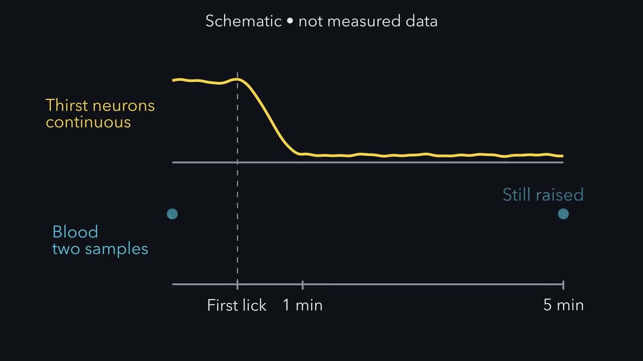
A thirsty mouse quiets its thirst neurons within a minute of drinking, while its blood osmolality is still raised five minutes later.
Pause when the reading reaches a film, and offer to play it. Saved on this device.
Watch the film now, or keep listening
Builds on:Synaptic Transmission and Quantal ReleaseTransmitter Systems and DrugsWhy these?
 ▶ Watch Correction, Threshold and Gain · 2:58
Read the transcript of “Correction, Threshold and Gain”
Film transcript: Correction, Threshold and Gain
A thirsty mouse starts to drink. Within a minute, its thirst neurons go quiet. Yet its blood osmolality is still raised. What are they reading? Start with a loop. For water, the thing held steady is plasma osmolality, the concentration of dissolved particles in plasma. The SFO and OVLT sit in the front wall of the third ventricle. They sample the blood directly, where the blood-brain barrier is incomplete. A third site behind the barrier, the MnPO, integrates their signals. Two effectors push back. Vasopressin saves water at the kidney. Thirst takes water in. Both lower osmolality. The response opposes the error, so this is negative feedback.
Is there a separate comparator that judges the error? The comparison is spread across the sensing and integrating neurons. We summarize the result with a threshold and a gain. Raise osmolality in a controlled way and record vasopressin. The plot is flat, then rises in a straight line. The knee is the threshold. It says when release starts. The slope is the gain. It says how much release grows. Suppose osmolality rises, but stays below threshold. Does vasopressin change?
No. Below threshold there is no osmotic release. The line does not go negative. Now two made-up people. Person P has the lower threshold. Person Q has the steeper gain. Just above P's threshold, who releases more? And far above both thresholds?
Just above P's threshold, P does. Q has not even started. Far above both, Q does. The steeper line wins. So one number, a set point, cannot describe the loop. It takes at least two. For osmotic vasopressin, the line is measured. Output is the gain, times how far osmolality is past threshold. But water-balance circuits do not read the blood alone. Think of them as acting on an estimate. Feedback reads the blood. Feedforward adds what was just swallowed, or a learned cue. The hypothalamus acts through nerves, often in seconds. It also acts through hormones, whose effects usually build over minutes to hours. Threshold says when. Gain says how much.
Keep the word estimate. The thirst neurons will show why.
From the film
- The osmotic vasopressin response needs two numbers: a threshold, where release starts, and a gain, how much release grows above it.
- Below threshold there is no osmotically driven release, so the response is flat and then rises in a straight line.
- Water-balance circuits act on an estimate: feedback reads the blood, and feedforward adds what was just swallowed or a learned cue.
- Nerves often act within seconds, while hormone effects usually build over minutes to hours.
A thirsty mouse that starts to drink quiets a population of its thirst neurons within a minute, yet five minutes later its blood osmolality is still raised.
These neurons drive thirst, so their quieting means the brain has judged that water is coming before any change in the blood is detected. If the brain only corrected errors it had measured in the blood, that would be hard to explain. To see what the neurons are reading, you need the idea this lesson is built on and the ways the hypothalamus sends its orders out.
The idea needs four terms. A regulated variable is a quantity the body holds within a range. For water it is plasma osmolality, the concentration of osmotically active particles per kilogram of water in plasma, the liquid part of the blood. A threshold is the value at which a corrective response starts. The gain is how much the response grows for each unit the variable moves past threshold. That distance past threshold is the error.
The one idea to carry through this lesson. The osmotic vasopressin response needs two numbers, not one set point: a threshold, where its osmotic component starts, and a gain, its slope above threshold. Above threshold, AVP = gain × (osmolality − threshold); below it, no osmotically driven release. Water-balance circuits also integrate more than the blood, and it helps to think of them as acting on an estimate of fluid balance: the blood, corrected for what is already on its way. Feedback builds the estimate from the blood. Feedforward updates it from what has just been swallowed, or from a learned cue, before the blood changes.
The hypothalamus acts through three kinds of output: pituitary hormones in the blood, autonomic nerves, and behaviour such as drinking.
The loop, built on water
Water is the running example. The loop has three parts you can point to.
What is held steady. Plasma osmolality. When you lose water, it rises.
What senses it. Osmosensitive neurons, mainly in the lamina terminalis, the front wall of the third ventricle. Two of its parts, the subfornical organ (SFO) and the organum vasculosum (OVLT), are circumventricular organs: their blood-brain barrier is incomplete, so their neurons sample the blood directly. The third part, the median preoptic nucleus (MnPO), sits behind the barrier and integrates their signals. The magnocellular vasopressin neurons, described next, are also osmosensitive themselves.
What pushes back. Two effectors: vasopressin saves water at the kidney, and thirst takes water in. Both lower osmolality, so the response opposes the error. That makes the loop negative feedback.
The hypothalamus is a small region at the base of the forebrain, below the thalamus, built of nuclei, clusters of neuron cell bodies inside the brain. Vasopressin is the first of its outputs you need. Large, magnocellular neurons in the supraoptic nucleus (SON) and the paraventricular nucleus (PVN) make vasopressin (AVP) and oxytocin in their cell bodies, carry them down their axons, and release them from terminals in the posterior pituitary into the systemic blood. The posterior pituitary stores and releases vasopressin; it does not make it.
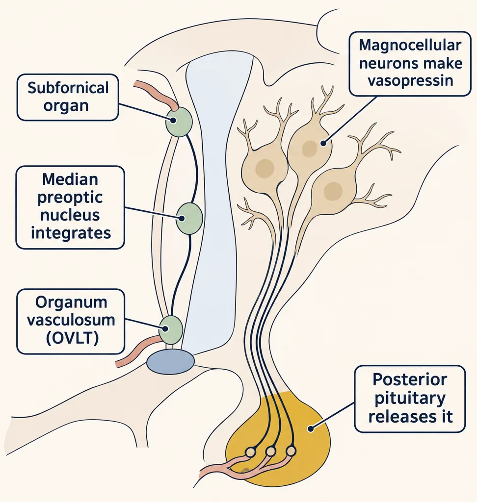
In the kidney, vasopressin acts on V2 receptors of collecting-duct cells and, through cAMP (cAMP and PKA), increases the aquaporin-2 water channels in their membranes, so more water returns to the blood (membrane transport covers osmosis).
An aside on the comparator. Textbook loops often add a comparator, a part that decides how far the variable is from where it should be. Here the comparison is a function, not a separate part: it is spread across the sensing and integrating neurons, the MnPO among them. The threshold you measure summarizes the whole response and need not equal any one neuron’s firing threshold, so measure both threshold and gain.
Threshold and gain
Picture the experiment. Raise plasma osmolality in a controlled way in healthy adults and measure vasopressin as you go. Plot vasopressin up the side and osmolality along the bottom. The plot is flat at first, then rises in a straight line. The knee is the threshold, when release starts; the slope above it is the gain, how much it grows.
Endotext’s review summarizes such data from healthy adults under controlled hypertonic stimulation with a regression line for the straight part:
AVP (pmol/L) = 0.43 × (Posm − 284), for Posm above 284 mOsm/kg
Here Posm is plasma osmolality, 284 mOsm/kg is the threshold and 0.43 pmol/L per mOsm/kg is the gain. Treat both as illustrative averages: thresholds and slopes vary between individuals. In this line the estimate is plasma osmolality and the output is the plasma vasopressin concentration. It predicts a hormone level, not how fast osmolality changes.
- Regulated variableplasma osmolality (horizontal axis)
- Sensorosmosensitive neurons of the lamina terminalis
- Effectorvasopressin (vertical axis); thirst is not drawn
- line from the sliders
- cursor
- threshold
- 294 mOsm/kg reference
- person P
- person Q
- crossing of P and Q
- Excess over threshold
- AVP at the cursor
- Change from the 294 mOsm/kg reference
- Person P at the cursor
- Person Q at the cursor
Rule to keep: the threshold is where release starts and the gain is how fast it grows above it, so a “set point” is not one fixed number; below threshold the output is zero, never negative. In this sketch: a static steady-state line, no time course and no rehydration, mechanism only. The defaults, 284 mOsm/kg and 0.43 pmol/L per mOsm/kg, are illustrative averages from an endocrinology reference for healthy adults under controlled hypertonic stimulation, as the lesson says; other slider settings are not data; thresholds and slopes vary between people, and P and Q are made up.
Predict. Plasma osmolality rises from 282 to 286 mOsm/kg. Does vasopressin change?
Check your prediction
Only for the last part of the rise. From 282 to 284 the person is below threshold. Plugging 282 into the formula would give a negative concentration, which is impossible: the line describes release only above threshold, so it predicts no osmotically driven release there. With little vasopressin the kidney passes dilute urine, so dilute blood is corrected by the hormone’s absence. Once osmolality passes 284, vasopressin rises by 0.43 pmol/L for each mOsm/kg beyond it, so at 286 it is 0.43 × 2 = 0.86 pmol/L.
Worked problem. A hypothetical adult follows this line. Plasma osmolality is 294 mOsm/kg, and after drinking it is 290 mOsm/kg. Assume the same relation holds at both times and that AVP has reached steady state for each value.
Step 1: the first error (distance above threshold). 294 − 284 = 10 mOsm/kg above threshold. AVP = 0.43 × 10 = 4.30 pmol/L.
Step 2: the second error. 290 − 284 = 6 mOsm/kg. AVP = 0.43 × 6 = 2.58 pmol/L.
Step 3: the change. 2.58 − 4.30 = −1.72 pmol/L. Check it another way: 0.43 × (290 − 294) = 0.43 × (−4) = −1.72 pmol/L. Less error, less hormone, less water saved.
Predict. Compare two made-up people. Person P has a threshold of 280 mOsm/kg and a gain of 0.43. Person Q has a threshold of 284 and a gain of 0.86. Who releases more vasopressin at 286 mOsm/kg? Who releases more at 290?
Check your prediction
At 286, P does; at 290, Q does.
| Posm (mOsm/kg) | P: 0.43 × (Posm − 280) | Q: 0.86 × (Posm − 284) |
|---|---|---|
| 282 | 0.86 | 0 (below threshold) |
| 284 | 1.72 | 0 (at threshold) |
| 286 | 2.58 | 1.72 |
| 288 | 3.44 | 3.44 |
| 290 | 4.30 | 5.16 |
Solve 0.43 × (Posm − 280) = 0.86 × (Posm − 284) and the lines cross at 288 mOsm/kg. Between 280 and 288 the lower threshold wins; above 288 the steeper gain wins. At or below 280 both release none. Overlay P and Q in the simulation to see the crossing.
Two people can give the same output by different routes, so one number cannot describe the loop.
Two ways out: blood and wires
Read the transcript of “Two Ways Out: Blood and Wires”
Film transcript: Two Ways Out: Blood and Wires
The hypothalamus has a correction to send. How does it leave the brain, and how fast? Nerves often act within seconds. Hormone effects usually build more slowly, over minutes to hours. Start with vasopressin. Large neurons in the supraoptic and paraventricular nuclei make it. Their axons release it in the posterior pituitary, straight into the blood. So the posterior lobe stores and releases vasopressin. It does not make it. The anterior lobe is reached by blood instead. Neurons release peptides into capillaries at the median eminence. Portal veins carry them down the stalk to a second capillary bed. They arrive concentrated, before the general circulation dilutes them.
Take the stress axis. The paraventricular nucleus releases corticotropin-releasing hormone, CRH. That drives adrenocorticotropic hormone, ACTH, from the pituitary. ACTH drives cortisol from the adrenal cortex. Cortisol feeds back on the hypothalamus and pituitary, and turns the axis down. Same loop, different variable: here, a hormone. Now the wires. The usual autonomic chain has two neurons and two synapses: one in a ganglion, one on the target. Rule one. The first synapse is the same everywhere: acetylcholine, acting mainly on nicotinic receptors. Rule two. The second synapse usually names the division. Parasympathetic: acetylcholine on muscarinic receptors.
Most sympathetic: noradrenaline on adrenergic receptors. Atropine blocks muscarinic receptors. Give a large dose. What happens to the heart rate? And to sweating? At a large dose, the heart rate rises. The vagal brake on the heart's own pacemaker was on all the time, even at rest. Sweating stops. The sympathetic fibres to sweat glands release acetylcholine onto muscarinic receptors. That is the first exception. Separate tests show steady sympathetic drive too. So the heart sits at a balance, like a sink filled from two taps. The second exception is the adrenal medulla. There, the second neuron has become a gland that releases adrenaline and noradrenaline into the blood.
The paraventricular nucleus uses every route: portal blood, the posterior lobe, and descending autonomic pathways. Two routes out. Nerves often work in seconds. Hormone effects usually build over minutes to hours.
From the film
- The posterior pituitary is reached by axons and the anterior pituitary by blood, through portal veins that carry releasing hormones.
- In the stress axis, CRH drives ACTH, ACTH drives cortisol, and cortisol feeds back to turn the axis down.
- The usual autonomic chain has two neurons: acetylcholine on nicotinic receptors at the ganglion, then at the target usually acetylcholine on muscarinic receptors for parasympathetic and noradrenaline for most sympathetic.
- Sweat glands and the adrenal medulla are the exceptions to the usual sympathetic pattern.
The hypothalamus receives visceral sensory information (so do brainstem centres), and it reaches the body’s organs in two ways. One is by blood: the pituitary gland below it, whose posterior lobe you have just met. The other is by wires: descending pathways to the autonomic nervous system. These are distinct outputs with their own targets, not one signal sent two ways. Nerves often act within seconds; hormone effects usually build over minutes (vasopressin at the kidney) to hours (the cortisol and thyroid axes).
The blood channel: the anterior pituitary
The two pituitary lobes are controlled in completely different ways. The posterior lobe is reached by axons. The anterior pituitary is reached by blood. Neurons in hypothalamic nuclei send axons to the median eminence at the base of the hypothalamus and release peptides into capillaries there. Those capillaries drain into the long hypophysial portal veins, which run down the stalk and open into a second capillary bed in the anterior pituitary. A portal system is two capillary beds joined in series, so a releasing hormone reaches the gland at high concentration before it is diluted in the general circulation.
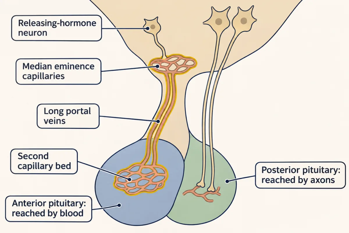
Two axes show the pattern, and both are negative-feedback loops with a hormone as the regulated variable.
- In the HPA axis, corticotropin-releasing hormone (CRH), made mainly in the paraventricular nucleus (PVN), stimulates corticotroph cells of the anterior pituitary to release adrenocorticotropic hormone (ACTH). ACTH stimulates the adrenal cortex to make and release cortisol. Cortisol then inhibits the axis at both the hypothalamus and the pituitary.
- In the HPT axis, thyrotropin-releasing hormone (TRH) stimulates the pituitary to release thyroid-stimulating hormone (TSH). TSH is the main regulator of the thyroid. Thyroid hormones feed back at both levels.
Picture each axis as two arrows down (hypothalamus to pituitary, pituitary to gland) and one inhibitory arrow back up from the gland’s hormone, branching to both the hypothalamus and the pituitary. With other inputs held constant, a rise in cortisol or thyroid hormone turns down the drive from above. The shared shape does not mean these axes follow the vasopressin line. Cortisol and thyroid hormone act on intracellular receptors (nuclear receptors). The HPA axis in stress belongs to emotion and stress.
Predict. A patient takes a high dose of a cortisol-like drug for months. What happens to CRH, to ACTH and to the adrenal cortex?
Check your prediction
The drug acts like cortisol at the hypothalamus and pituitary, so the feedback arrow is pressed hard: CRH and ACTH are both suppressed. With little ACTH, the ACTH-dependent zones of the adrenal cortex shrink. That is why such drugs are withdrawn slowly: the axis needs time to recover.
The wired channel: two synapses, two rules, two exceptions
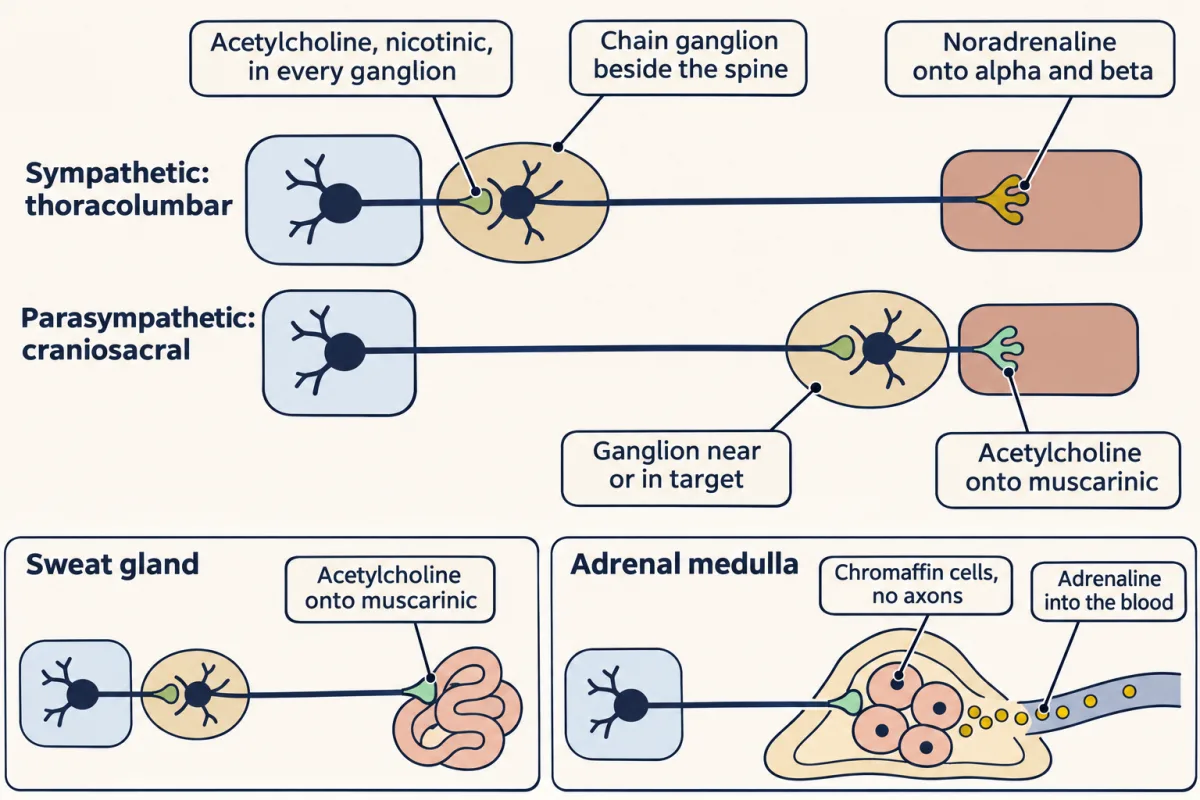
The autonomic nervous system controls cardiac muscle, smooth muscle and glands. Descending pathways, many from the PVN, reach its preganglionic neurons, whose cell bodies lie in the brainstem or spinal cord. Each preganglionic axon runs out to a ganglion, a cluster of neuron cell bodies outside the central nervous system, and synapses there on a postganglionic neuron, whose axon reaches the target. So the usual chain has two neurons and two synapses: one in the ganglion, one on the target. The adrenal medulla, below, is the exception.
The two divisions differ in where the chain starts and where the ganglion sits.
- The sympathetic division leaves the spinal cord from the thoracic and upper lumbar segments, so it is called thoracolumbar. Its preganglionic axons are relatively short. They end in the paravertebral chain of ganglia beside the vertebral column or in prevertebral ganglia in the abdomen, and the postganglionic axons are relatively long.
- The parasympathetic division is conventionally called craniosacral; whether the sacral outflow belongs here is disputed (see the anecdote “Is the sacral outflow parasympathetic?”). Its preganglionic neurons lie in brainstem nuclei, whose axons leave in cranial nerves, and in the sacral cord. Its preganglionic axons are long and end in ganglia near or inside the target, so its postganglionic axons are short.
You can work out the transmitter at the main autonomic synapses from two rules and two exceptions; the transmitters themselves are in transmitter systems.
Rule 1: the first synapse is the same everywhere. Every preganglionic neuron, sympathetic or parasympathetic, releases acetylcholine (ACh). In the ganglion it acts mainly on nicotinic receptors, ligand-gated cation channels like those at the neuromuscular junction, though of a neuronal subtype, so the postganglionic neuron is excited fast. Ganglia also use slower muscarinic and peptide transmission, hence mainly.
Rule 2: the second synapse usually names the division. Parasympathetic postganglionic neurons release acetylcholine onto muscarinic receptors on the target. Most sympathetic postganglionic neurons release noradrenaline onto alpha and beta adrenergic receptors. Muscarinic and adrenergic receptors are G-protein-coupled receptors (G proteins), so the target responds more slowly than the ganglion does.
Exception 1: sweat glands. The sympathetic fibres to eccrine sweat glands release acetylcholine onto muscarinic receptors. They are sympathetic by anatomy, leaving the thoracolumbar cord and relaying in the chain ganglia, and cholinergic by chemistry.
Exception 2: the adrenal medulla. The adrenal medulla is a modified sympathetic ganglion in which the second neuron has become a gland. Preganglionic fibres end directly on its chromaffin cells, modified postganglionic neurons with no axons, which release adrenaline and noradrenaline into the blood. This one sympathetic wire ends in a hormone.
| Synapse | Parasympathetic | Sympathetic, most targets | Sympathetic, sweat glands | Adrenal medulla |
|---|---|---|---|---|
| In the ganglion | ACh → nicotinic | ACh → nicotinic | ACh → nicotinic | ACh → nicotinic, on chromaffin cells |
| On the target | ACh → muscarinic | noradrenaline → alpha, beta | ACh → muscarinic | none: adrenaline and noradrenaline go into the blood |
So “sympathetic” does not mean “noradrenaline at every synapse”.
Predict. Atropine blocks muscarinic receptors. The vagus nerve carries the parasympathetic fibres to the heart. Someone is given a large dose of atropine. What happens to their heart rate, and to their sweating?
Check your prediction
The heart rate rises and sweating stops. The vagus slows the heart’s own pacemaker through muscarinic receptors, and it does so all the time, even at rest; remove that brake and the rate climbs. Sweating stops because the sympathetic sweat-gland fibres act on muscarinic receptors, which is Exception 1 at work. The skin becomes hot and dry.
Both divisions are active at rest, at a tonic level, a steady background rate of firing. Atropine revealed the vagal brake. Separate tests, such as blocking beta receptors, show steady sympathetic drive to the heart as well. So the heart sits at a balance between a steady parasympathetic brake and steady sympathetic drive, like a sink filled from a cold tap and a hot tap. Turning either tap changes the temperature, and in a resting person the cold tap is the stronger. “Fight or flight” and “rest and digest” describe which tap is turned up, not two positions of a switch, and many organs are set by the balance of the two at once.
Some targets have only one tap. Not every organ has dual innervation, a supply from both divisions. Most arteries, the sweat glands and the adrenal medulla receive sympathetic fibres and no parasympathetic ones. The nervous system controls them by turning that one input up or down. In many arteries, more sympathetic firing narrows the vessel and less lets it widen; local signals in the vessel wall also set its width.
One nucleus, three channels
The PVN uses every route in this section. Its parvocellular (small-celled) neurons release CRH into portal blood, its magnocellular neurons release vasopressin from the posterior lobe, and its pre-autonomic neurons descend to the brainstem and spinal cord. Of the three, the autonomic outflow is usually the fastest.
Thirst: acting before the blood changes
Read the transcript of “Acting Before the Blood Changes”
Film transcript: Acting Before the Blood Changes
Back to the thirsty mouse. Its thirst neurons go quiet within a minute. No change in the blood has been detected. So what has changed? Researchers recorded subfornical organ neurons in awake mice, using an optical fibre and a calcium indicator.
Picture the neurons and the blood on one time axis. A line marks when water arrives. The neurons' activity starts to fall at the first lick. Within a minute, it is down. The blood was sampled, not traced. Plasma osmolality was still raised five minutes after water access. What could turn the neurons down? The sight of water? The licking? Or the water itself?
Seeing water it could not reach did not lower the activity. Licking an empty bottle did not either. So it is something that comes with fluid going in. Drinking concentrated saline lowers it at first, like water. After about a minute, the fall reverses. A fast signal counts fluid going in. A slower one reports what the fluid contains. Next, bypass the mouth. Put water straight into the stomach. In dehydrated mice, the activity falls in about two minutes. Separate blood measurements found no detectable change in osmolarity, so the response comes first. Hypertonic saline in the stomach raises the activity instead. Then researchers used light to hold the neurons active.
Now water in the gut no longer brings its usual satiation. Suppose the brain read only the blood. How much would a thirsty animal drink? Too much, if it drinks faster than water is absorbed. Water reaches the blood gradually, so a blood-only brain keeps drinking.
So the estimate must count what is on its way. The brain can also learn. In trained mice, vasopressin neurons dipped when a water bowl arrived, before any drinking. So feedforward can sense fluid going in, or predict it from a learned cue. Keep the limits. This is population calcium, recorded in mice. It is not the feeling of thirst. Feedback reads the blood. Feedforward reads what is coming. The estimate is the blood, corrected for what is on its way.
From the film
- In awake mice, subfornical organ neuron activity fell within a minute of drinking, while plasma osmolality was still raised five minutes after water access.
- Seeing unreachable water or licking an empty bottle did not lower the activity, so the signal comes with fluid going in.
- Water put directly into the stomach of dehydrated mice lowered the activity in about two minutes, before any detectable change in blood osmolarity.
- Feedback reads the blood and feedforward reads what is coming; this is population calcium in mice, not the feeling of thirst.
Now back to the mouse. A pure feedback loop reacts to an error it has measured. Feedforward control acts on a disturbance that has been sensed or predicted before it changes the regulated variable. Mouse experiments show that thirst uses both.
Start with what the subfornical organ (SFO) can do. In 2015 Oka, Ye and Zuker drove excitatory SFO neurons with light in water-satiated mice, and the mice drank; driving inhibitory SFO neurons in thirsty mice cut licking sharply (numbers in Oka’s profile). That shows the cells are enough to push drinking up or down. It does not show they are the only route, or that the SFO is a “thirst centre”.
The next question was what these neurons do during ordinary drinking. Christopher Zimmerman, Zachary Knight and colleagues used fibre photometry, an optical fibre that collects the fluorescence of a calcium indicator from a whole population, to record SFO neurons marked by the gene Nos1, in awake mice. Nos1 gives a genetic handle for targeting these neurons; the enzyme it encodes is not the point here.
The picture
Picture their result (Nature, 2016) from water-restricted mice on one time axis, as a schematic. A vertical line marks the moment water becomes available. The top trace is SFO Nos1 population activity, recorded continuously: it starts to fall at the first lick and is down within one minute. The blood was not traced. Plasma osmolality was measured in separate samples by freezing-point osmometry, so picture it as points: it is still raised five minutes after water access.
So a fall in the blood is unlikely to be what turns the neurons off; something that comes with drinking does. The controls narrow down what.
Predict. Concentrated (hypertonic) saline is salt water more concentrated than plasma. It is a fluid, but drinking it would raise plasma osmolality, not lower it. If a mouse drinks it, does SFO activity fall?
Check your prediction
Yes, at first, just as with water. But that fall reverses after about a minute. The fast signal counts fluid going in; a slower signal reports what the fluid contains.
| Condition | What it isolates | SFO Nos1 activity |
|---|---|---|
| Water-restricted mice drink water | the whole act of drinking | falls at the first lick, down within a minute |
| Salt-challenged mice see water but cannot drink | the sight or expectation of water | does not fall |
| Salt-challenged mice lick an empty bottle | the licking movement | does not fall |
| Mice drink concentrated saline | fluid going in, but salty enough to raise osmolality | falls at first, then reverses after about a minute |
| Fasted mice eat | a meal, which can later draw water out of the blood | rises |
Read the rows in turn. Seeing water fails, so it is not the sight or expectation of water alone in this paradigm. Air-licking fails, so it is not the licking movement. Eating raises activity, as if the neurons anticipate the water that a meal will draw out of the blood (into the gut, and as absorbed food raises osmolality). An oral clue appears in the anecdote “Cold metal on the tongue”.
Design problem
The fast signal comes with fluid going in. But the blood was sampled at set times, not continuously, so a small early change could have been missed. What experiment would show where the signal comes from, whether it travels by a nerve, and whether the fall in SFO activity matters for ending thirst? (Hint: where else could fluid enter the body?)
Saved in this browser only.
One way to do it
Bypass the mouth. Infuse water or hypertonic saline straight into the stomach while you record, and sample blood at the same times. If gut water lowers activity and gut saline raises it before blood osmolality moves, the gut is reporting what came in. Then cut the abdominal vagus nerve in one group and sham-operate another, to ask whether the signal travels that way. Finally, test whether the fall in SFO activity matters: hold the neurons active with light during the infusion and see whether the water still ends thirst.
What the gut experiment found
Zimmerman and colleagues did these experiments in 2019. In dehydrated mice, water infused into the stomach inhibited the neurons after 107 ± 15 s (mean ± SEM), about two minutes; in hydrated mice the latency was 105 ± 13 s. Hypertonic saline activated them. Blood osmolarity, measured separately in hydrated mice from samples begun three minutes into the infusion, showed no detectable change, so the response comes ahead of any detectable systemic correction. Subdiaphragmatic vagotomy attenuated the satiating effect of gut water, and holding the neurons active prevented that satiation. So the gut reports what has come in, ahead of the blood.
Why does the brain need that? Swallowed water is absorbed gradually, so the blood lags behind fast drinking. An animal that read only its blood could keep drinking while water was still on its way, and drink well past its deficit. A toy model (Going deeper) shows that this overshoot appears when drinking is fast compared with absorption, and vanishes when the estimate also counts swallowed water not yet absorbed. That is the estimate in the one idea: the blood, corrected for what is on its way.
A cue the brain learns
The gut and mouth signals sense fluid entering. Feedforward can also predict. Kim and colleagues (2021) found that in trained, water-restricted mice the arrival of a water bowl lowered vasopressin-neuron activity before the mice drank; the anecdote “A cue the brain learns” gives the details and the circuit. The rise with eating in the table is best read as a prediction too. Feedback reads the blood; feedforward reads what is coming.
Keep the limits. Photometry gives population calcium, not spikes and not the feeling of thirst. The work is in mice. Other sites, including the median preoptic nucleus (MnPO), integrate oral, gut and blood signals, so the result is a circuit, not a single centre.
Prediction of this kind returns for feeding in motivation and reward and for daily timing in sleep and rhythms.
Going deeper
What the threshold-gain line does not cover
The line fits only the roughly straight part of the osmotic vasopressin response above threshold. The mouse recordings show that thirst circuits integrate anticipatory signals as well as the blood; they are not fitted to this equation, and no equation of this kind is given here for thirst, the endocrine axes or autonomic outflow. Nor do the experiments establish a common two-parameter law, or an optimal estimator, for hypothalamic responses in general; the estimate is a useful way to think, not a measured quantity. The line does not cover vasopressin released by falls in blood volume or pressure, which follows a steeper, non-linear relation, or vasopressin released by nausea; these can release vasopressin below the osmotic threshold. It does not describe the circadian and pulsatile rhythms of the HPA axis. The wiring that carries the outputs is anatomy; the idea says how that wiring is used, not why it is built that way.
The enteric nervous system and the origin of ganglion neurons
Many textbooks also count the enteric nervous system in the gut wall as a third autonomic division; this lesson covers the other two. Autonomic ganglion neurons come from the neural crest, as neural crest explains; in the adult they are mature relay stations.
Vasopressin also reaches the anterior pituitary
Vasopressin is not only a posterior-pituitary hormone. Parvocellular paraventricular nucleus (PVN) neurons also release vasopressin into portal blood, where it amplifies the effect of corticotropin-releasing hormone (CRH) on adrenocorticotropic hormone (ACTH) release.
Fever in threshold language
Fever offers a contrast to the water loop. In the standard model, fever raises the thresholds at which heat-producing and heat-saving responses switch on, so a person shivers and feels cold while body temperature is still normal. Textbooks often call this a raised thermoregulatory set point, but whether there is a single thermoregulatory set point is itself contested. Most work on the prostaglandin E2 mechanism behind that shift comes from rodents.
A model of drinking with and without feedforward
A simple model makes the case for feedforward. Let D be the body-water deficit and G the swallowed water not yet absorbed, both in litres, with time in minutes. Start with D = 1 and G = 0, and ignore ongoing losses. Water leaves the gut at G/τ per minute, where τ is the absorption time constant, so D falls at G/τ and G changes at u − G/τ, where u is the drinking rate.
- Blood-only estimate: u = k × D, and drinking stops when D first reaches zero. With k = 1 per minute and τ = 5 minutes, drinking stops at about 4.1 minutes, after 2.48 L. With τ = 30 minutes it stops at about 9.1 minutes, after 5.70 L. The extra is still in the gut when drinking stops.
- Estimate that counts swallowed water: u = k × (D − G), the predicted deficit. The predicted deficit shrinks steadily toward zero, and total intake approaches 1.00 L for any k and τ.
Blood-only control also approaches 1.00 L when k × τ is 1/4 or less, that is, when drinking is slow compared with absorption. These are illustrative outputs of a toy model of the logic, not a fit to any animal’s data; other gains and absorption rates give other overshoots.
Your notes
Select any text in the lesson to highlight it or add a note to it. Your highlights and notes are saved in this browser and collected in your notebook.
This browser is not letting the site save anything (a private window or strict privacy settings), so highlights and notes will be lost when you leave the page.
How we measure it
Fibre photometry
A genetically encoded calcium indicator such as GCaMP is expressed in a chosen cell type, and an optical fibre over the region collects its fluorescence in an awake animal. The signal tracks the summed calcium activity of the population over seconds; it does not resolve single cells or spikes, and it does not measure a feeling such as thirst.
Optogenetic activation and silencing
A light-gated channel or pump is expressed in a defined cell type, and light delivered through a fibre excites or inhibits those cells on command. Driving cells tests whether their activity is enough to produce a behaviour; silencing them tests whether it is needed, in that preparation and task.
Infusion of hypertonic solutions
Small volumes of concentrated solutes are infused into an artery or the stomach while urine flow, a hormone or neural activity is recorded. Comparing solutes that do and do not cross cell membranes tells you whether a sensor responds to osmotic shrinkage or to a particular substance.
Pituitary stalk section and grafting
Cutting the stalk, or moving pituitary tissue to a new site, separates the gland from its neural and vascular links to the hypothalamus. Comparing the hormone output of grafts at different sites tests which link carries the control.
Practice questions on this lesson (3) → MCAT-style, original, each with an explanation
Where this lesson fits
Before thisReview first
- Synaptic Transmission and Quantal ReleaseThis course · Lesson 5
You need how a synapse releases transmitter onto receptors to follow the two-synapse autonomic chain.
- Transmitter Systems and DrugsThis course · Lesson 6
Naming acetylcholine and noradrenaline at each autonomic synapse uses their nicotinic, muscarinic and adrenergic receptors from there.
After thisStudy next
- Circadian Rhythms and SleepThis course · Lesson 21
Daily timing is another prediction made before the body changes, building on acting ahead of the error.
- Emotion and StressThis course · Lesson 20
It uses the PVN's CRH output and the HPA axis with cortisol feedback introduced here.
Elsewhere on the siteFilms, simulations and method films in other courses
- Step through a feedback loopSimulation · Biology
It steps through a feedback loop with a regulated variable, sensor and effector, as used for water balance here.
- Read the Pair, Not One NumberFilm · Physiology
It shows how to read hypothalamic-pituitary-adrenal axis hormones in pairs, extending the CRH, ACTH and cortisol feedback chain.
- Sweat Cools Only When It EvaporatesFilm · Physiology
Cholinergic sympathetic sweat-gland fibres drive the cooling response discussed as an autonomic exception.
Suggested by an AI model from the lessons themselves, then checked. How we use AI
Finish line
Test yourself
8 questions
Practice and MCAT-style, with explained answers.
Next · Lesson 19
Feeding, Motivation and Reward
A well-fed mouse starts eating within minutes when one small group of hypothalamic neurons is switched on with light. What are those neurons for?
Continue →