Cell Signaling · Unit 6: Channels, the nucleus, and life or death
Nuclear Receptors: Receptors That Are Transcription Factors
What does a cell do with a hormone that goes straight through its membrane?
Pause when the reading reaches a film, and offer to play it. Saved on this device.
Watch the film now, or keep listening
Builds on:A Field Guide to ReceptorsBinding: Affinity, Occupancy and Dose–ResponseTGF-β and SmadsWhy these?
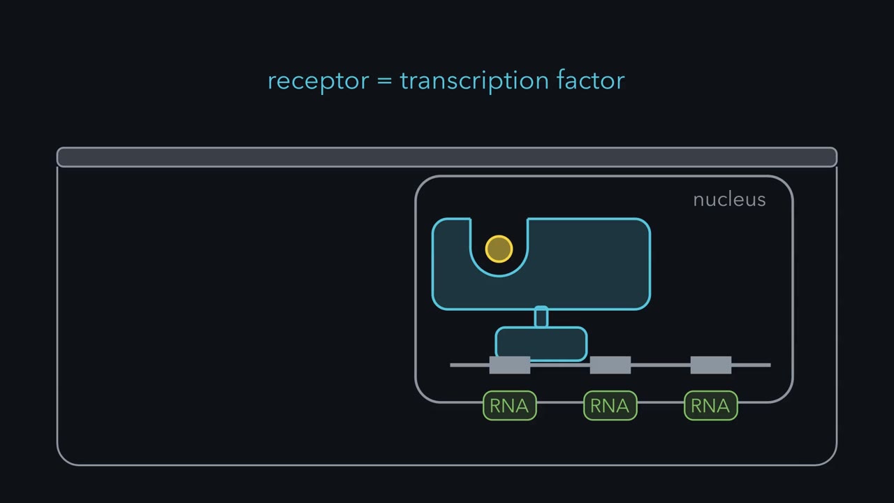 ▶ Watch The Receptor Is the Gene Switch · 2:47
Read the transcript of “The Receptor Is the Gene Switch”
Film transcript: The Receptor Is the Gene Switch
Nuclear receptors are proteins inside the cell that catch chemical signals and control whether genes are copied into RNA. The receptor itself is a transcription factor. That matters because one receptor can reset many genes at once. New RNA and protein take time to make, so the response builds over hours. But the new proteins remain after the hormone has gone. A steroid hormone crosses the membrane to reach its receptor. The receptor is one chain with two grips. One region binds the hormone. Another recognizes a specific short stretch of DNA. Most nuclear receptors work in pairs, and their DNA binding site is called a response element. The glucocorticoid receptor pairs with another copy of itself after hormone arrives.
The thyroid hormone receptor waits on DNA with a different partner, called RXR. Hormone recognition and DNA recognition are separate jobs in the same protein. Hormone binding changes the receptor's shape and which helper proteins it holds. The thyroid hormone receptor releases a co-repressor and brings in a co-activator. These helpers act on chromatin, the packaged form of DNA. Co-activators bring enzymes that add acetyl groups to histones, the proteins DNA wraps around. That loosens the packing and helps RNA polymerase start. Co-repressors bring enzymes that remove those groups. The cell's own mix of helpers also matters. The family reaches beyond familiar hormones.
Some receptors sense the cell's own lipids. PPARs bind fatty acids. LXR binds oxidized cholesterol, and FXR binds bile acids. PPAR-gamma drives the making of fat cells. So an intracellular signal can connect to gene control in metabolism, too. That same route meets medicine. Prednisone and dexamethasone act through the glucocorticoid receptor to calm inflammation. Long treatment also raises blood glucose and thins bone. Tissue context matters, too. Tamoxifen blocks estrogen in the breast but imitates it in the uterus and, after menopause, in bone. Its receptor still binds DNA, and the switching surface called AF-1 can still work. What happens next can depend on the helper proteins each tissue makes.
The signal, the receptor and the cell's helpers together shape gene control.
Inject a rat with radioactive estradiol, and almost every tissue lets the hormone go again. The uterus and the vagina keep it. Elwood Jensen reported that result from the University of Chicago in 1958. It meant that the tissues estrogen acts on contain something built to catch it. Every pathway so far in this course started at a protein in the cell membrane. This one starts inside the cell and has almost no middle, because the protein that catches the hormone is the same protein that switches the genes on.
The idea: the hormone goes in, and its receptor reads DNA
Estradiol, cortisol and testosterone are steroids: four fused carbon rings made from cholesterol (their synthesis is in the lesson on lipid synthesis). A steroid dissolves in fat, so it crosses the lipid bilayer on its own. Nothing on the outer surface of the cell has to receive it. The receptor field guide set out the receptor architectures and why speed follows architecture. This is the slow one, and here is how it works.
Jensen’s radioactive label, tritium (a heavy form of hydrogen), was still attached to unchanged estradiol when he recovered it. That was the awkward part. Many biochemists then expected estradiol to work by being oxidized and reduced over and over, passing hydrogen between coenzymes. An intact hormone held in place pointed to a binding protein instead. In 1966 David Toft and Jack Gorski, at the University of Illinois, extracted an estrogen-binding protein from rat uterus. In 1980 Jensen and Geoffrey Greene made monoclonal antibodies against the human receptor (antibodies of a single kind, each binding one spot on its target). In March 1986 the laboratories of Pierre Chambon in Strasbourg and of Greene in Chicago published the full sequence of its cDNA (a DNA copy of a messenger RNA) within weeks of each other.
The sequence, and the experiments it made possible, showed one chain with two grips. One region closes around the hormone. Another takes hold of a specific short stretch of DNA. No second messenger is made and no kinase cascade runs. A transcription factor is a protein that binds DNA and controls whether a gene is copied into RNA. In the lesson on TGF-β and Smads the messenger was the transcription factor. Here the receptor is, and the family is called the nuclear receptors.
Two cautions keep this exact. Thyroid hormone, retinoic acid (made from vitamin A) and the active form of vitamin D use receptors of the same family, but thyroid hormone carries charges and enters cells through transporter proteins. A transporter is a door; the receptor is still inside. And some steroid effects appear within seconds to minutes. They do not run through new RNA, and this lesson leaves them aside.
One plan, forty-eight receptors
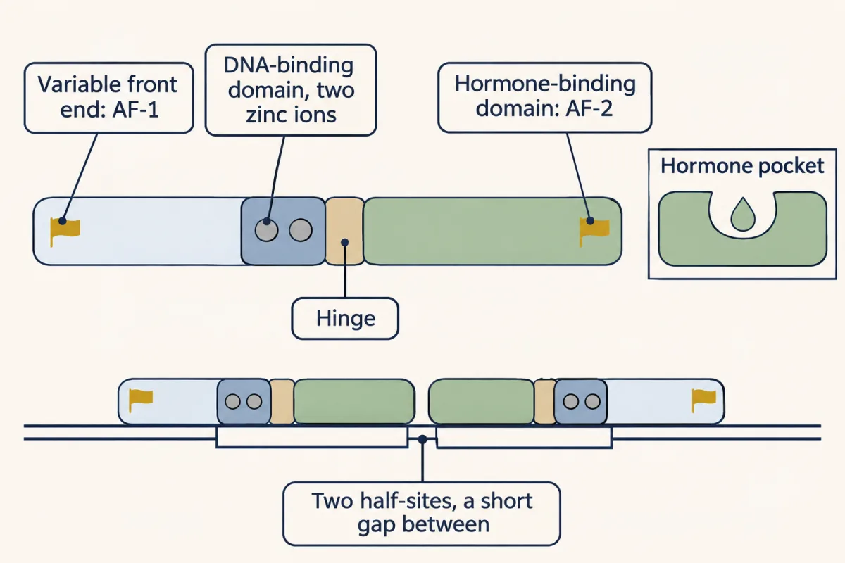
Once receptors had sequences, the sequences could be compared. Ronald Evans’s laboratory published the sequence of the human glucocorticoid receptor in 1985, and the estrogen receptor, months later, resembled it. Both resembled v-erbA, a cancer gene carried by a chicken virus. By the end of 1986 the cell’s normal version of that gene was known to encode a receptor for thyroid hormone. The human genome holds 48 genes of this family, and most share one plan:
- A variable front end. It differs most between receptors. It carries one surface for switching genes on, called AF-1, that can work without the hormone-binding domain.
- A DNA-binding domain of about 70 amino acids, folded around two zinc ions. Four cysteines hold each zinc. The fold sets one helix into the major groove of DNA, where it reads six bases.
- A hinge.
- A hormone-binding domain: about twelve helices packed around a pocket lined with hydrophobic side chains. It also carries the surface that pairs two receptors, and a second switching surface, AF-2.
Most of these receptors work in pairs, so their binding site on DNA, called a response element, has two six-base halves. For the estrogen receptor:
AGGTCA nnn TGACCT (two halves that read the same on opposite strands, three bases apart; n is any base)
Most of the 48 were found by their resemblance to known receptors before anyone knew what they bind, and were named orphan receptors. Several have since been matched to a ligand (the small molecule a receptor binds): the PPARs bind fatty acids, LXR binds oxidized cholesterol and FXR binds bile acids. So this family also reports on the cell’s own lipids. Others still have no agreed ligand.
Two ways to wait for a hormone
Read the transcript of “A Receptor Waits Off DNA or On It”
Film transcript: A Receptor Waits Off DNA or On It
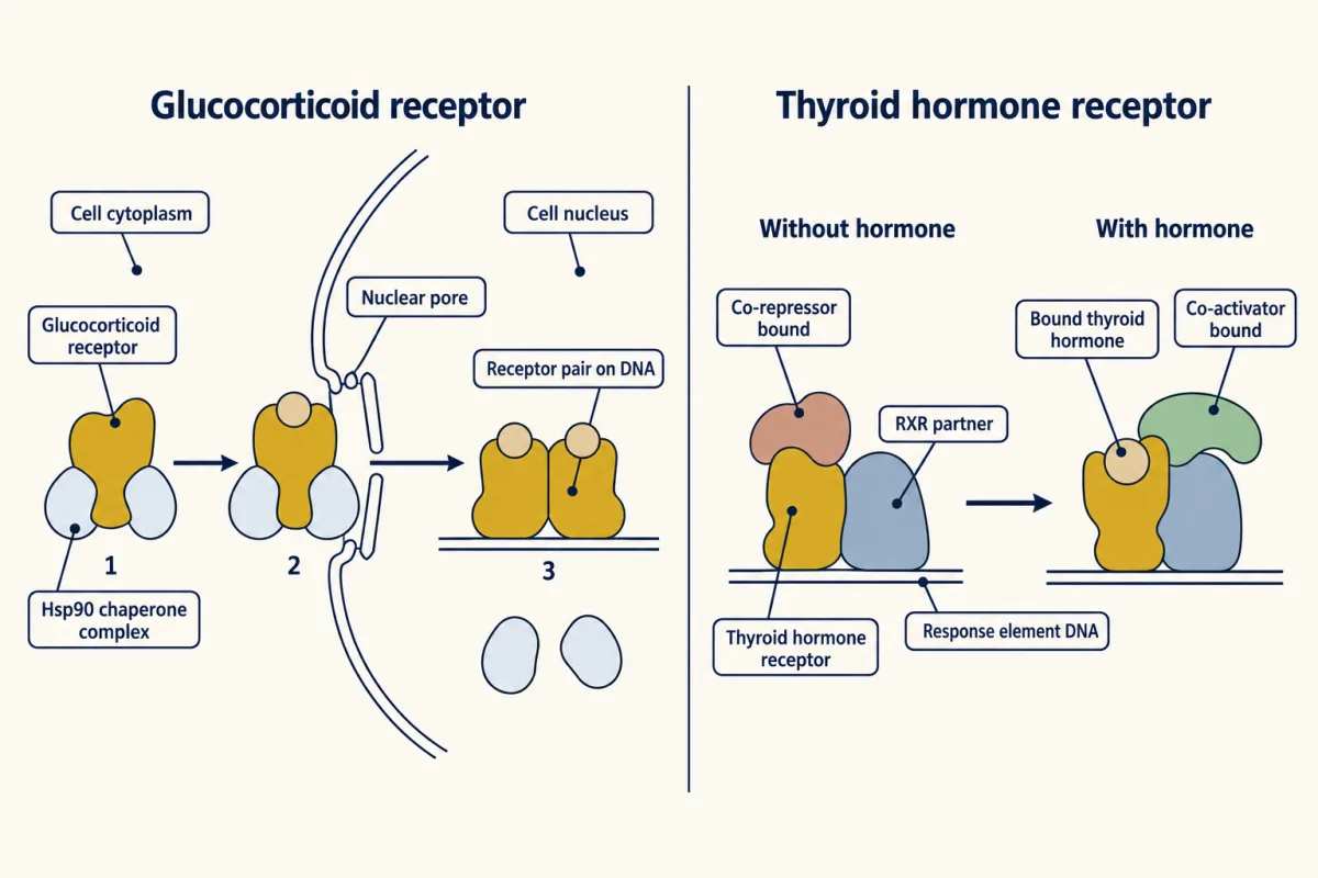
What does a receptor do before its hormone arrives? The family has two answers. The first is a chaperone. The glucocorticoid and androgen receptors wait mostly in the cytoplasm, held by chaperones built around Hsp90. A chaperone is a protein that helps other proteins hold their shape. Here it keeps the pocket ready for hormone, and keeps the receptor off DNA. When hormone binds, the receptor is released and moves into the nucleus. There it pairs with a second copy of itself and binds its response elements. The second answer is to wait on the DNA. The receptors for thyroid hormone, retinoic acid and vitamin D stay in the nucleus, and so do the PPAR receptors.
They bind DNA paired with a partner from the same family called RXR. Their two half sites point the same way. The gap between them helps decide which pair fits: one base for PPAR, three for vitamin D, four for thyroid hormone, five for retinoic acid. Without hormone, the thyroid hormone and retinoic acid receptors hold co-repressors, proteins that keep the gene off. Hormone releases the co-repressor and brings in its place a co-activator, a protein that helps switch the gene on. An empty receptor of this kind is an active off. A gene carrying an empty thyroid hormone receptor is quieter than the same gene with no receptor at all. So a receptor can wait off the DNA, or wait on it.
What does a receptor do before its hormone arrives? The family has two answers.
Waiting with a chaperone. The glucocorticoid and androgen receptors wait mostly in the cytoplasm, held by a group of chaperones built around Hsp90. A chaperone is a protein that helps other proteins hold their shape. Here it keeps the pocket ready for hormone and keeps the receptor off DNA. When hormone binds, the chaperone group rearranges, the receptor moves into the nucleus and sheds its chaperones, then pairs with a second copy of itself and binds its response elements. The estrogen receptor is held by the same chaperones, but experiments in 1984, on enucleated cells and with antibody staining, placed most of it in the nucleus even before hormone arrives.
Waiting on the DNA. The receptors for thyroid hormone, retinoic acid and vitamin D, and the PPARs, stay in the nucleus. They bind DNA paired with a partner from the same family called retinoid X receptor (RXR). Their two half-sites point the same way, and the gap between them helps decide which pair fits: one base for PPAR, three for vitamin D, four for thyroid hormone, two or five for retinoic acid. Without hormone, the thyroid hormone and retinoic acid receptors hold co-repressors, proteins that keep the gene off. Hormone releases the co-repressor and brings in its place a co-activator, a protein that helps switch the gene on. An empty receptor of this kind is an active “off”: a gene carrying an empty thyroid hormone receptor is quieter than the same gene with no receptor at all. This repression is best documented for those two receptors, so do not assume it for every partner of RXR.
Co-repressors and co-activators act largely on chromatin, the packaged form of DNA. DNA in the nucleus is wound around histone proteins, and tightly packed DNA is hard to transcribe. Co-activators bring enzymes that add acetyl groups to histones, which loosens the packing and helps RNA polymerase start; co-repressors bring enzymes that take the acetyl groups off.
What the hormone changes: one helix closes
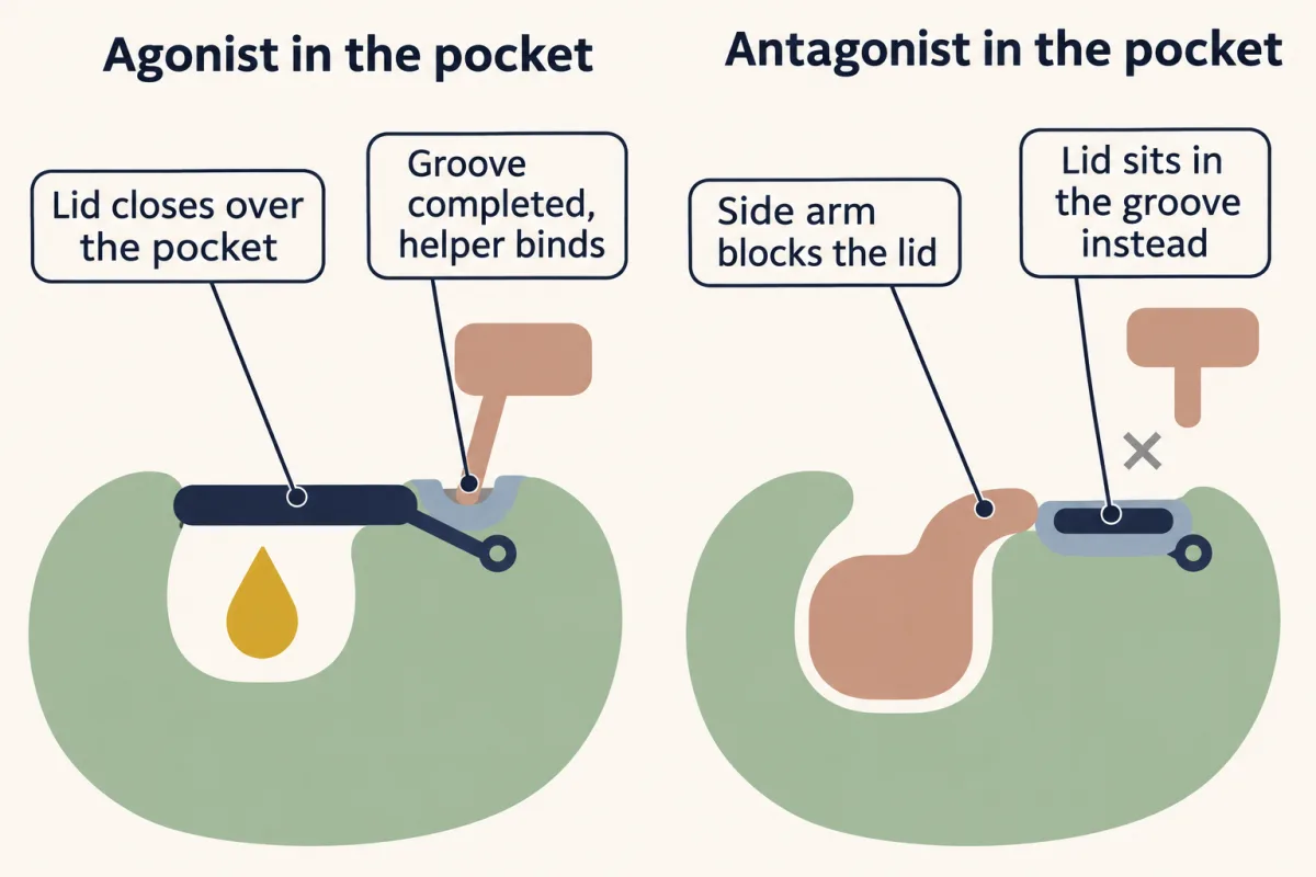
The main moving part is the last helix of the hormone-binding domain, helix 12. With an agonist (a molecule that switches the receptor on) in the pocket, helix 12 folds across the mouth of the pocket like a lid. In that position it completes a shallow groove on the surface of the domain. Co-activators carry a short helix with the sequence LXXLL, where L is leucine and X is any amino acid, and that helix fits the groove. This groove is the main surface through which the hormone brings in co-activators.
An antagonist, a molecule that occupies the receptor without switching it on, often fills the pocket and leaves the helix in the wrong place. The liver converts tamoxifen to forms that bind the estrogen receptor much more tightly, such as 4-hydroxytamoxifen. That molecule fits the pocket, but one side chain is too long to stay inside and sticks out where the lid should close. In a 1998 crystal structure from the laboratories of Greene and David Agard, helix 12 lies in the co-activator groove itself. The groove is taken, so the co-activator has nowhere to bind.
Design problem
Tamoxifen blocks the action of estrogen in the breast and imitates it in the uterus and, after menopause, in bone. One receptor and one drug give opposite answers. Propose what could differ between the tissues to explain this, using the parts in this lesson. Then design one experiment that would test your proposal.
Saved in this browser only.
One way to do it
Start with what tamoxifen leaves alone. The receptor still pairs and still binds DNA, and AF-1 at the front end does not depend on helix 12. So a tamoxifen-bound receptor sits on the gene with one of its two switching surfaces blocked. What happens next could depend on the helper proteins each tissue makes.
Proposal: a tissue with a high level of a co-activator that can work through AF-1 reads tamoxifen as “on”. A tissue with relatively more co-repressor reads it as “off”.
Experiment: take a breast cell line in which tamoxifen keeps an estrogen-responsive gene off, and a uterine cell line in which tamoxifen turns a gene on. Measure the co-activators in each. Then change them: lower one in the uterine cells and raise it in the breast cells. If the proposal is right, tamoxifen loses its estrogen-like effect in the first and gains one in the second, while estradiol works in both. A receptor with AF-1 removed should lose tamoxifen’s estrogen-like effect in both.
In 2002 Yongfeng Shang and Myles Brown reported that tamoxifen brought co-activators to a subset of genes in a uterine cell line, that this required a high level of the co-activator SRC-1 (steroid receptor co-activator 1, unrelated to the kinase Src), and that in breast cells tamoxifen brought co-repressors. Other differences are possible too. There are two estrogen receptor genes, and ERK, from the lesson on Ras and MAPK, can phosphorylate AF-1.
A drug of this kind is called a selective estrogen receptor modulator. It is neither an agonist nor an antagonist until you name the tissue and the gene. Raloxifene is a second example: it acts like estrogen in bone, does not stimulate the uterus, and is used against osteoporosis.
Why it takes hours, and where it meets medicine
A signal that works through new RNA and new protein cannot be fast. The first new messenger RNAs appear within tens of minutes of the hormone, and the change in the cell’s proteins builds over hours. Adrenaline acting through cyclic AMP (the lesson on cAMP and PKA) takes seconds. The slow route has its own strength: the new proteins remain after the hormone has gone, and one receptor can reset many genes at once.
This family meets medicine in many places. Here are three.
Glucocorticoids. Prednisone and dexamethasone are synthetic relatives of cortisol, used to calm inflammation. The hormone-bound receptor can bind NF-κB (from the lesson on Toll-like receptors and NF-κB) at inflammatory genes and blunt it without binding DNA itself, and in some cells it also raises production of the inhibitor IκB. It also switches on genes of its own whose proteins damp inflammation. Nearly every cell has this receptor, so long treatment also raises blood glucose and thins bone.
PPAR-γ. This receptor drives the making of fat cells. The thiazolidinediones, such as pioglitazone, are agonists for it and make the body more sensitive to insulin. The drugs were found by their effect in diabetic animals, and in 1995 their target turned out to be PPAR-γ.
Androgen insensitivity. The gene for the androgen receptor is on the X chromosome. A person with one X and one Y whose only copy of the gene cannot work has testes that make testosterone and cells that cannot read it. In the complete form of the condition the body develops along female lines. Blood testosterone is in or above the usual male range, which is itself a clue in someone with a female body form. The fault is in the receptor, and in this pathway the receptor is nearly the whole pathway.
Your notes
Select any text in the lesson to highlight it or add a note to it. Your highlights and notes are saved in this browser and collected in your notebook.
This browser is not letting the site save anything (a private window or strict privacy settings), so highlights and notes will be lost when you leave the page.
How we measure it
Radioactive hormone binding
Make the hormone radioactive without changing its chemistry (Jensen used tritium, a heavy form of hydrogen), give it to an animal or a tissue extract, and count where it stays. Binding that reaches a ceiling and is displaced by an excess of unlabelled hormone marks a limited number of specific sites, which is how a receptor behaves.
cDNA cloning and sequence comparison
Copy a cell's messenger RNAs into DNA, find the copy that encodes your protein with an antibody or with a probe designed from a piece of its amino acid sequence, and read it. Lining up the sequences of different receptors showed the domains they share and turned up relatives with no known hormone.
Domain exchange with a reporter gene
Join the hormone-binding part of one receptor to the DNA-binding part of another and put the hybrid into cells that carry a reporter, a gene whose product is easy to measure, placed behind a chosen response element. Which hormone turns on which reporter tells you which part of the protein does which job.
X-ray crystallography of the hormone-binding domain
Crystallize the domain once with an agonist and once with an antagonist, and solve both structures. The difference between them shows what the hormone changes, which here is the position of helix 12.
Practice questions on this lesson (4) → MCAT-style, original, each with an explanation
Where this lesson fits
Before thisReview first
- A Field Guide to ReceptorsThis course · Lesson 4
It sets out receptor types and why speed follows design, which this lesson uses to place the slow nuclear receptor.
- Binding: Affinity, Occupancy and Dose–ResponseThis course · Lesson 3
You need affinity and occupancy from there to follow how agonists, antagonists and tamoxifen compete for one pocket.
- TGF-β and SmadsThis course · Lesson 23
Smads are the contrasting case where a messenger is the transcription factor, which this lesson refers back to.
After thisStudy next
- Signaling to the Nucleus: CREB, AP-1 and Immediate Early GenesThis course · Lesson 32
It takes the step after receptors reach the nucleus: how signals switch on immediate early genes.
- The Shape of a Signal: Size, Duration, Frequency and PlaceThis course · Lesson 35
It uses the slow, hours-long timing of gene-based signals to ask how cells read a signal's duration.
- Drugs Against a PartThis course · Lesson 42
It extends drug action on one receptor part, as with tamoxifen and the PPAR-γ drugs here.
Suggested by an AI model from the lessons themselves, then checked. How we use AI
Finish line
Test yourself
9 questions
Practice and MCAT-style, with explained answers.
Next · Lesson 32
Signaling to the Nucleus: CREB, AP-1 and Immediate Early Genes
A resting cell starts transcribing c-fos within minutes of a signal, even with its ribosomes stopped. What was waiting in the nucleus?
Continue →