Cell Biology · Unit 2: Membranes and compartments
New to this? Review it at high school level: Crossing the Membrane
Transport Across Membranes
Rewritten on : new text, flashcards and film. Your notes and highlights stay, but the text around them may have moved.
Which way will potassium flow if you hold a cell at −70 millivolts? Its concentrations alone will not tell you.
Pause when the reading reaches a film, and offer to play it. Saved on this device.
Watch the film now, or keep listening
Builds on:Membrane Organization and TopologyCrossing the MembraneWhy these?
 ▶ Watch Two Pushes, One Hill · 3:18
Read the transcript of “Two Pushes, One Hill”
Film transcript: Two Pushes, One Hill
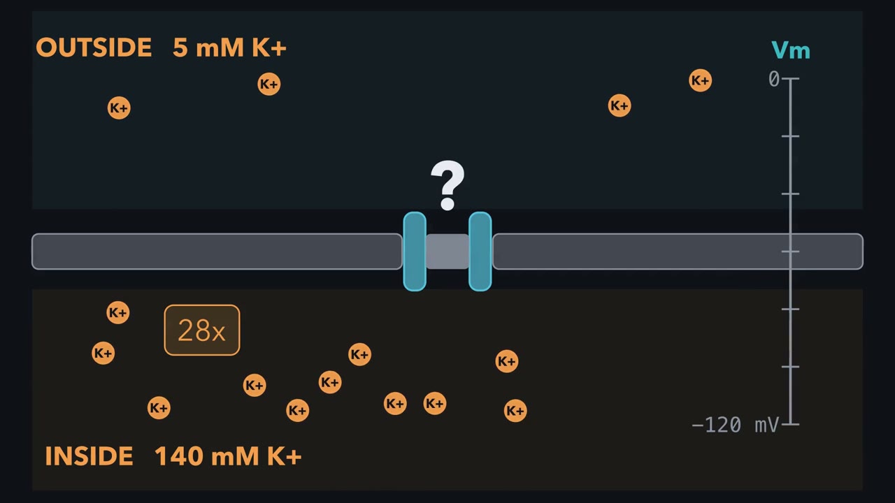
Why would potassium flow into a cell, against a 28-fold concentration difference? Take a cell with 140 mM K+ inside and 5 mM outside. Hold it at minus −70 mV and open potassium channels. Potassium leaves. Hold it at minus −100 and potassium comes in.
An ion feels two pushes. The first is concentration: potassium drifts from where it is crowded to where it is scarce. The second is voltage. The membrane potential, is the inside minus the outside, and a negative inside pulls positive ions in. Add the two pushes into one number. Picture the trip into the cell as a hill with two slopes. The concentration slope makes entering uphill. The voltage slope makes it downhill. Net flow always goes the way the total runs downhill. For each ion, one voltage makes the two slopes cancel. That is its equilibrium potential. For potassium, with these teaching values at 37 °C, the Nernst equation puts E_K at −89.1 mV. There, ions still cross, but equal numbers go each way.
So compare the voltage with that green mark. At minus −70 mV, which way does potassium go? And at minus −100 Decide before the picture moves. Minus −70 sits above the green mark. The voltage slope is shallower than the concentration slope, so entering is uphill, and potassium leaves. Minus −100 sits below it. Entering is downhill, so potassium comes in. That is the rule for passive flow. Find the equilibrium potential, then compare Vm with it. For a positive ion, Vm above the mark sends net flow out. Below the mark, net flow goes in. For a negative ion, the rule flips. The channel did the same thing in both cases. It opened a path, and it chose which ion could use it, and how fast.
The slopes chose the direction. Channels and passive carriers never decide which way. How does a cell move anything uphill? It couples that trip to one running further downhill. The sodium pump splits ATP to push 3 Na+ out and bring 2 K+ in. Then SGLT1 lets 2 Na+ run downhill into the cell, and that run pays for glucose to climb. Every crossing follows the same hill. A channel or passive carrier only opens the way. Add up the slopes of everything that moves together, and the total must run downhill.
From the film
- An ion feels two pushes: its concentration difference and the membrane voltage.
- Net flow goes whichever way the two pushes together run downhill.
- At its equilibrium potential the pushes cancel, and ions cross equally both ways.
- For a positive ion, a voltage above that potential sends net flow out; below it, in.
- Channels and passive carriers choose which solute and how fast, never which way; climbing needs a steeper downhill partner.
The first film used a voltage clamp, an amplifier that fixes the membrane voltage at a chosen value whatever current flows, to show potassium reversing direction while its concentrations stayed fixed. It left open how measurements reveal the route a solute takes.
The wall and the one rule
A lipid bilayer has an oily core about as thick as two lipid tails. Gases such as oxygen and carbon dioxide and small nonpolar molecules dissolve in that core and cross it by simple diffusion, needing no protein. Water crosses too, though slowly. Ions and larger polar molecules such as glucose cross almost not at all, because leaving water for oil costs them too much energy. You met the chemistry behind this in membrane structure and function.
So ions and sugars cross the bilayer in useful amounts only through proteins, and the proteins are specific: a potassium channel passes potassium and almost no sodium, and the glucose carrier GLUT1 passes glucose and not fructose. A cell decides what crosses by choosing which proteins to build, where to put them and when to open them. The sidedness of those proteins in the membrane, covered in membrane organization and topology, fixes which face of each protein looks into the cell. For a pump that orientation sets which way it pushes; for a channel or a passive carrier, the gradients decide.
The one idea to carry through this lesson is the film’s hill. Any solute, charged or not, flows on its own only downhill in its own electrochemical energy, the sum of a concentration push and a voltage push. Channels and passive carriers decide which solute crosses and how fast, never which way. Uphill transport is coupled to something running further downhill: ATP splitting (a pump), light or electron flow in some pumps, or another ion’s gradient (a symporter or antiporter, such as sodium-glucose cotransporter 1 (SGLT1)). The coupled total must still be downhill.
The rule sets the direction of net flow at a given moment. Here, concentrations stand in for activities, meaning effective concentrations. Separate questions are how fast transport runs and where a cell permeable to several ions will rest (that is the resting membrane). Water follows the rule too: aquaporins open a path, and water’s own energy difference, set mainly by the solutes on each side (and by pressure, in walled cells), decides the direction.
Two pushes, one number
Now calculate the hill for moving one mole of solute from outside to inside. The concentration push runs from high concentration to low. The voltage push depends on Vm, inside minus outside: a negative Vm favours entry for cations and opposes it for anions. Their sum is one number:
In words Delta G in equals R T times the natural log of inside over outside concentration, plus z F times the membrane potential.
- ΔGin“delta G in”
- the free energy change for moving one mole of X from outside to inside. Negative means downhill.J/mol
- RT × ln([X]in / [X]out)“the concentration push”
- the ln measures the concentration difference on a log scale; negative when X is moving to the side where it is scarcerJ/mol
- zF × Vm“the voltage push”
- charge z (with its sign) times F times Vm. Zero for an uncharged solute.J/mol
- R“R, the gas constant”
- 8.314 J per mol per KJ/(mol·K)
- T“T”
- the absolute temperature, in kelvin (37 °C = 310.15 K)K
- F“F, the Faraday constant”
- the charge on one mole of +1 ions, 96 485 C per molC/mol
- Vm“V m”
- the membrane potential, inside minus outsideV
Sanity check Equal concentrations and Vm = 0 give ΔGin = 0: nothing pushes either way.
Try one number
Sodium entering a cell at −70 mV, with teaching values of 145 mM outside and 12 mM inside, at 37 °C.
Concentration push: RT × ln(12/145) = 2579 J/mol × (−2.49) = −6.4 kJ/mol.
Voltage push: (+1) × 96 485 × (−0.070) = −6.75 kJ/mol.
Total: about −13.2 kJ/mol. Both slopes run inward, so sodium is steeply downhill into the cell. Keep this number; SGLT1 spends it below.
Need a refresher? e and ln · units and prefixes
For each ion there is one voltage at which the two pushes cancel exactly. That is its equilibrium potential, EX. Set ΔGin to zero and solve for Vm, and you have the Nernst equation:
In words E sub X equals R T over z F, times the natural log of the outside concentration over the inside concentration.
- EX“E sub X”
- the equilibrium potential of ion XV or mV
- R, T, F“R, T and F”
- as in the card above: the gas constant, the absolute temperature and the Faraday constant—
- z“z”
- the ion’s charge, with its sign: +1 for K+, −1 for Cl−no unit
- ln“natural log”
- the logarithm to base e. ln(1) = 0; a ratio below 1 gives a negative ln, above 1 a positive one.no unit
- [X]out“X out”
- the ion’s concentration outside the cellmM
- [X]in“X in”
- the ion’s concentration inside the cellmM
Sanity check If the concentrations are equal, the ratio is 1, ln(1) = 0, and EX = 0: with no concentration push, no voltage is needed to balance it. For a cation that is more concentrated inside, like K+, the ratio is below 1, the ln is negative, and EX is negative. Flip the sign of z (an anion) and EX flips sign too.
Try one number
Where the “61.5 mV per tenfold” shortcut comes from. At 37 °C, RT/F = (8.314 × 310.15) / 96 485 = 26.73 mV.
A tenfold ratio has ln(10) = 2.303. So for a +1 ion, a tenfold ratio gives 26.73 mV × 2.303 = about 61.5 mV. That is why E = 61.5 mV × log10(ratio) gives the same answer as the ln form.
Where it comes from optional
Step 1, at equilibrium nothing is downhill: 0 = RT × ln([X]in / [X]out) + zF × EX.
Step 2, move the concentration term across: zF × EX = −RT × ln([X]in / [X]out) = RT × ln([X]out / [X]in), because flipping a ratio flips the sign of its ln.
Step 3, divide by zF.
Need a refresher? subscripts and Greek letters · e and ln · log scales
At EX the ion’s net passive flux is zero. Ions still cross; equal numbers cross each way.
For an ion you can fold the two pushes into one gap. The gap between Vm and EX is the driving force, and multiplying it by the charge gives the same ΔGin as before:
In words Delta G in equals z F times the gap between the membrane potential and the ion’s equilibrium potential.
- ΔGin“delta G in”
- the free energy change for moving one mole of the ion from outside to inside. Negative means downhill.J/mol
- z“z”
- the ion’s charge, with its signno unit
- F“F, the Faraday constant”
- 96 485 C per molC/mol
- Vm“V m”
- the membrane potential, inside minus outsideV
- EX“E sub X”
- the ion’s equilibrium potential, from the Nernst equationV
Sanity check If Vm equals EX, ΔGin is zero: no net flow, which is what equilibrium potential means. If ΔGin is negative, net passive flow is inward; if positive, outward. Units: coulombs per mole times volts gives joules per mole.
Try one number
Keep z’s sign. Take a cation (z = +1) with Vm − EX = +10 mV.
ΔGin = (+1) × 96 485 × 0.010 = +965 J/mol, about +0.96 kJ/mol. It is positive, so net flow of the cation is outward.
Where it comes from optional
Step 1, from the Nernst derivation, the concentration push equals −zF × EX.
Step 2, substitute it into the two-push form: ΔGin = −zF × EX + zF × Vm.
Step 3, take out the common factor: ΔGin = zF × (Vm − EX).
Need a refresher? subscripts and Greek letters · units and prefixes
So comparing Vm with EX is the downhill rule, written for one ion.
Worked problem. Take a membrane permeable only to K+, at 37 °C (310.15 K), with [K+]in = 140 mM and [K+]out = 5 mM. These are teaching values close to those of a typical mammalian cell.
Step 1: find EK. RT/F = 26.73 mV (from the Nernst card). The ratio 5/140 = 0.0357, and ln(0.0357) = −3.332 (a calculator’s ln key; see e and ln). Here z = +1, so RT/zF = RT/F. So
EK = 26.73 mV × (−3.332) = −89.1 mV
Predict. Clamp the membrane at −70 mV and open the channels. Does net K+ flow in or out? Now clamp at −100 mV.
Check your prediction
At −70 mV, out. Vm − EK = −70 − (−89.1) = +19.1 mV, so ΔGin = (+1)(96 485)(+0.0191) = +1.84 kJ/mol. Moving K+ in is uphill. In two-push terms, the concentration push on entering K+ is +8.59 kJ/mol and the voltage push only −6.75 kJ/mol: the inside is not negative enough to hold potassium in.
At −100 mV, in. Vm − EK = −10.9 mV, so ΔGin = −1.06 kJ/mol. The voltage push has grown to −9.65 kJ/mol and now outweighs the same +8.59. This is the reversal promised at the top.
One channel. Suppose a single K+ channel has a conductance γ (how easily current passes) of 20 picosiemens (pS) and behaves ohmically: its current is proportional to the driving force, Vm − EK, and is zero at EK. By convention, positive current is outward movement of positive charge. At Vm = −40 mV its current is
i = γ × (Vm − EK) = 20 pS × 49.1 mV = 0.98 pA, outward
A current of 0.98 pA is 0.98 × 10⁻¹² coulombs per second; dividing by the charge on one ion (1.602 × 10⁻¹⁹ C) gives about 6.1 × 10⁶ ions per second through one pore.
Predict. Now take chloride (z = −1) with the same gap, Vm − ECl = +10 mV. Which way does net chloride flow?
Check your prediction
Inward. ΔGin = (−1) × 96 485 × 0.010 = −965 J/mol, negative. Because z is negative for an anion, the same Vm − EX gives ΔGin the opposite sign.
The physical reading, for the usual case of chloride more concentrated outside and a negative inside: the negative inside pushes chloride out. When Vm is less negative than ECl, that outward electrical push is weaker than chloride’s inward concentration push, so chloride comes in.
Do not conclude that a cell rests at EK. A real membrane is permeable to several ions, and the resting potential usually lies between their equilibrium potentials, pulled toward each according to its share of the membrane’s conductance (see the resting membrane). Many cells rest less negative than EK for exactly this reason.
The machinery, sorted by the rule
The first film used the simplest case; here is the fuller machinery. Two kinds of protein carry solutes across: channels and carriers. (“Carrier” and “transporter” mean the same thing here. A pump is a carrier with its own power supply.)
Downhill only: channels and uniporters
Channels form an aqueous pore that, when open, connects both sides at once. Ions flow through, often in single file, down their electrochemical gradient. A channel can switch between open and closed (gating) in response to voltage, a ligand or stretch, but while open it has no cycle to complete, so it is fast: millions of ions per second through one pore. Aquaporins are water channels. Ligand-gated channels that act as receptors belong to ion channels as receptors in the signaling course.
Carriers work differently. A carrier binds its solute on one side, changes shape, and releases it on the other; in the ideal cycle its binding site is never open to both sides at once, though some carriers also have a small channel-like leak. This is alternating access. Because each crossing needs a shape change, a carrier works far more slowly than a channel, and because it has a limited number of binding sites, its rate saturates as solute rises, like an enzyme with Michaelis-Menten kinetics.
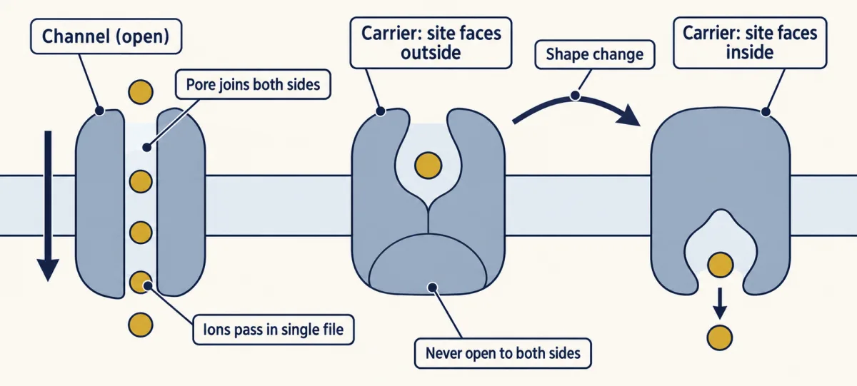
A uniporter is a carrier that moves one solute, as GLUT1 moves glucose into red blood cells. Glucose has no charge, so only the concentration push counts: GLUT1 can speed glucose across, but with glucose alone, only until inside and outside are equal; it can never concentrate it.
Uphill by coupling: pumps, symporters and antiporters
Pumps couple transport to a direct energy source, usually ATP hydrolysis, and so move ions uphill. This is primary active transport. Pumps work by alternating access too. The Na+/K+-ATPase in the plasma membrane of animal cells exports three Na+ and imports two K+ for each ATP it splits. At −70 mV both moves are uphill, but together they cost less than one ATP supplies (Going deeper, “The pump’s bill”). It moves one net charge out per cycle, so it makes the inside more negative directly. In most cells that direct effect is small, a few millivolts, but in some it is large. Mostly it builds the sodium and potassium gradients that other proteins spend.
Carriers that move two or more solutes can do the same with no ATP. A symporter moves them the same way. An antiporter (or exchanger) moves them opposite ways, as the Na+/Ca2+ exchanger brings sodium in and sends calcium out. Spending one solute’s gradient to move another is secondary active transport.
Sodium-glucose cotransporter 1 (SGLT1) in intestinal cells brings glucose in with sodium. You will see SGLT1 again in digestion, and the same logic with protons instead of sodium in the proton-motive force.
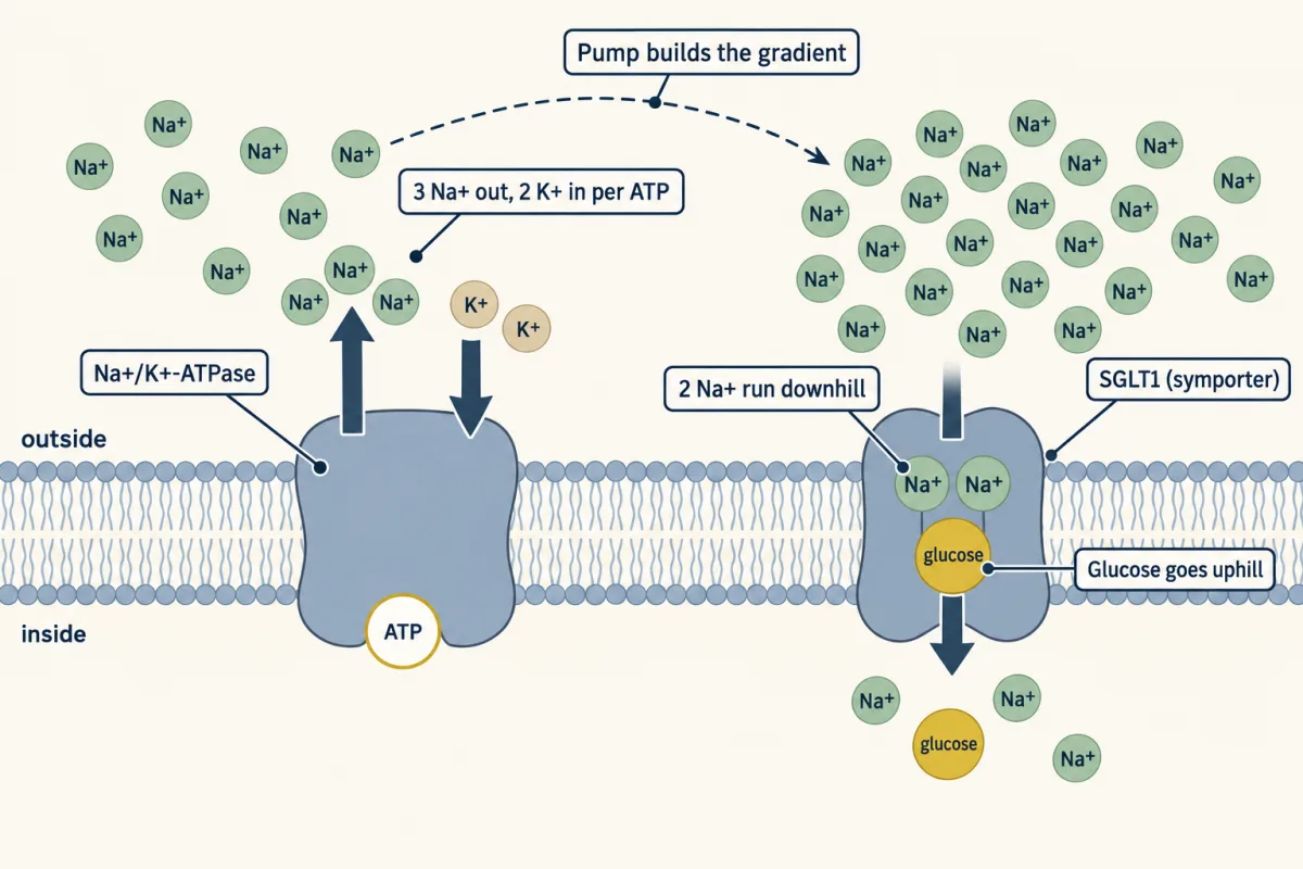
Predict. Block the pump with a drug. What happens to glucose uptake through SGLT1, and does it happen at once?
Check your prediction
Uphill glucose uptake fails, but not at once. SGLT1 needs only that the coupled total, sodium’s payment plus glucose’s cost, is still downhill. With the pump blocked, the sodium gradient runs down as sodium keeps leaking in, the payment shrinks, and SGLT1 can no longer pull glucose uphill.
How far uphill can that payment carry glucose?
In words n times the free energy for one mole of sodium entering, plus R T times the natural log of the glucose ratio, must be zero or less.
- n“n”
- the number of Na+ carried with each glucose: 2 for SGLT1no unit
- ΔGin(Na+)“delta G in for sodium”
- the free energy for one mole of Na+ entering: about −13.2 kJ/mol in the two-push exampleJ/mol
- RT × ln(...)“glucose's push”
- concentration only, since glucose has no charge. Positive when glucose piles up inside.J/mol
Sanity check With n = 0 the glucose term alone must be zero or less, so glucose can never be concentrated inside: that is the uniporter.
Try one number
At 37 °C, each tenfold step uphill costs glucose RT × ln(10) = 5.94 kJ/mol.
With 2 Na+ per glucose, the payment is 2 × 13.2 = 26.4 kJ/mol. That buys 26.4 / 5.94 = 4.44 tenfold steps: 104.44, about 27,500-fold.
With 1 Na+ per glucose, 13.2 / 5.94 = 2.22 steps: about 166-fold.
One extra sodium per trip multiplies the ceiling by about 166. These are thermodynamic ceilings, not measured ratios: leaks, and glucose leaving the cell by other carriers, keep real cells far below them.
Need a refresher? e and ln · log scales
Downhill flow cannot tell a channel from a carrier, since both carry it; net uphill flow points to a coupled carrier. The second film picks up here: for downhill flow, read the traffic pattern to tell a channel from a carrier.
Read the transcript of “Read the Traffic”
Film transcript: Read the Traffic
Chapter one showed that the energy hill sets which way a solute moves. So how can data reveal whether it crossed through a channel or a carrier? Watch the traffic, not the protein's name. An open channel is a continuous pore between both sides. Ions stream through without waiting for a transport cycle. A channel can switch between open and closed; a carrier must finish each alternating-access cycle. A carrier opens to one side, closes around its solute, then opens to the other. That shape change limits each trip. A single channel gives the detector square current steps. Millions of ions per second can cross one pore, producing measurable current.
Each jump marks an opening. The step height is the unitary current through one open channel. The fraction of recording time spent open is its open probability. Those are different measurements. Now a drug makes the openings last longer, but every step keeps the same height. What changed: current through one open channel, or open probability?
Decide from the trace. Open probability increased: the channel spent more of its time open. Unchanged step height means unchanged unitary current at this voltage. A whole-cell current alone mixes channel number, unitary current, and open probability, so it cannot separate them. Carriers leave a different signature.
Raise substrate concentration and their limited binding sites become occupied. The rate bends toward a ceiling called Vmax. A pore or simple diffusion can stay nearly proportional over the tested range. Channels can saturate too, usually only at much higher concentrations.
Saturation points toward a carrier, but it is evidence, not proof. Tracer experiments add a detectable tag to some substrate molecules. The counter sees tagged molecules entering, not the total amount inside. Take carrier vesicles with substrate already equal inside and outside. Add tagged substrate outside. Tagged molecules enter as untagged ones leave, so the total inside stays the same. A chemical assay of total substrate supplies what the tracer counter lacks. That is exchange, not net uptake.
Label has its own concentration difference, so it can run in while total substrate stays level. A rising count alone never proves accumulation. So measure the total, and combine exchange with rate, saturation, and single-channel steps instead of trusting one clue. The hill from chapter one tells you which way traffic can run. The pattern of that traffic reveals the route. Square steps point to a pore. Saturation and exchange point toward a carrier. Mechanism lives in the measurements, not the family name.
From the film
- An open channel forms a continuous pore, while a carrier completes an alternating-access cycle.
- A single-channel trace separates unitary current from open probability.
- A rate that saturates at low substrate concentrations points toward a carrier.
- Tracer entry can be exchange rather than net uptake.
- Mechanism comes from combined functional evidence, not a protein family name.
Telling a channel from a carrier from the data
The chapter gave the signatures: rate, single-channel steps, saturation and exchange. The numbers and controls below show their limits and put them to work.
Rate
The worked problem’s one open K+ channel carried about 6.1 million ions per second. Carriers typically cycle from tens to a few thousand times a second, and even the fastest known reach only tens of thousands, far below a million, so millions per second through one protein points to a pore.
Watching one channel
In 1976 Erwin Neher and Bert Sakmann recorded square steps of a few picoamperes (trillionths of an ampere) from a small patch of frog muscle membrane, each step the current through one open channel. The 1981 gigaseal methods made such records routine; a gigaseal is an extremely high-resistance seal between glass and membrane. Each opening lasts a random time and openings come at irregular intervals. In a patch with one simple channel every opening has the same height, though some channels also show lower sub-levels.
The unitary current, i, the step height, is set by the channel’s conductance and the driving force. The open probability, Po, is the fraction of time the channel is open. A drug that changes Po without changing i alters gating; one that changes i alters conduction or driving force. Across N identical channels, the mean current combines the chapter’s two readouts with channel number:
In words The total current equals the number of channels, times the current through one open channel, times the fraction of time each is open.
- I“capital I”
- the mean current through all the channelspA
- N“N”
- the number of channelsno unit
- i“small i”
- the unitary current: the step height through one open channelpA
- Po“P sub o”
- the open probability: the fraction of time a channel is open, from 0 to 1no unit
Sanity check If Po is 0, there is no current however many channels there are.
Try one number
Round values: 100 channels, a 1 pA unitary current, and Po = 0.5. I = 100 × 1 pA × 0.5 = 50 pA.
Fifty channels that are always open (Po = 1) give the same 50 pA: 50 × 1 × 1 = 50. That is why a whole-cell current alone cannot separate N, i and Po.
Need a refresher? subscripts and Greek letters
The sandbox below is the worked problem’s 20 pS K+ channel. Move the clamp voltage first: the step height follows Vm − EK and flips to inward below −89.1 mV. Then move Po: only the time spent open changes.
- Unitary current i = γ × (Vm − EK)
- Ions per second through the open channel
- Direction of K+ flow
- Fraction of time open: measured here, set
- Mean current, I = N × i × Po: expected, from this record
The step height i comes from the driving force and sets which way ions move; the open probability Po is read from how long the channel stays open. In this sketch, Po is set separately from Vm; in a real voltage-gated channel Vm can change both. Assumes the lesson’s ohmic 20 pS K+ channel; EK = −89.1 mV (140 mM in, 5 mM out, 37 °C). Dwell times are drawn at random. Time is in relative units, so this record is a sketch of the logic, not a prediction for a real channel.
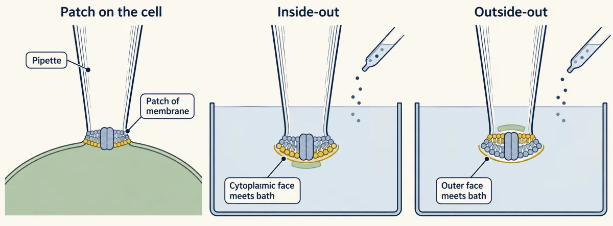
Orientation matters. In an inside-out patch, pulled off the cell, the cytoplasmic face meets the bath; in an outside-out patch, the outer face does. A substance that cannot cross, such as ATP, added to each bath shows which side it acts from. Closures briefer than the recording can resolve vanish from the trace, so openings look longer than they are.
Counting molecules: tracer flux
One carrier moves too few molecules per second to show up as a current step, so carriers are often studied by counting. In a tracer-flux experiment you add radiolabelled substrate outside cells or vesicles, stop uptake at timed points by filtering and washing, and count the label inside. The early, straight part of the time course gives the initial rate. Apply the chapter’s signatures to these two invented teaching experiments.
Does the rate level off? Two unknown proteins, A and B, are each put into matched vesicles. You measure the initial rate of uptake at six substrate concentrations and subtract the rate into protein-free vesicles.
| Substrate outside (mM) | A: rate (pmol/min) | B: rate (pmol/min) |
|---|---|---|
| 0.5 | 20 | 2.5 |
| 1 | 33 | 5 |
| 2 | 50 | 10 |
| 5 | 71 | 25 |
| 10 | 83 | 50 |
| 20 | 91 | 100 |
Look at the last two rows first: doubling substrate from 10 to 20 mM doubles B’s rate but raises A’s by about a tenth.
Predict. Which protein behaves like a carrier?
Check your prediction
A. Its rate climbs steeply at first, then levels off toward a ceiling near 100 pmol/min (its Vmax), and it reaches half that ceiling at 2 mM (its Km). This is the Michaelis-Menten curve, v = Vmax[S] / (Km + [S]): once the few binding sites are nearly always occupied, more substrate cannot speed them up. B’s rate stays proportional to concentration across the whole range, which is what a pore or simple diffusion does. Channels can saturate too, but usually only at tens to hundreds of millimolar, while carriers often level off at micromolar to low millimolar substrate.
Is label going in the same as substrate building up? Now a third protein, C. Preload one batch of C vesicles with 10 mM unlabelled substrate and leave another batch unloaded. Add 1 mM labelled substrate outside both. After a minute, measure the label inside and, by chemical assay, the total substrate inside.
| Vesicles | Label inside | Total substrate inside |
|---|---|---|
| Unloaded | little | little |
| Preloaded with 10 mM | a lot | unchanged |
Predict. Label pours into the preloaded vesicles. Are they taking up substrate?
Check your prediction
No. The total inside did not rise. C is swapping: labelled molecules come in as unlabelled ones go out, one for one overall, so the total stays put. At first, label counts only inward traffic; the outgoing molecules were unlabelled, so the counter missed them. The unloaded vesicles take up little label because C moves quickly only when it has something to carry back on the return trip. That is the mark of an exchanger (though preloading can also speed up some uniporters; Going deeper, “Trans-stimulation and exchange”).
Did the label enter against a gradient, 1 mM outside and 10 mM inside? At first, no. Labelled molecules have a gradient of their own, 1 mM outside and none inside, and they ran down it. Given time, an exchanger can even pile label up inside well above 1 mM, the classic counterflow test for a carrier, because each labelled molecule that enters is paid for by an unlabelled one leaving down its own gradient.
A carrier’s Km is not a simple binding constant (Going deeper, “Km is not a binding constant”).
Family does not decide mechanism
Cystic fibrosis transmembrane conductance regulator (CFTR), the chloride channel mutated in cystic fibrosis, belongs to the ABC family, most of whose members are ATP-driven transporters, yet it is a channel. It still binds and splits ATP, but to open and close its gate, not to push chloride uphill: chloride flows through it downhill. The reverse surprise came in the ClC family of chloride channels: its bacterial member ClC-ec1, from E. coli, proved in reconstituted vesicles to be a carrier that exchanges protons for chloride. Only functional tests like those above decide.
Design problem
You inject RNA for an unknown membrane protein, Q, into Xenopus oocytes, and the oocytes become more permeable to chloride. Oocytes have chloride channels of their own. Is Q itself a chloride pore, or a protein that switches on an endogenous channel? And how would you tell whether Q is a channel or a carrier?
Saved in this browser only.
One way to do it
Start with controls: compare Q-injected oocytes with oocytes injected with the same volume of water, from the same frog and on the same day, so that any change can be traced to expressing Q. That alone does not make Q the pore: high expression of many foreign membrane proteins switches on the oocyte’s own chloride current.
Then take Q out of the oocyte. Purify it, put it into lipid vesicles and fuse them with a planar bilayer. If chloride current appears with purified Q and no other protein, Q is enough to form the pathway. This is how Christine Bear, John Riordan and colleagues showed in 1992 that purified CFTR is itself a regulated chloride channel.
Next, look for single-channel events: square current steps of fixed height whose reversal potential (the voltage at which the current changes direction) tracks ECl as you change chloride on one side. Then mutate residues you suspect line the pore. If a mutation changes the unitary conductance or which anions pass, Q most likely forms the pore. A mutation that changes only Po is consistent with Q being a regulator, or a channel whose gating you hit, so it decides less.
To separate a channel from a carrier, weigh rate, saturation, discrete unitary events, and exchange (does chloride entry need another ion leaving?). Remember ClC-ec1: a family name will not answer this for you.
Going deeper
58 or 61.5 mV per tenfold
Using base-10 logs gives the same EK as the worked problem: 61.5 mV × log10(5/140) = 61.5 × (−1.447) = −89.0 mV, the small difference being rounding. Many books quote 58 mV per tenfold change in concentration. That figure holds near room temperature. At 37 °C it is 61.5 mV.
Resting with several ions
With two ions, in the simple conductance picture (passive currents only, no pump current), the resting potential sits nearer the equilibrium potential of the ion with the larger conductance. With three or more, that can fail: if a second ion’s conductance nearly matches potassium’s and a third ion pulls the same way, the voltage can end up nearer the second ion’s equilibrium potential than EK, as the resting membrane works through.
The pump’s bill
At −70 mV, with the lesson’s teaching values, pushing Na+ out costs about +13.2 kJ/mol (the two-push example, reversed) and bringing K+ in +1.84 kJ/mol. Three Na+ out and two K+ in cost about 3 × 13.2 + 2 × 1.84 ≈ +43 kJ/mol. Splitting ATP in a cell releases roughly 50 to 60 kJ/mol, so the coupled total is downhill, with a margin, as the rule requires.
Km is not a binding constant
A carrier’s Km depends on the conditions on the far side (trans conditions), on the membrane voltage and on any coupled ion. Different protocols, such as zero-trans uptake (nothing inside to start) and equilibrium exchange (the same substrate on both sides), give different values for the same protein.
Trans-stimulation and exchange
The preloaded-vesicle test above read fast label entry as exchange. A uniporter whose empty return step is slow also speeds up when preloaded, so faster label entry into preloaded vesicles alone does not prove an obligate exchanger. Exchange and net flux can even be pulled apart within one protein: in 2023 a study of the E. coli carrier DgoT found a mutant, E133Q (glutamate 133 replaced by glutamine), that kept robust substrate exchange although Glu133 is needed for net flux.
Your notes
Select any text in the lesson to highlight it or add a note to it. Your highlights and notes are saved in this browser and collected in your notebook.
This browser is not letting the site save anything (a private window or strict privacy settings), so highlights and notes will be lost when you leave the page.
How we measure it
Patch clamp
A glass pipette sealed onto a cell isolates a small patch of membrane, and an amplifier records the current through it at a voltage you set. With one channel in the patch you see its openings as square steps; excised patches let you change the solution on the cytoplasmic face (inside-out) or the outer face (outside-out).
Radiotracer flux
You add a radioactively labelled substrate, stop uptake at timed points by filtering and washing, and count the label retained by cells or vesicles. Subtracting a matched control and dividing by specific activity gives an amount; a time series gives a rate. Tracer entry alone does not show net accumulation, since a carrier can exchange labelled for unlabelled substrate.
Reconstitution into vesicles and planar bilayers
A purified membrane protein is placed in artificial lipid vesicles, which can be assayed directly or fused with a flat bilayer for electrical recording. If the activity appears with the purified protein and nothing else, that protein is enough to carry it.
Expression in Xenopus oocytes
RNA injected into large frog oocytes (immature eggs) is translated into protein, and many membrane proteins made this way reach the surface. Comparing injected oocytes with matched control oocytes, by swelling in dilute solution or by electrical recording, reveals what the new protein adds.
Practice questions on this lesson (3) → MCAT-style, original, each with an explanation
Where this lesson fits
Before thisReview first
- Membrane Organization and TopologyThis course · Lesson 4
It explains which face of each membrane protein looks into the cell, which sets the direction a pump pushes.
- Crossing the MembraneBiology · High school
It introduces diffusion, channels and carriers across the bilayer, which this lesson makes quantitative with the Nernst equation.
After thisStudy next
- Organelles as Distinct EnvironmentsThis course · Lesson 6
It uses pumps and the downhill rule to explain how each compartment holds its own ion and proton levels.
- Epithelia and Tissue OrganizationThis course · Lesson 23
It uses coupled carriers like SGLT1 and the sodium pump to explain how a cell sheet moves glucose in one direction.
- Endocytosis, Recycling and DegradationThis course · Lesson 13
Its tracer uptake and binding-versus-entry experiments reuse the flux reasoning you practiced here.
Elsewhere on the siteFilms, simulations and method films in other courses
- The length constantSimulation · Neuroscience
It shows how potassium and sodium leaks set the resting voltage from the same two pushes.
- Run the 1952 equationsSimulation · Neuroscience
It uses Nernst potentials and voltage-driven flow to explain how sodium channels carry the spike.
- A Path Is Not a PumpFilm · Biochemistry
It shows how an electrochemical gradient sets an ion's direction of flow, and why a path is not a pump.
Suggested by an AI model from the lessons themselves, then checked. How we use AI
Finish line
Test yourself
8 questions
Practice and MCAT-style, with explained answers.
Next · Lesson 6
Organelles as Distinct Environments
A lysosome near pH 4.7 and a mitochondrial matrix near pH 8 share one cytosol near 7.2. What holds each at its own number?
Continue →