Organismal Biology · Unit 4: Water, salts and wastes
The Kidney
Rewritten on : new text, flashcards and film. Your notes and highlights stay, but the text around them may have moved.
Your kidneys filter about 180 litres of plasma a day. Why filter so much only to take almost all of it back?
Pause when the reading reaches a film, and offer to play it. Saved on this device.
Watch the film now, or keep listening
Builds on:Osmoregulation and Nitrogenous WasteBlood Vessels, Blood and LymphTissues, Organs and HomeostasisWhy these?
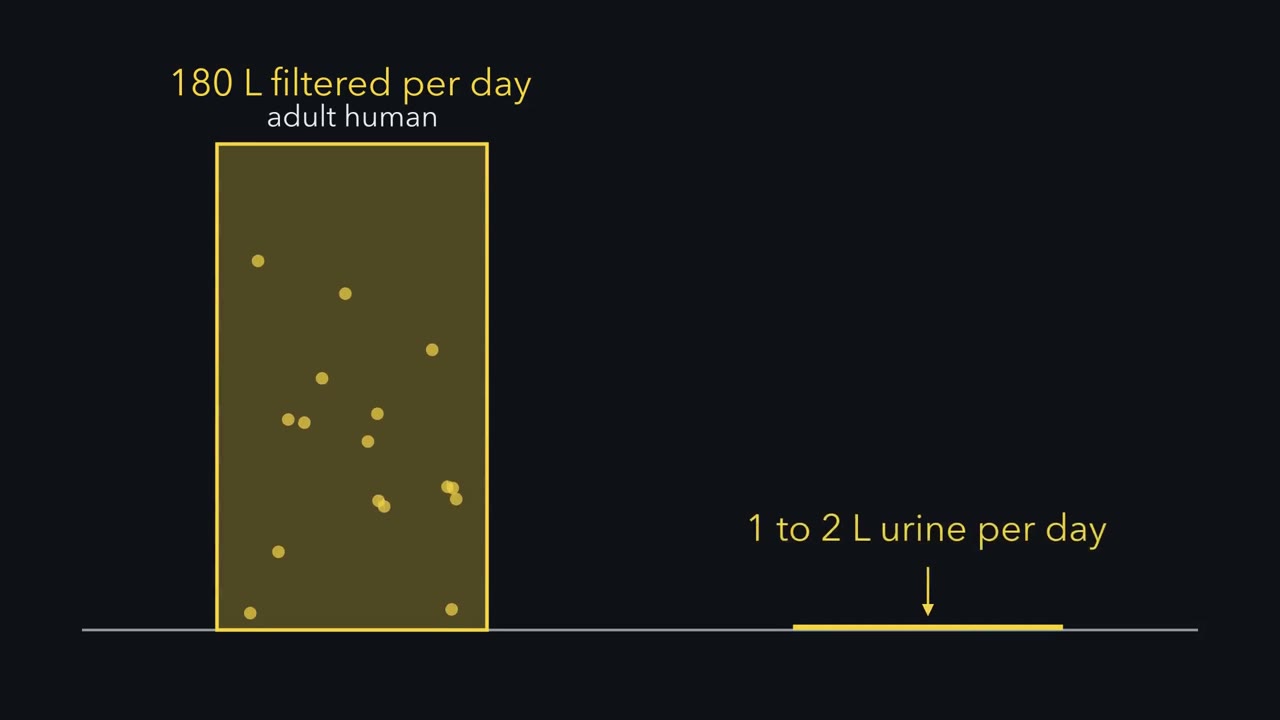 ▶ Watch Filter Far More, Take Most Back · 3:17
Read the transcript of “Filter Far More, Take Most Back”
Film transcript: Filter Far More, Take Most Back
Every day, your kidneys filter about 180 litres of plasma. Yet you pass only one or two litres of urine. Why would an organ filter so much more than it lets go? The kidney's working unit is the nephron: a filter attached to a long tube. Blood enters a knot of capillaries, the glomerulus, through the afferent arteriole. It leaves through the efferent arteriole. Blood pressure pushes plasma through a barrier into Bowman's capsule, the cup around the capillaries. This fluid is the filtrate. It carries water, salt, glucose and urea, but no blood cells and very little protein. From here on, the tube does two jobs. It takes substances back into the blood.
That is reabsorption. It also adds substances from the blood. That is secretion. So keep a ledger. What leaves in urine is what was filtered, minus what was taken back, plus what was added. Run three substances through the same ledger. First, glucose. It is filtered at about its level in plasma. The proximal tubule, the first stretch of tube, normally takes almost all of it back. Almost none is left to leave. Next, sodium. The proximal tubule takes back about 60 to 70% percent of the filtered sodium, and water follows it. Later stretches of the tube take back more. Only a small share is left to leave. Last, some drugs. They are filtered, and the proximal tubule also secretes them into the tube. So more of the drug can leave than was filtered.
Now water, the biggest entry of all by volume. Of everything filtered each day, about 99% % comes back to the blood. So the filter does not set how much urine you make. The take-back does. Suppose the tube took back 98% % of the water instead of 99%. Would urine rise by about 1%, % or by much more? Pause and decide.
Much more: urine about doubles. What leaves is a thin sliver of a huge flow. Shift the take-back slightly, and that sliver changes a lot. A healthy kidney does not leave this to chance. When filtration rises, the proximal tubule takes back about the same share. Hormones make the fine adjustments in the last stretches, mainly the collecting duct. So the kidney filters a lot, and the take-back largely sets how much water you lose. Glucose, sodium, a drug and water all fit one ledger: filtered, minus taken back, plus added.
From the film
- What leaves in urine is what the kidney filtered, minus what its tubules took back, plus what they added.
- An adult human filters about 180 litres of plasma a day and passes one or two litres of urine, so about 99% of the filtered water comes back.
- The filtrate carries water and small solutes such as salt, glucose and urea, but no blood cells and very little protein.
- Because so much is filtered, a small change in take-back makes a large change in urine; in a healthy kidney hormones make the fine adjustments in the last stretches of the tube, mainly the collecting duct.
The first film’s 180 litres a day is about four times the volume of water in the whole body. This lesson builds on osmoregulation.
The one idea. What leaves in urine is what the kidney filtered, minus what its tubules took back, plus what they added: excreted = filtered − reabsorbed + secreted. Because the kidney filters far more than it lets go, the take-back, not the filter, largely sets how much water you lose.
Scope: a mammalian kidney, one substance, over a set time. If tubule cells make a substance and release it into the tubule, as with ammonium, count that as secreted; if the kidney makes or uses up a substance any other way, that needs its own term. No pump moves water across the tubule wall: water taken back moves passively, mainly by osmosis down gradients that solute transport creates, mostly with salt and, deep in the kidney, also with urea. Filtration is different: at the glomerulus, Starling forces push water through the filter in bulk (blood and vessels).
Where each term happens
An adult human kidney holds roughly a million nephrons, each a filter (the glomerulus, a knot of capillaries in the cup of Bowman’s capsule) attached to a long tube. Blood enters and leaves the glomerulus by the afferent and efferent arterioles (afferent in, efferent out), then runs through the peritubular capillaries around the tube and, for nephrons that reach deep into the kidney, through long hairpin vessels, the vasa recta. The filtrate then flows through four stretches, the last shared by many nephrons. In mammals:
| Stretch | Takes back (reabsorbed) | Adds (secreted) |
|---|---|---|
| Proximal tubule | About 60 to 70% of filtered sodium, with water following so the fluid stays at nearly plasma’s concentration; almost all filtered glucose and amino acids | Hydrogen ions, ammonium, some drugs |
| Loop of Henle (descending, then ascending limb) | Water from the descending limb, salt from the ascending limb; together they build much of the medulla’s gradient (the second film) | |
| Distal tubule | More salt | |
| Collecting duct | Sodium (principal cells); water, when antidiuretic hormone is present | Potassium (principal cells); acid or bicarbonate (intercalated cells, part of acid-base balance, gas exchange) |
The late distal tubule and the collecting duct set the final water and salt content of urine under hormonal control. How epithelial cells move ions is in epithelia and membrane transport.
Micropuncture: is the first step really a filter?
The first film took the filter for granted, but it was long disputed. A rival camp held that the glomerulus supplies mostly water and tubule cells secrete the solutes of urine.
Predict. If the rival camp were right, what should fluid in Bowman’s capsule lack? And how could you show, in the same animal, that the tubule takes something back?
Check your prediction
It should lack plasma solutes such as sugar. To show reabsorption, compare it with bladder urine: a solute in the capsule but not the bladder was taken back.
In 1924 Joseph Wearn and Alfred Newton Richards, at the University of Pennsylvania, used micropuncture: under a microscope, they drew a tiny sample from Bowman’s capsule of a frog kidney with a fine glass pipette. It held sugar and chloride but no protein their tests could find; the frog’s bladder urine held no sugar.
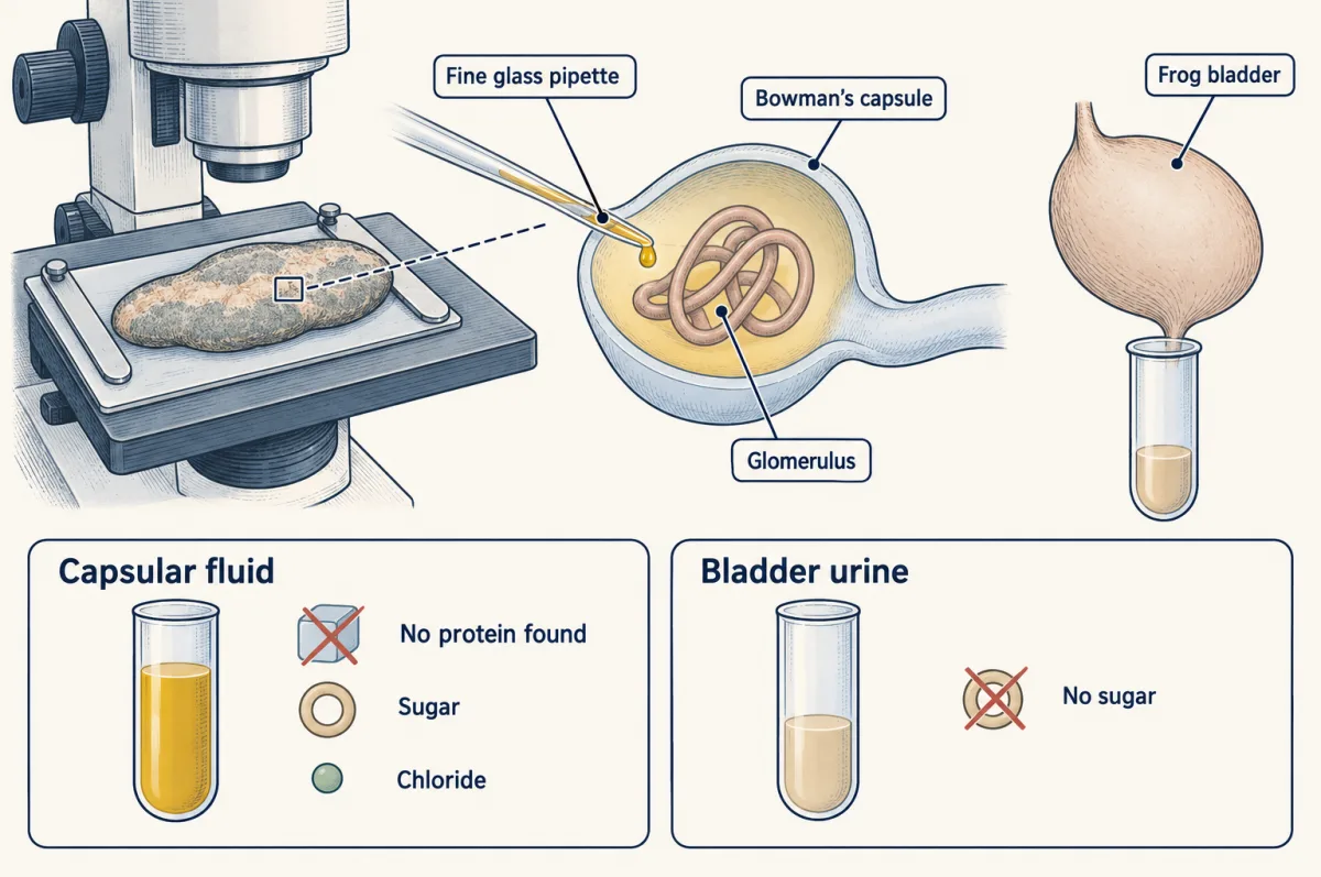
That fitted a filter followed by reabsorption, in the frog. It did not say where along the tubule or give mammalian rates, and frog nephrons lack a loop of Henle. Later micropuncture in rats and other mammals placed glucose reabsorption mainly in the proximal tubule.
Clearance: reading the ledger from outside
Can blood and urine alone, with no pipette, read the ledger? Urine concentration U times urine flow V is the excretion rate, the ledger’s “excreted” term. Divide by plasma concentration P and you get the volume of plasma that would hold that much each minute: the clearance. No plasma is really wiped clean.
In words Clearance equals urine concentration times urine flow, divided by plasma concentration.
- C“C”
- clearance, a volume of plasma per minutemL per min
- U“U”
- concentration in urinemg per mL
- V“V”
- urine flowmL per min
- P“P”
- concentration in plasmamg per mL
Sanity check U × V alone is mg per min, the excretion rate. Dividing by mg per mL leaves mL per min: a volume, not an amount.
Need a refresher? rates and “per” · units and prefixes
Inulin, a plant sugar polymer, is freely filtered and neither reabsorbed nor secreted, so excreted equals filtered and its clearance equals the glomerular filtration rate (GFR), the plasma volume filtered per minute.
Worked problem: clearance and filtered load
Illustrative numbers, for an adult human at steady state.
- Clearance. 120 mL of urine in 60 min gives V = 2.0 mL per min. With inulin at U = 30 mg per mL and P = 0.50 mg per mL, C = 30 x 2.0 / 0.50 = 120 mL per min. Over a day: 120 x 1,440 = 172,800 mL, about 173 L.
- Filtered load. Glucose is freely filtered. With plasma glucose at an illustrative 5.0 mmol per L, filtered load = GFR x P = 0.120 L per min x 5.0 mmol per L = 0.60 mmol per min, or 864 mmol per day (about 156 g). With no glucose in the urine, all of it was reabsorbed.
- Reading other clearances. For a freely filtered substance, a clearance above GFR means net secretion: at 300 mL per min the substance leaves faster than filtration alone could carry it. One of 1 mL per min means it is mostly reabsorbed. Creatinine, a muscle waste, is used to estimate GFR, but a little is secreted, so creatinine clearance tends to overestimate GFR.
- Urine flow V
- Clearance C
- C over a day (1,440 min)
- C against GFR (120 mL per min)
- Filtered glucose load, GFR × P
In this sketch: adult human at steady state, illustrative numbers, GFR taken as the inulin clearance of 120 mL per min, the test substances freely filtered, and the sketch holds urine glucose at 0 at every plasma glucose. Schematic tube, not to scale; the stack lists segments in the order the filtrate meets them.
Clearance C = U × V / P is a volume of plasma per minute. For a freely filtered substance, C equal to GFR fits no net handling (inulin); C above GFR means net secretion; C below GFR means net reabsorption. Creatinine, a muscle waste, is used to estimate GFR, but a little is secreted, so creatinine clearance tends to overestimate GFR.
Making urine more concentrated than blood
The last litres of water are the hardest to take back: they must come out of fluid that, with antidiuretic hormone (ADH) present, ends up more concentrated than blood. The second film shows how.
Read the transcript of “Making Urine More Concentrated Than Blood”
Film transcript: Making Urine More Concentrated Than Blood
The first film showed the kidney taking back almost all filtered water. The last litres are the hardest. A water-deprived adult can make urine about four times as concentrated as plasma. Yet no pump in the kidney moves water. So how does it pull water out of such urine? Concentration here means osmolality, the number of dissolved particles in each kilogram of water. Water moves by osmosis, toward the more concentrated side. So the tissue outside must be a little more concentrated than the urine. Salt supplies most, and deep down, urea helps. Picture a slice of kidney. The outer layer is the cortex. Beneath lies the medulla, an outer zone above an inner zone.
It ends at the papilla, where urine drains. A hairpin tube, the loop of Henle, dips into it. Start with the loop's upward arm. In the outer medulla it is thick, the thick ascending limb. It moves salt out, but its wall lets almost no water through. Salt leaves, water stays, and the tissue gets saltier. Now the thin descending limb, the downward arm. In rabbit tubules, it lets water through but holds its salt.
Passing salty tissue, it loses water by osmosis, so its fluid concentrates on the way down. The two arms carry fluid in opposite directions. Concentrated fluid rounds the bend, so the ascending limb works from a saltier start. Level by level, a modest difference adds up. This is the countercurrent multiplier. Deeper down, how the rise continues is still debated. Suppose the thick ascending limb let water through as easily as the descending limb. Would the multiplier still work well? Pause and decide.
Badly weakened. Water would follow the salt out, and most of the difference across the wall would collapse. Salt moving without water is the trick. Is real tissue graded like this? Wirz, Hargitay and Kuhn measured the freezing point of thin rat kidney slices from different depths. More particles mean a lower freezing point. Osmolality rose from cortex toward papilla. The collecting duct runs from cortex to papilla through ever more concentrated tissue. Antidiuretic hormone, ADH, acts on its cells. They insert stored water channels, aquaporin-2, facing the urine. Water leaves the urine by osmosis into the concentrated medulla. The urine comes out concentrated.
Take ADH away, the channels withdraw, and dilute urine flows out. So the kidney never pumps water. It moves solute first, building a medulla more concentrated than blood. Water follows where ADH opens the way.
From the film
- No pump in the kidney moves water: it moves solute, mostly salt, and water follows by osmosis.
- In the outer medulla, the thick ascending limb moves salt out but holds its water, so the tissue there becomes saltier than blood.
- Because fluid flows down one limb and up the other, a modest difference at each level adds up along the loop; rat kidney slices show osmolality rising from cortex to papilla, and how the inner medulla keeps rising is still debated.
- With ADH, the collecting duct inserts water channels and water leaves into the concentrated medulla, so urine becomes concentrated; without ADH, dilute urine flows out.
In numbers: adult human plasma is about 290 mOsm per kg (osmolality, dissolved particles per kilogram of water); a water-deprived adult can make urine near 1,200. The second film’s medulla has two zones: the outer medulla, where thick ascending limbs run, and the inner medulla below it, down to the papilla, which only thin limbs, collecting ducts and vessels reach.
Why a modest difference adds up
The second film’s multiplier works in the outer medulla.
Predict. In an idealized outer-medullary loop, keep salt transport at each level, flow, wall leakiness and blood washout the same, and make the loop twice as long. Could the deepest tissue end up more concentrated?
Check your prediction
Yes, in this idealized model: a longer countercurrent path gives the difference more steps to add up. That is a direction, not a number, and real long loops also reach the inner medulla, whose mechanism is unresolved. Hence the comparison below of loop length and medullary thickness.
Testing each limb on its own
The second film’s descending limb comes from Kokko (1970), who held single thin descending limbs from rabbit kidneys between glass pipettes and perfused them, setting both lumen and bath (the isolated perfused tubule method). Sodium permeability was low, and no active salt transport could be detected. The thick ascending limb takes up sodium chloride mainly through a cotransporter called sodium-potassium-chloride cotransporter 2 (NKCC2), with the sodium-potassium pump supplying the energy. Not every descending segment is water-permeable in every species (Going deeper).
Keeping the gradient
The vasa recta run in hairpins too: blood going down gains salt and loses water, and blood coming up does the reverse. So blood leaves carrying reabsorbed water but little of the salt: this countercurrent exchange limits washout of the gradient.
The inner medulla, and urine without ADH
The thick ascending limb does not reach the inner medulla, yet osmolality keeps rising there toward the papilla, by means still under study (Going deeper). Urea is part of it: there, urea leaves the collecting duct through urea transporters, by facilitated diffusion rather than an active pump, and adds to the gradient there.
Without ADH, urine is dilute, not merely plasma-strength: the thick ascending limb has already removed salt without water, and a collecting duct mostly closed to water keeps it that way.
Hormones tune the reabsorbed term
| Hormone | Released when | Ledger term it changes | Urine |
|---|---|---|---|
| ADH | Plasma osmolality rises or blood volume falls | Water reabsorbed rises (collecting duct) | Less, more concentrated |
| Angiotensin II and aldosterone | Blood pressure or salt is low | Sodium reabsorbed rises | Less sodium |
| ANP | The atria are stretched | Filtered rises; sodium and water reabsorbed fall | More sodium and water |
ADH is made in the hypothalamus and released from the posterior pituitary (endocrine system; hypothalamus and autonomic control). In collecting-duct cells it acts through V2 receptors and cAMP (G proteins, cAMP and PKA). Its water channels are aquaporin-2 (Nielsen and colleagues, 1995, in perfused rat ducts).
The renin-angiotensin-aldosterone system answers falling pressure or salt by holding on to sodium, through a chain:
- Renin, an enzyme, is released by juxtaglomerular cells in the wall of the afferent arteriole when renal perfusion pressure falls, when less salt reaches the macula densa (cells at the end of the thick ascending limb, where it touches its own glomerulus’s arterioles), or when sympathetic nerves stimulate beta-adrenergic receptors.
- Renin cuts angiotensinogen, a plasma protein made by the liver, into angiotensin I; angiotensin-converting enzyme turns that into angiotensin II.
- Angiotensin II raises sodium reabsorption in the tubule and stimulates the adrenal cortex to release aldosterone, a steroid that raises sodium reabsorption through epithelial sodium channel (ENaC) channels in principal cells and, coupled to it, potassium secretion (nuclear receptors).
Atrial natriuretic peptide (ANP), from heart muscle cells of the stretched atria (as after a rise in blood volume), pushes the other way. It raises glomerular filtration rate (GFR) and suppresses the release of renin and aldosterone. In anesthetized rats it inhibited sodium reabsorption in the inner-medullary collecting duct, and in perfused rat ducts it reduced ADH-stimulated water permeability. It works through cGMP (cGMP signaling).
Kidneys compared: the ledger in other animals
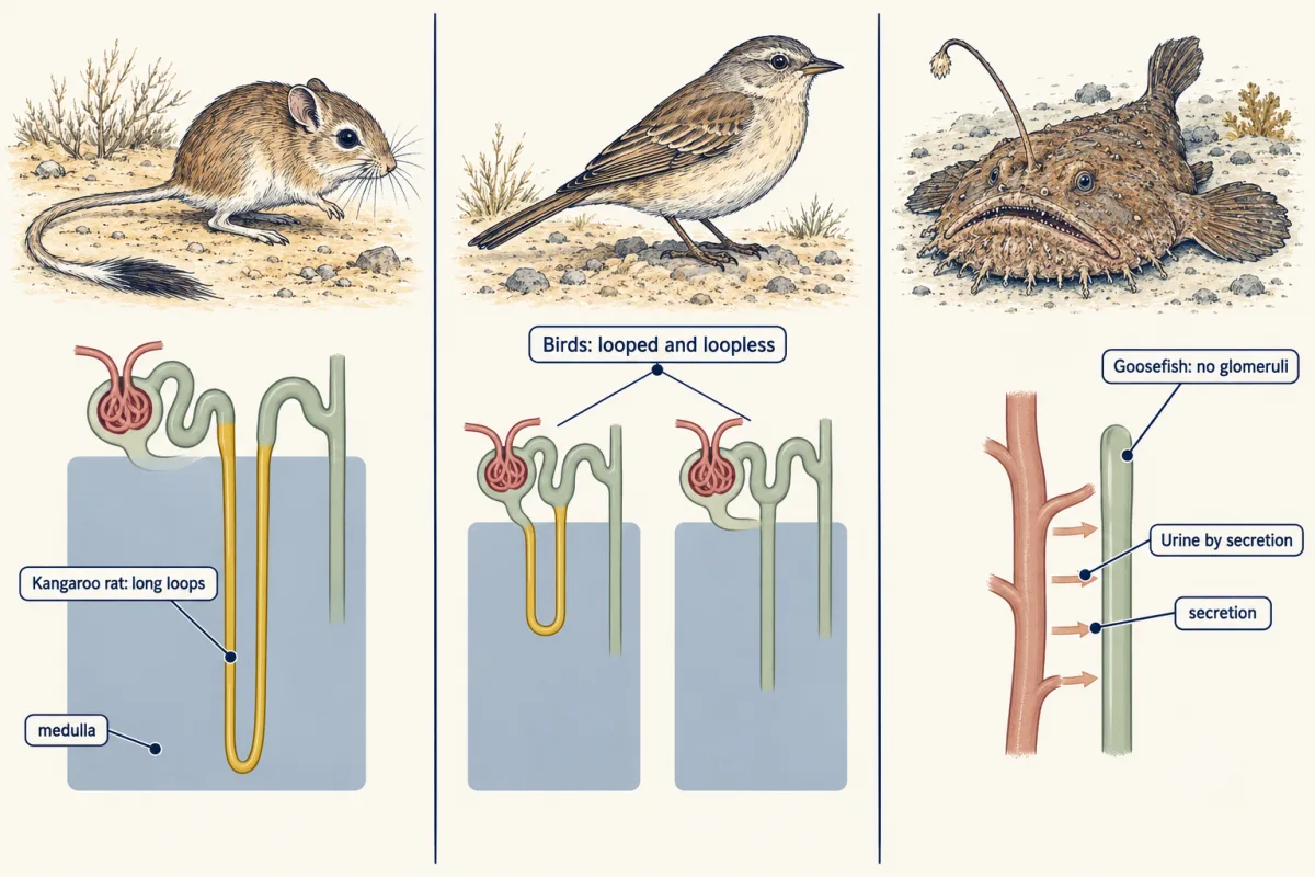
Among vertebrates, loops of Henle occur only in mammals and birds, and only these make urine more concentrated than their plasma. Amphibians and most fish have no loop, and their urine is at most about as concentrated as their plasma.
Across many mammal species, Beuchat (1990) found that relative medullary thickness tracks maximum urine concentration. The kangaroo rat Dipodomys merriami, a seed-eater of North American deserts, has long loops and a thick inner medulla and makes urine several times more concentrated than an adult human can (osmoregulation tells how it balances water).
Predict. To excrete an illustrative 600 mOsm of solute, how much water does urine at plasma’s level (about 300 mOsm per kg) take? At the human maximum, near 1,200? At four times that?
Check your prediction
About 2.0, 0.5 and 0.13 L: load divided by the concentration the kidney can reach.
Birds have a mix of loopless and looped nephrons and concentrate urine only modestly, to a small multiple of plasma osmolality; much of their nitrogen leaves as uric acid, which takes little water.
Some marine teleosts, including the goosefish Lophius and the toadfish Opsanus, have kidneys with no glomeruli. On the ledger, filtered is zero: everything in their urine reached the tubule by secretion. Tubule cells move ions such as magnesium and sulfate into the tubule, and water follows. Plants move water with no filter at all (plant transport).
Design problem (optional, harder)
Urea comes from protein breakdown (protein turnover). Fenton and colleagues (2004) made mice lacking UT-A1 and UT-A3, the urea transporters of the collecting duct. Predict how maximal urine osmolality compares between knockout and normal mice on a 20% protein diet and on a 4% protein diet, after water restriction. Then predict inner-medullary sodium chloride under the passive model, which says urea leaving the collecting duct helps draw salt out of the thin ascending limb.
One way to do it
If urea leaving the inner-medullary collecting duct adds to the gradient, knockouts should concentrate less, most on high protein, since low protein supplies little urea in either genotype. In fact knockouts concentrated less on 20% protein, the gap almost vanished on 4%, and inner-medullary sodium and chloride did not differ significantly between genotypes.
Two readings fit the deficit. Knockouts had much less urea in the inner medulla, so urea transport helps urea build up there. And, as Fenton and colleagues stressed, urea that cannot leave the collecting duct holds water in the urine, an osmotic diuresis.
The passive model predicts less inner-medullary NaCl in knockouts; they did not have less, which argues against that dependency. Keep the limits: these are mice, and one knockout does not settle how the inner medulla concentrates; that question is still open.
The ledger once more: in a filtering kidney the take-back largely sets urine volume, while all three terms together set how much of each solute leaves.
Going deeper
The take-back keeps pace with filtration
The first film’s “about the same share” has a name, glomerulotubular balance. Its doubling of urine was ledger arithmetic, not what a healthy kidney lets happen.
The descending limb across species
Kokko’s water-permeable descending limb is the rabbit’s; in rats, some inner-medullary descending segments lack the water channel aquaporin-1.
Which particles draw water
“More concentrated” is shorthand: what draws water across a wall is the particles that cannot cross that wall freely (effective osmolality). While water flows, the tissue need be only slightly more concentrated than the urine.
The inner medulla, an open question
There the thin ascending limb has no active NaCl transport. The passive model of Kokko and Rector and of Stephenson relies on urea from the collecting duct, which the Fenton knockouts challenge; other proposed mechanisms remain under study.
Your notes
Select any text in the lesson to highlight it or add a note to it. Your highlights and notes are saved in this browser and collected in your notebook.
This browser is not letting the site save anything (a private window or strict privacy settings), so highlights and notes will be lost when you leave the page.
How we measure it
Micropuncture
Insert a fine glass pipette into one part of a single nephron in an anesthetized animal and draw off a small fluid sample for analysis. Comparing samples from different points shows what each segment adds or removes, in the species studied.
Isolated perfused tubule
Dissect one tubule segment, hold it between pipettes in a bath, and pump fluid of known composition through it. Because lumen and bath are both controlled, you can measure that segment's permeability to water and salt and whether it transports actively.
Clearance measurement
Collect urine over a timed period at steady state and measure a substance's concentration in urine and in plasma. C = U x V / P gives the volume of plasma cleared of that substance per minute, and with inulin it estimates the glomerular filtration rate.
Tissue cryoscopy
Measure the freezing point of thin kidney slices taken at known depths. A lower freezing point means more dissolved particles, so the readings map osmolality from cortex to papilla.
Practice questions on this lesson (6) → MCAT-style, original, each with an explanation
Where this lesson fits
Before thisReview first
- Osmoregulation and Nitrogenous WasteThis course · Lesson 15
It explains how water and salt balance is kept, which this lesson takes as its starting point.
- Blood Vessels, Blood and LymphThis course · Lesson 11
You need Starling forces and capillary flow from there to see how the glomerulus filters in bulk.
- Tissues, Organs and HomeostasisThis course · Lesson 6
You need negative feedback control from there to follow how hormones tune the kidney's take-back.
After thisStudy next
- Moving Water and Sugar in PlantsThis course · Lesson 23
It moves water by water potential with no filter, which this lesson sets against the kidney.
Elsewhere on the siteFilms, simulations and method films in other courses
- Random Moves, One-Way DriftFilm · Life Science
It shows water moving toward the side with more particles, which drives water leaving the limbs.
- Downhill for FreeFilm · Biology
It shows net water movement by osmosis across a membrane, as in the collecting duct.
Suggested by an AI model from the lessons themselves, then checked. How we use AI
Finish line
Test yourself
11 questions
Practice and MCAT-style, with explained answers.
Next · Lesson 17
Innate Immunity: Recognize and Respond
A cut lets in 100 bacteria that double every 20 minutes, and new antibodies take days. What does your body have to do in the first few hours to stay ahead?
Continue →