Physiology · Unit 1: The internal environment and its control
Endocrine Axes and How to Test Them
Rewritten on : new text, figures and flashcards. Your notes and highlights stay, but the text around them may have moved.
Two people have the same low morning cortisol. In one the adrenal has failed; in the other it is healthy but unsignalled. Which second number tells them apart?
Pause when the reading reaches a film, and offer to play it. Saved on this device.
Watch the film now, or keep listening
Builds on:Body Water and Its CompartmentsWhy these?
 ▶ Watch Read the Pair, Not One Number · 2:47
Read the transcript of “Read the Pair, Not One Number”
Film transcript: Read the Pair, Not One Number
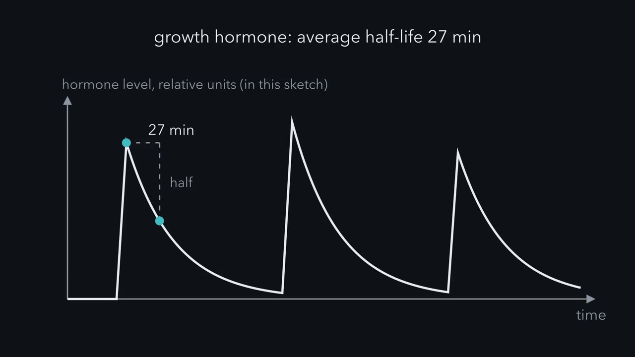
One blood sample gives you one number. What can it tell you? A hormone level is one point on a moving curve. Hormones are released in pulses, then cleared. Growth hormone left plasma with an average half-life of twenty-seven minutes. That was one small study's mean, after a large intravenous dose in five people. A sample drawn just before a pulse can read many times lower than one drawn on its peak. So read two hormones together. Take the cortisol axis. CRH, helped by vasopressin, drives ACTH from the pituitary. ACTH drives the adrenal cortex to make cortisol. Call ACTH the trophic hormone and cortisol the target hormone. Cortisol then feeds back on CRH and ACTH.
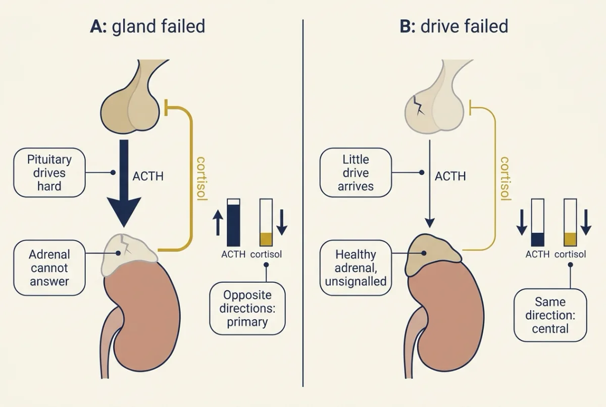
That is long-loop feedback, with strong evidence at both levels. Now break the chain at the gland. In this made-up example, adults on a normal sleep-wake schedule have blood drawn at 08:00. Person A has cortisol of 3 µg per dL and ACTH of 300 pg per mL. Low cortisol should make an intact pituitary drive hard, and it does. The adrenal does not answer: that is the primary pattern. Reference intervals differ by laboratory and assay. Person B has the same low cortisol, but ACTH is only 5 pg per mL. The drive itself is missing. That points to the pituitary, a secondary disorder, or to the hypothalamus, a tertiary one. The pair cannot say which, because either lesion lowers ACTH.
It can separate gland disease from central disease. Now change one input and watch the output. Person B receives synthetic ACTH. Thirty minutes later, cortisol is 16 µg per dL. Whether that is adequate depends on the assay, because criteria for newer, more specific assays sit below the historical one. An adequate response shows only that the adrenal can answer ACTH. After a recent loss of central drive, the adrenal cortex may not yet have atrophied. So a normal result does not exclude a recent pituitary or hypothalamic defect. The thyroid reads the same way. High TSH with low free T4 is the classical primary pattern. Low free T4 with low or inappropriately normal TSH points to central disease.
A hormone level is a point on a moving curve, and a criterion belongs to its assay. Read hormones as pairs, and know which assay made the number.
Two people have the same low morning cortisol. In one, the adrenal gland has failed. In the other, the adrenal is healthy, but nothing is telling it to work.
One number cannot tell them apart. Two numbers can: cortisol, and adrenocorticotropic hormone (ACTH), the pituitary hormone that drives the adrenal, read together. Two terms first. A target hormone is made by a gland the pituitary drives, such as cortisol from the adrenal cortex. A trophic hormone is the pituitary hormone that drives that gland, such as ACTH.
The one idea to carry through this lesson. Read the target hormone and its trophic hormone together. If the fault is in the target gland, feedback still works, so the pituitary pushes against it and the two hormones move in opposite directions. If the fault is in the drive from above (the pituitary or the hypothalamus), they move in the same direction.
The rule holds once the axis has settled, for free hormone, in a sample timed to the hormone’s rhythm, on an assay whose number means what you think. It does not apply to prolactin. Going deeper lists where else it is silent or fooled.
One chain, three links, one feedback loop
The endocrine system introduced the hypothalamus, portal vessels and anterior pituitary, and hypothalamus and autonomic control tells how the portal link was shown. The plan is a relay, like a chain of managers: a hypothalamic signal in portal blood, a pituitary hormone, a target tissue. Five axes follow it.
- Adrenocorticotropic hormone (ACTH). CRH (corticotropin-releasing hormone), helped by vasopressin from the same hypothalamic neurons, drives ACTH, and ACTH drives the adrenal cortex to make cortisol (the adrenal glands).
- Thyroid-stimulating hormone (TSH). Thyrotropin-releasing hormone (TRH) drives TSH, and TSH stimulation is required for the thyroid to make enough T4 and T3 (thyroid hormone).
- Growth hormone (GH). GHRH (growth hormone-releasing hormone) stimulates somatotrophs; somatostatin, in the same portal blood, inhibits them. GH makes the liver secrete IGF-1 (insulin-like growth factor 1). GH also acts directly through its own receptors, for example promoting breakdown of stored fat in adipose tissue, so IGF-1 does not carry every GH effect.
- Luteinizing hormone (LH) and follicle-stimulating hormone (FSH). Pulsatile gonadotropin-releasing hormone (GnRH) drives both (animal reproduction).
- Prolactin, the axis that runs on a brake. Hypothalamic dopamine tonically inhibits prolactin release through lactotroph D2 receptors. Several factors, TRH among them, can stimulate prolactin in experiments, but no releasing hormone dominates. Milk production belongs to pregnancy, birth and lactation.
For the MCAT: releasing hormones, pituitary hormones and dopamine are peptides or amines acting mostly on G protein-coupled receptors and second messengers such as cyclic AMP and calcium (GPCRs and G proteins, cAMP and PKA). GH and prolactin are the exceptions: their receptors signal through JAK2 and STAT5 (JAK-STAT). Cortisol and thyroid hormone travel mostly on carrier proteins and act on nuclear receptors. Target specificity is receptor expression: every cell sees ACTH, and only cells with ACTH receptors respond.
Trophic means two jobs. A trophic hormone tells its gland to secrete now. Over weeks it also keeps the gland’s secreting cells from shrinking: take ACTH away for weeks and the cortisol-making layers of the adrenal cortex shrink, which will matter for the stimulation tests.
Long-loop feedback closes the chain. The target hormone travels back and restrains the links above it: cortisol restrains CRH and ACTH, T4 and T3 restrain TRH and TSH, and IGF-1 restrains GH. The evidence for all three loops is strong, at both the hypothalamus and the pituitary for cortisol and thyroid hormone, and from a human infusion experiment for IGF-1. Shorter loops, from a pituitary hormone back to the hypothalamus or onto the cells that made it, have causal evidence in mice for prolactin and GH and weaker evidence elsewhere; how much they matter in humans is open (Going deeper).
Feedback is why the rule works: an intact pituitary keeps reading the target hormone, so the trophic hormone is a running report on its target. If the drive from above fails or runs too hard, the gland simply follows it.
Which link failed? Read the pair
A disorder is named by the link that fails. In a primary disorder the target gland itself fails. In a secondary disorder the pituitary fails. In a tertiary disorder the hypothalamus fails. Secondary and tertiary together are called central.
The film at the top of this page walks through persons A and B below. It opens with hormone pulses, which the section on timing takes up.
Three mornings
In this made-up example, three adults on a normal sleep-wake schedule have blood drawn at 08:00, near cortisol’s daily peak. None takes a glucocorticoid medicine, and all have normal cortisol-binding globulin. The numbers are illustrative, the adrenocorticotropic hormone (ACTH) values are invented, and reference intervals differ by laboratory and assay.
| Person | Cortisol (µg per dL) | ACTH (pg per mL) |
|---|---|---|
| A | 3 | 300 |
| B | 3 | 5 |
| C | 14 | 30 |
Cortisol is reported in two units. Its molar mass is about 362.5 g per mol, so 1 µg per dL = 10 µg per L = (10 x 10−6 g per L) / (362.5 g per mol) = 2.76 x 10⁻⁸ mol per L, or 27.6 nmol per L.
cortisol (nmol per L) = cortisol (µg per dL) x 27.6
A and B: 3 x 27.6 = 82.8 nmol per L. C: 14 x 27.6 = 386.4 nmol per L.
Predict. A and B have the same low cortisol. Where has each chain failed?
Check your prediction
A: low cortisol, very high ACTH. The two move in opposite directions. The pituitary senses too little cortisol and drives hard, and the adrenal does not answer. The fault is in the gland: primary adrenal insufficiency.
B: low cortisol, low ACTH. The two move in the same direction. The adrenal is getting almost no signal. The fault is above it: central. The pair cannot say whether it is the pituitary (secondary) or the hypothalamus (tertiary), because either lesion lowers ACTH.
C has cortisol and ACTH both in the usual morning range, so nothing has failed at rest. A resting pair cannot show how the axis would cope with stress, and a value of 14 µg per dL does not by itself prove normal reserve if adrenal insufficiency is suspected. That is a question for the tests in the next section.
”Inappropriately normal”
Suppose B’s ACTH had been 25 pg per mL, inside a typical reference interval. Would that clear B’s pituitary? No. With cortisol at 3 µg per dL, an intact pituitary should be driving hard, as A’s is. An ACTH that sits in the normal range while cortisol is low has failed to rise. Endocrinologists call it inappropriately normal, and it reads the same as low. A reference interval comes from people whose target hormone is normal, so judge a trophic hormone against its own target, not against the population.
The thyroid reads the same way
High thyroid-stimulating hormone (TSH) with low free T4 is the classical primary pattern. Low free T4 with low or inappropriately normal TSH points to central disease. The pituitary is very sensitive to thyroid hormone: a small fall in free T4 brings a much larger proportional rise in TSH. That is why TSH alone is usually the first screen for primary thyroid disease. It is also why a TSH-only screen misses central hypothyroidism; when pituitary disease is suspected, measure free T4 too.
Excess reads the same way
The rule does not care whether the target hormone is too low or too high.
| Trophic hormone high | Trophic hormone low | |
|---|---|---|
| Target hormone high | Same direction: excess drive. Examples: an ACTH-secreting pituitary tumour (Cushing disease); a TSH-secreting pituitary adenoma. | Opposite: the gland is working on its own. Examples: a cortisol-secreting adrenal adenoma; Graves disease, where an antibody stimulates the thyroid’s TSH receptor. |
| Target hormone low | Opposite: the gland has failed. Examples: primary adrenal insufficiency (A); primary hypothyroidism. | Same direction: missing drive. Example: secondary or tertiary insufficiency (B). |
Predict. A patient has high cortisol and an ACTH so low the assay can barely detect it. Is the problem in the pituitary or the adrenal?
Check your prediction
The adrenal. Cortisol is high and ACTH is suppressed, so the two move in opposite directions. The pituitary is obeying feedback and shutting down. If no glucocorticoid medicine is involved, the cortisol is coming from the adrenal itself, for example an adenoma.
Change one input: stimulation and suppression tests
A dynamic test changes one input and watches the output. Stimulation tests ask about reserve: can this gland respond? Suppression tests ask about autonomy: does secretion still fall when the signals that normally turn it down are applied?
Take B. B receives a dose of synthetic adrenocorticotropic hormone (ACTH) (cosyntropin, ACTH 1-24), and cortisol 30 minutes later is 16 µg per dL, or 16 x 27.6 = 441.6 nmol per L.
Predict. Suppose B’s pituitary stopped making ACTH months ago. What should the adrenal do when given ACTH? What if it stopped last week?
Check your prediction
Months ago: without its trophic support the cortex has shrunk, so one dose of ACTH usually gives a subnormal response. That is how the test detects long-standing central insufficiency, even though the adrenal itself is not diseased. It misses some cases, especially partial deficiency, where enough ACTH remains to keep the cortex from shrinking.
Last week: the cortex has not had time to shrink, so it can answer normally.
So a normal response shows only that the adrenal can answer ACTH today. It does not exclude a recent pituitary or hypothalamic defect.
Is 16 µg per dL a normal response? That depends on the assay that made the number. The historical criterion, 18 µg per dL (about 500 nmol per L), was carried over from older, less specific immunoassays. Newer, more specific assays read lower on the same blood, and the criteria derived for them in 2021 are lower, about 14 to 15 µg per dL. Measured on a newer assay, B’s 16 passes. Judged against the historical number, it would fail.
So: A is primary. B is central, with an adrenal that, on a newer assay, can still answer ACTH. That fits a loss of drive too recent for the cortex to have shrunk, or a partial one, and the test does not clear B. C shows no failure at rest.
The other dynamic tests
- Dexamethasone suppression. A night-time dose of dexamethasone, a synthetic glucocorticoid, normally lowers ACTH and next-morning cortisol. Failure to suppress suggests secretion escaping feedback, but it does not locate the source. Faster dexamethasone clearance or more binding globulin can confound it.
- Glucose suppression of growth hormone (GH). Oral glucose normally suppresses GH. Failure to suppress helps when GH excess is in question.
- GH stimulation. Insulin-induced hypoglycemia, glucagon, or macimorelin (a ghrelin-receptor agonist) test whether the GH axis can release GH. Several act through the hypothalamus rather than on somatotrophs directly.
Like cosyntropin, any test that stimulates a gland directly cannot say whether a weak response reflects the gland itself or a long-standing loss of drive from above.
Test the wiring: a compressed stalk
A non-secreting mass presses on the pituitary stalk, and portal delivery from the hypothalamus falls.
Predict. What happens to ACTH, thyroid-stimulating hormone (TSH) and GH? What happens to prolactin?
Check your prediction
Hormones driven by releasing hormones tend to fall (ACTH, TSH, GH, LH and FSH), in combinations that vary from patient to patient, and with them cortisol, free T4, IGF-1 and sex steroids. TSH may be low or inappropriately normal rather than high. Prolactin rises, usually modestly, because the signal it loses is inhibitory dopamine.
This is why prolactin sits outside the pair rule: it has no target hormone to pair with, and its hypothalamic control is a brake. A drug that blocks D2 receptors, as many antipsychotics do, also raises prolactin, with no lesion at all, and a D2 agonist lowers prolactin whether the cause is a stalk lesion or a prolactinoma. So a drug response does not locate the problem. What points to the stalk is a modest prolactin rise together with low levels of other pituitary hormones.
Condition 1: catch the right moment
Many pituitary hormones are released in pulses, and several follow a daily rhythm, so when the blood was drawn can matter as much as what is wrong.
Rhythms. In young adults the night’s large growth hormone (GH) surge comes at the onset of deep (slow-wave) sleep. Takahashi, Kipnis and Daughaday (1968) delayed sleep by about three hours in four young adults, and in the three who had a surge it moved with sleep: the surge was tied to sleep onset, not clock time (sleep staging is in circadian rhythms and sleep). The HPA axis keeps a daily rhythm with pulses on top. In humans under normal conditions on a usual sleep-wake schedule, it peaks between 06:00 and 10:00 and reaches its nadir between 22:00 and 02:00, with adrenocorticotropic hormone (ACTH) and cortisol pulses on that curve; the rodent pulse experiments are in emotion and stress. That is why the three mornings were drawn at 08:00.
Pulses and clearance. Pulses matter for measurement because GH is cleared fast: a 1962 study measured an average half-life of 27 minutes after large intravenous doses, and later estimates are shorter (Going deeper). Either way the scale is the same: a pulse decays to a quarter or less within an hour, and a sample drawn just before a pulse can read many-fold lower than one drawn on its peak.
Predict. A single GH sample, drawn at a random time in an adult, comes back low. Does it show GH deficiency?
Check your prediction
No. The sample may simply have fallen between pulses. A pulse that peaked at 12 ng per mL and then stopped would be down to about 2.6 ng per mL an hour later, at a 27-minute half-life (12 x 2−60/27). A random GH value is weak evidence, and in adults a single random GH or IGF-1 value is not considered enough, in most patients, to establish GH deficiency. That is why the stimulation tests above exist.
- Single sample reads (relative units)
- 0
- 24-hour mean (relative units)
- 0
- Lowest and highest over the 24 h
- 0
- Same sample drawn 10 min later
- 0
- Slow readout at the sample time (relative)
- 0
- The reading, step by step
- 0
In this sketch, the status appears here.
In this sketch: the half-life is the only measured number, and two measured averages are offered: 27 minutes after large intravenous doses (1962, five people; individual values 23 to 38) and about 15 minutes from later deconvolution of naturally secreted GH; neither is a constant. The slow readout is a made-up smoothing to show the idea (each minute it moves 1/600 of the way toward the GH trace, starting at the 24-hour mean); real IGF-1 also depends on nutrition, liver function and age, and its kinetics are not modelled. Everything else is made up for the sketch: a daytime pulse is 1 relative unit and the sleep-onset pulse is 3; pulses come every interval from 07:00 until sleep onset and none follow it; release is instantaneous (real secretion is not instantaneous; in the 1968 study of eight young adults, seven had a major peak at the onset of deep sleep that lasted 1.5 to 3.5 hours); a later onset also adds daytime pulses; there is no baseline secretion and nothing carries over from the day before.
Keep this: with a half-life of 15 to 27 minutes, a single sample mostly reflects the last hour, so one reading can depend on when it was drawn as much as on how much was secreted, and a mean from frequent sampling over 24 hours is steadier.
An integrated readout. IGF-1 serves as a readout of GH secretion over many hours because it circulates mostly bound to binding proteins and is considered to have a much longer half-life than GH, so it averages many pulses. It also depends on nutrition, liver function and age.
So time the sample to the rhythm, and where a single value swings too much, read an integrated measure or run a dynamic test.
Condition 2: know what the assay measured
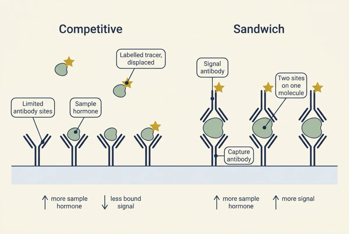
Two antibody designs. Competitive radioimmunoassay is explained in detecting a signal. In a competitive assay, labelled and sample hormone compete for a limited antibody, so bound signal falls as hormone rises. In a two-site (sandwich) assay, a capture antibody and a signal antibody bind two sites on one molecule, so signal rises with hormone. Sandwich assays suit large peptides such as thyroid-stimulating hormone (TSH). Small molecules such as free T4 are usually measured competitively, because a small molecule is too small for two antibodies to bind it at once.
What the antibody sees. Both designs measure immunoreactivity, what an antibody recognizes, not bioactivity, what a receptor responds to. Macroprolactin, a large prolactin-IgG complex, is detected by immunoassays but has low biological activity, so a high reported prolactin can mean little at the receptor. Antibodies also cross-react with related molecules, a problem for steroids. LC-MS/MS (liquid chromatography-tandem mass spectrometry) separates the hormone from its relatives and identifies it by mass and fragments, so it reports a defined chemical species. Better specificity changes the numbers, so a criterion from an older assay can mislabel a newer result. That is what happened to B’s cosyntropin result: measured on a newer specific assay, 16 µg per dL passes that assay’s criterion, yet it would be called a failure if read against the historical 18.
Free and total. Roughly 99.97 percent of circulating T4 is protein-bound in commonly quoted figures, and published values vary a little with method. Free hormone is the unbound fraction, considered the part available to tissues (binding and dose-response). Total T4 moves with binding protein: more thyroxine-binding globulin, as with oral estrogen, raises total T4 while free T4 stays near its usual range and TSH stays normal. (Pregnancy raises the binding globulin too, but adds its own effects: hCG lowers TSH in the first trimester.) Read the pair with total T4 and the rule seems broken; read it with free T4 and it holds. Physical separation by equilibrium dialysis followed by isotope-dilution LC-MS/MS is a reference approach for free thyroid hormones; routine free-hormone immunoassays are method-dependent estimates.
When the assay fakes a pattern. Some immunoassays capture the antibody complex with biotin and streptavidin, and a biotin supplement can block that step. On a susceptible platform TSH then reads falsely low and free T4 falsely high: the opposite-direction pattern of an overactive thyroid, in a healthy person. Back to the rule: before reading a pair, ask what the assay measured.
Design problem
A patient had pituitary surgery 10 days ago. Morning cortisol is low, adrenocorticotropic hormone (ACTH) is low-normal, and a cosyntropin test is normal. Lay out the reasoning, and say what each result can and cannot show about whether the axis is intact.
Saved in this browser only.
One way to do it
Read the pair first. Low cortisol with an ACTH that has not risen is a same-direction pattern: the low-normal ACTH is inappropriately normal, so the drive from above looks inadequate.
Then weigh the cosyntropin result against the time since surgery. Ten days is too short for the adrenal cortex to have shrunk, so a normal response shows only that the adrenal works today. It cannot clear the pituitary.
Check the conditions: a morning sample, no glucocorticoid medicine suppressing ACTH, and a cortisol criterion that matches the assay.
So the low morning pair is the stronger evidence now. A stimulation test repeated weeks later, once the cortex would have had time to shrink if drive is truly missing, can detect the loss, though it can still miss a partial deficiency. Until then, the normal cosyntropin result is not reassurance.
Going deeper
Short and ultra-short loops
Loops are named by how far the signal travels back: long-loop from the target-gland hormone, as above; short-loop from a pituitary hormone back to the hypothalamus. Ultra-short feedback is a hormone acting back on the very cells that make it, classically a hypothalamic releasing hormone acting on its own neurons. The term is sometimes also used for a pituitary hormone acting on its own cells.
| Arrow | Type | Evidence |
|---|---|---|
| Cortisol on CRH and ACTH | Long | Strong, at both levels |
| T4 and T3 on TRH and TSH | Long | Strong, at both levels |
| IGF-1 on GH | Long | Strong, human infusion |
| Prolactin on dopamine neurons | Short | Causal, knockout mice |
| GH on GHRH neurons | Short | Causal, cell-specific knockout mice |
| TSH or ACTH on hypothalamus | Short | Less established |
| Prolactin on lactotrophs | Ultra-short | Suggested by cell culture, of proliferation not secretion |
Prolactin. Prolactin is thought to stimulate the dopamine neurons that inhibit it. Schuff, Low and colleagues (2002) compared mice lacking D2 receptors, mice lacking prolactin receptors and compound mutants, used D2 agonist and antagonist challenges, and cultured pituitary cells. Females lacking either receptor developed hyperprolactinemia and large prolactinomas, but prolactin receptor knockouts had more hyperprolactinemia than age-matched D2 knockouts and had lost much of their dopamine tone. Here is the step that matters. If prolactin restrained lactotrophs only by driving dopamine, losing its receptor could at most remove dopamine’s brake, which the D2 knockout already does. Doing worse than the D2 knockout therefore points to a second restraint that does not run through dopamine, though the two knockouts also differ in other ways (prolactin receptor-null females are infertile, with an altered steroid milieu). The culture result supports the direct action: prolactin slowed proliferation of normal female lactotrophs. So the loop runs through the hypothalamus and also acts at the pituitary independently of dopamine, which fits an ultra-short action on lactotroph proliferation, shown in cultured cells. Global, lifelong deletions cannot assign each phenotype to one cell population, and the results are mainly in female mice.
Growth hormone (GH). Deleting GH receptors only in GHRH-expressing mouse cells raised pulsatile GH secretion and pulse amplitude, yet acute GH or IGF-1 did not change GHRH-neuron firing in recordings (Gusmao and colleagues, 2025): short-loop feedback, since GH is a pituitary hormone acting on hypothalamic neurons, and it need not mean fast electrical inhibition. Thyroid-stimulating hormone (TSH) and adrenocorticotropic hormone (ACTH) short loops rest on older, thinner evidence, and their role in humans is not settled.
The IGF-I infusion experiment
The GH long loop was tested in people. Bermann, Barkan and colleagues (1994) gave six normal fed men a saline infusion followed, in the same admission, by a 48-hour infusion of recombinant IGF-I, sampling GH every 10 minutes for 24 hours in each period. Overall GH fell by 85 plus or minus 3 percent and pulse amplitude by 77 plus or minus 4 percent (mean plus or minus SE). The authors favoured increased hypothalamic somatostatin, an inference from hormone responses; nobody sampled portal blood.
The 1968 sleep study
Takahashi, Kipnis and Daughaday (1968) asked what times the night-time GH rise. They studied eight young adults over 38 nights, drawing repeated blood samples through an indwelling catheter while electroencephalogram (EEG) and eye-movement recordings staged sleep. If a clock or the overnight fall in glucose set the rise, delaying sleep should leave the peak in place. In seven of the eight, a major GH peak appeared at the onset of deep (slow-wave) sleep and lasted 1.5 to 3.5 hours. Besides the delay result above, three subjects woken for two to three hours showed another peak on falling asleep again, and the peak did not correlate with concurrent changes in glucose, insulin or cortisol. That argues against a simple glucose or insulin trigger. It does not show the hypothalamic mechanism (more GHRH or less somatostatin), that the pattern holds at every age, or that glucose never regulates GH.
How fast GH clears
Parker, Utiger and Daughaday (1962) injected 5 or 10 mg of human GH intravenously into five people and found that plasma levels fell with an average half-life of 27 minutes (individual values 23 to 38). That is one small study’s mean after a large dose, not a constant. Later deconvolution of naturally secreted GH in healthy adults gave shorter values, about 15 minutes.
The biotin study
In a 2020 study, ten healthy adults took a biotin supplement daily for a week, at a lower and later a higher amount, and their thyroid tests were run on three platforms. Two hours after the last dose of the first regimen, one platform reported lower TSH and higher free T4 than baseline, a pattern that looks hyperthyroid, while the tested assays on another showed no significant change.
The standard explanation: excess sample biotin occupies the streptavidin, less antibody complex is captured, and signal falls. In a sandwich assay (TSH) less signal reads as less hormone; in a competitive assay (free T4) it reads as more. Both errors point toward “hyperthyroid”.
Ways to separate a real change from an artefact follow from that. Rerun the same sample on a platform without a biotin capture step: a biological change should appear on both, an artefact only on the susceptible one. Repeat after stopping biotin: in this study the interference lasted about one to two days after the higher regimen on the susceptible systems, so a repeat after that should return to baseline if the change was analytical, though this small study does not set a universal washout. Look for physiological agreement: a real hyperthyroid state usually also moves heart rate and symptoms, though older adults may show few, and T3 measured on a platform without a biotin step, because T3 assays on the susceptible platform can be biased too. The study did not test larger doses, impaired kidney function, other analytes or current versions of every assay.
When the pair rule is silent or fooled
- During transitions. The rule describes a settled axis. After a hyperthyroid patient is treated, TSH can stay suppressed for weeks to months while free T4 is already normal or low. The cosyntropin limit above is the same problem.
- Bioinactive TSH. The exception to the central thyroid pattern: in hypothalamic hypothyroidism, immunoreactive TSH can be mildly high but poorly bioactive, which mimics the opposite-direction pattern of thyroid failure. The assay reports the antibody’s view, not the receptor’s.
- Where, not just which way. “Same direction” says the drive is at fault, not where. It cannot split pituitary from hypothalamus, or pituitary ACTH from an ectopic ACTH-secreting tumour elsewhere in the body.
- A raised set point. If the pituitary becomes less sensitive to feedback, it holds both hormones higher. That reads as “same direction, drive”, which correctly places the fault in the regulator rather than the gland. It cannot say whether the regulator is a tumour or a receptor that resists the hormone, as in resistance to thyroid hormone, so hormone-resistance syndromes and hormones taken as medicines need their own reading.
- The receptor needs a partner. Strictly, the ACTH receptor (MC2R) also needs an accessory protein, MRAP, to reach the cell surface. So “the gland cannot answer ACTH” includes the receptor machinery, and a fault there reads as primary: high ACTH, low cortisol.
- A model check. Suppose gland output is proportional to the trophic hormone, and pituitary output falls as the target hormone to the power 1.6 (an assumed feedback strength; real axes, especially the thyroid, are steeper). A partial loss of drive that lowers the target to 0.82 times normal then leaves the trophic hormone at 0.82 times normal, possibly inside a reference range. An intact pituitary facing the same target level would sit at 0.82−1.6, about 1.37 times normal. That is “inappropriately normal” in numbers.
Your notes
Select any text in the lesson to highlight it or add a note to it. Your highlights and notes are saved in this browser and collected in your notebook.
This browser is not letting the site save anything (a private window or strict privacy settings), so highlights and notes will be lost when you leave the page.
How we measure it
Frequent blood sampling with sleep staging
Place an indwelling venous catheter so blood can be drawn every 10 to 30 minutes with little disturbance to the subject, and record EEG and eye movements to stage sleep. The paired records separate short secretory pulses from the daily rhythm and link release to a state such as sleep onset.
Competitive and two-site (sandwich) immunoassays
In a competitive assay, sample hormone and a labelled hormone compete for a limited antibody, so bound label falls as hormone rises. In a sandwich assay, a capture antibody and a signal antibody bind two sites on one hormone molecule, so signal rises with hormone. Both report what an antibody recognizes, which is not always what a receptor responds to.
Liquid chromatography-tandem mass spectrometry (LC-MS/MS) with equilibrium dialysis
Chromatography separates the hormone from related molecules, and two stages of mass spectrometry identify it by its mass and fragments and quantify it. Dialysing serum across a membrane first, until free hormone equilibrates, lets the method measure the free fraction directly rather than estimate it.
Gene knockout mice, global and cell-specific
Delete a receptor gene in every cell, or only in one cell type with a Cre recombinase driven by that cell's promoter, and ask which feedback arrow disappears. Lifelong deletion can let other parts of the axis adapt, so a phenotype is evidence about a loop, not a direct reading of its strength in adults.
Practice questions on this lesson (3) → MCAT-style, original, each with an explanation
Where this lesson fits
Before thisReview first
- Body Water and Its CompartmentsThis course · Lesson 1
It sets up the body's internal environment, which hormone axes regulate, though this lesson uses it only loosely.
After thisStudy next
- Thyroid Hormone: Tissues Decide What T4 BecomesThis course · Lesson 19
It applies the paired TSH and free T4 reading to where T4 becomes T3 in tissues.
- The Adrenal Cortex and MedullaThis course · Lesson 20
It takes the ACTH and cortisol axis further, including how blocking an enzyme changes the feedback loop.
- Insulin, Glucagon and Blood GlucoseThis course · Lesson 21
It uses a challenge test, the glucose drink, to read how a hormone system responds.
Elsewhere on the siteFilms, simulations and method films in other courses
- RadioimmunoassayMethod film
It shows how competitive hormone assays work, which the lesson needs to say what a measured number means.
- Receptor binding and dose-response analysisMethod film
It covers binding and dose-response, the basis of how assays and challenge tests read a hormone.
- When the Body Pushes BackFilm · Biology
It shows how feedback pushes a variable back toward its range, the loop behind the long-loop feedback here.
- Leak and Push-BackFilm · Organismal Biology
It shows a control loop with a sensor and push-back, the same wiring as an endocrine axis.
Suggested by an AI model from the lessons themselves, then checked. How we use AI
Finish line
Test yourself
10 questions
Practice and MCAT-style, with explained answers.
Next · Lesson 3
Skeletal Muscle: Length, Load and Speed
A muscle lifting nothing and a muscle holding a load it cannot move both do zero external work. Where is the power?
Continue →