Neuroscience · Unit 5: Homeostasis and affect
Circadian Rhythms and Sleep
Rewritten on : new text, figures, flashcards and film. Your notes and highlights stay, but the text around them may have moved.
Stay up all night and you feel worst near dawn, then better by the afternoon, still without sleep. How can more time awake make you less sleepy?
Pause when the reading reaches a film, and offer to play it. Saved on this device.
Watch the film now, or keep listening
Builds on:Autonomic Control and the HypothalamusThe RetinaTransmitter Systems and DrugsWhy these?
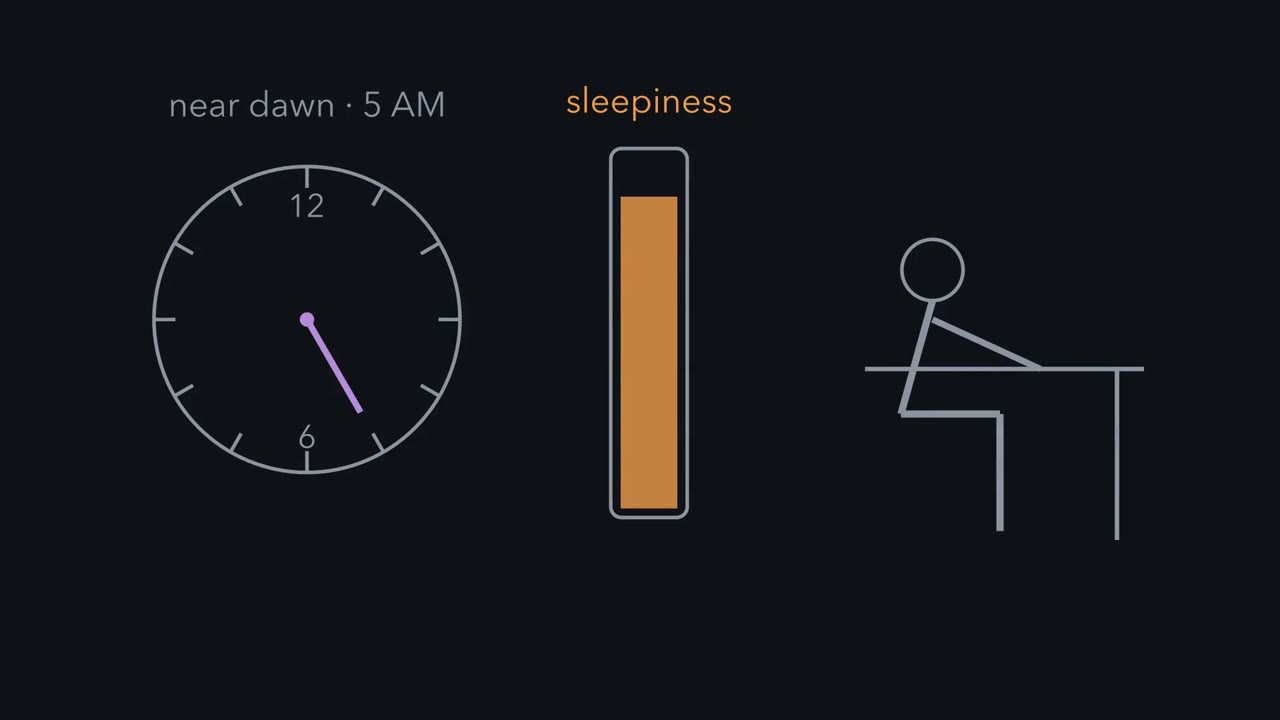 ▶ Watch Pressure and the Clock · 2:45
Read the transcript of “Pressure and the Clock”
Film transcript: Pressure and the Clock
Stay up all night. Near dawn, you can barely keep your eyes open. By early afternoon, still without sleep, you feel better. How can more time awake make you less sleepy? If time awake were all that mattered, the afternoon would feel worse. Something else changed: a clock. The first process is sleep pressure. It builds while you are awake, quickly at first, then more slowly. While you sleep, it drains. The second process is a clock in your brain. It keeps a cycle of about a day, even with no windows. The clock's main push is toward waking. It is hardest in the late afternoon and evening, weakest in the early morning. Put both on one graph. An upper wavy line is the level pressure must reach before you fall asleep.
A lower wavy line is the level it must drain to before you wake. Both lines rise and fall with the clock. Here is the one idea, in the simple model of an adult's undisturbed sleep. Sleep pressure climbs while you are awake and drains while you sleep. You fall asleep when it climbs to an upper level. You wake when it drains to a lower one. The clock raises both levels by day and lowers them by night. Why two levels? Replace them with one line midway between, and the model chatters on and off at that line. The gap keeps this model's sleep in one long block. Back to the all-nighter. Staying up means refusing sleep after pressure crosses the upper level.
Near dawn, the clock has pulled that level low. So pressure sits far above it, and you feel very sleepy. By early afternoon, pressure is higher still. But the clock has lifted the level, so the excess has shrunk. When you finally sleep, will you get back every hour you lost?
No. Recovery sleep is longer than usual, but nowhere near hour for hour. Much of the payback comes as deeper sleep. Can the two processes be pulled apart? In rats, destroy the clock: the suprachiasmatic nucleus, just above the optic chiasm. Sleep scatters across day and night. Yet after a spell without sleep, these rats still slept more, and more deeply. Keep this. Pressure climbs and drains, and the clock sets the levels.
From the film
- In the simple model, sleep pressure builds while you are awake and drains while you sleep.
- You fall asleep when pressure climbs to an upper level and wake when it drains to a lower one, and the clock raises both levels by day and lowers them by night.
- Two levels with a gap keep the model's sleep in one long block, and recovery sleep after a missed night is longer than usual but nowhere near hour for hour.
Stay up all night and something odd happens. Near dawn you can barely keep your eyes open. By early afternoon, still without sleep, you feel better, though you have been awake hours longer.
If sleepiness depended only on how long you had been awake, the afternoon should feel worse than the dawn. Something besides time awake changed between them. That something is a clock.
Two processes decide when you sleep
The first process is sleep pressure, often called Process S. It builds all the time you are awake and drains while you sleep. It builds quickly at first, then more slowly as it nears a ceiling. In people, one sign of it is the strength of the slow brain waves of deep sleep, measured on the EEG, at the start of the night, which is larger the longer you were awake beforehand.
The second process is the circadian clock, Process C. Circadian means “about a day”: the clock keeps a cycle near 24 hours even in a room with no windows or clocks. The clock’s main push is toward waking, and that push rises and falls once a day. In people it is strongest in the late afternoon and evening, a few hours before your usual bedtime, and weakest in the early morning, near the daily low point of body temperature.
Put the two together in a picture, with time of day along the bottom and sleep pressure up the side.
- During the day, pressure climbs as a rising curve.
- Above it runs a wavy line: the level pressure must reach before you fall asleep.
- Below it runs a second wavy line: the level pressure must drain to before you wake.
- Both wavy lines rise and fall with the clock.
Over several days the pressure curve is a sawtooth: a slow climb by day to the upper line, then a faster fall by night to the lower one.
The one idea to carry through this lesson. In the simple model of an adult’s undisturbed sleep: sleep pressure climbs while you are awake and drains while you sleep. You fall asleep when it climbs to an upper level, and you wake when it drains to a lower one. The clock raises both levels by day and lowers them by night.
Two features of this rule do the work.
- There are two levels, with a gap between them. Suppose one level, set midway between the two, did both jobs. In the model you would fall asleep the moment pressure touched it, drain a little, wake, refill a little and fall asleep again, flickering at the line. The gap keeps the model’s night of sleep in one long block.
- The clock moves the levels against the pressure. As pressure climbs through the afternoon, the clock lifts the upper level ahead of it, so sleep is harder to start and you stay alert into the evening. After you fall asleep, the clock lowers the lower level through the early morning, so you stay asleep while pressure drains. Toward morning the lower level rises again and meets the falling pressure, and you wake.
Predict. Use the rule to explain the all-nighter: why is dawn worse than early afternoon? Then predict: when you finally sleep, will you get back every hour you missed?
Check your prediction
Staying up means preventing sleep even after pressure crosses the upper level. Here we use the excess above that level as a rough model measure of sleepiness, not a measurement of how a person feels. Near dawn, pressure is high and the clock has pulled the upper level close to its lowest, so pressure sits far above it. By early afternoon, pressure is higher still, but the clock has lifted the upper level, so the excess has shrunk. You are less sleepy without having slept at all.
Recovery sleep is longer than usual, but nowhere near hour for hour. In the simple model (The two-process model in numbers, under Going deeper), pressure drains faster than it built and the rising morning level still ends the night, so a whole night missed buys under an hour of extra sleep. In real recovery nights, much of the payback comes as deeper slow-wave sleep rather than as extra hours.
Can the two processes be pulled apart?
The clock sits in the suprachiasmatic nucleus (SCN), a small paired cluster of neurons just above the optic chiasm, where some fibres from each optic nerve cross to the opposite side. A nucleus here is a compact group of neuron cell bodies in the brain. If the rule is right, removing the clock should take away the timing of sleep but leave the pressure in place.
Predict. Destroy the SCN in a rat, then keep the rat awake for some hours. Will it make up the lost sleep afterwards?
Check your prediction
It does. In rats whose SCN had been destroyed, sleep was no longer gathered into the daily rest phase but was scattered across day and night. After sleep deprivation, these rats still slept more, and more deeply, as intact rats do (Tobler, Borbély and Groos, 1983; Mistlberger and colleagues, 1983). Without the clock, sleep loses its timing, but the pressure still builds and is still repaid.
The simple model shares the key feature: with the clock’s daily swing set to zero, it still alternates, one block of sleep about every 22 hours, out of step with the day.
What the rule covers. It describes when the main sleep episode starts and ends in adults, and why it comes as one block. Its numbers are illustrative, and it does not show the many short bouts in which rats and mice sleep. It does not say which stage of sleep you are in, and it does not say how neurons make the flip. S and C are quantities in a model: the slow-wave activity of the electroencephalogram (EEG) is a marker of S, not S itself, and what S is made of in the brain is not settled.
The rest of the lesson takes the rule’s questions in turn. Where is the clock, and is it really a clock? How does a cell keep time near 24 hours? How does light keep it on the 24-hour day? What does pressure look like across a night? And what holds the switch?
Where the clock is
Read the transcript of “A Clock You Can Transplant, and How Light Sets It”
Film transcript: A Clock You Can Transplant, and How Light Sets It
Destroy one tiny pair of nuclei above the optic chiasm, and a rat's daily rhythms of drinking and running vanish. Is that a clock, or a relay? The rats still drank, but not on a daily schedule. Yet a lesion also cuts passing fibres, so it cannot tell a clock from a relay. In hamsters, a mutation called tau makes the clock run fast, which labels whose clock is running. Destroy a host's suprachiasmatic nucleus, or SCN. Then graft in SCN tissue from a donor of a different genotype. Whose period comes back?
The donor's, whatever the host's genotype. A relay would pass on the host's timing. A clock imposes its own. How does a cell keep time? Two proteins, CLOCK and BMAL1, pair up and switch on the genes for PER and CRY. PER and CRY proteins build up, bind each other, and enter the nucleus. There they stop CLOCK and BMAL1, so PER and CRY switch off their own genes. That is the brake. Only as PER and CRY are broken down does the brake come off. Then a new cycle begins. Why does it cycle, instead of settling? Think of a shower with a long pipe. You turn toward hot, and nothing changes. You turn further. When the hot water arrives, you have overshot.
Feedback that is delayed enough, and strong enough, keeps overshooting. In the clock, these steps together set a cycle of about a day. About a day, not exactly. So light resets it each day. Some retinal ganglion cells sense light themselves, using a pigment called melanopsin. Their axons reach the clock through the retinohypothalamic tract. When the light arrives matters. Light in your biological evening says the day is not over yet. The clock runs later. Light in your biological morning says the day began early. Which way does that move the clock?
Earlier. The switchover falls near the daily low of body temperature, in the early morning hours. In one study, the human clock averaged a little longer than a day. For a clock like that, the morning advance must outweigh the evening delay. Keep this. The SCN imposes its own period. A gene loop keeps its time. Daily light keeps it on the day.
From the film
- In hamsters, a grafted suprachiasmatic nucleus imposed the donor's period on the host, so the nucleus acts as a pacemaker and not just a relay.
- The clock is a delayed negative feedback loop: CLOCK-BMAL1 switch on Per and Cry, and PER and CRY block CLOCK-BMAL1 until they are degraded.
- The loop keeps a period near 24 hours, not exactly, so light in the biological evening delays the clock and light in the biological morning advances it.
Four terms first.
- A rhythm’s period is the time one cycle takes.
- Its phase is where in the cycle it is at a given moment, such as the hour daily activity starts.
- A rhythm is free-running when every time cue is removed. The period it keeps then is its free-running period.
- A clock is entrained when a repeating cue, usually daily light, locks it to that cycle with a steady phase. For the light-dark day, that means a period of exactly 24 hours.
A circadian rhythm free-runs near a day, so the timer is inside the animal. The suprachiasmatic nucleus (SCN) sits in the anterior hypothalamus (see the hypothalamus and autonomic control).
Two experiments made the SCN the master clock. In 1972 Friedrich Stephan and Irving Zucker made bilateral electrolytic lesions of the SCN in albino rats, destroying it on both sides with an electric current. The rats still drank, but no longer on a daily schedule: SCN lesions eliminated circadian drinking and locomotor rhythms. That shows the region is needed, not that it generates time. A lesion also cuts passing fibres and could remove a relay.
Martin Ralph, Russell Foster, Fred Davis and Michael Menaker answered that in hamsters in 1990. They used the tau mutation as a label. It shortens the hamster’s free-running period: wild-type hamsters run near 24 hours, hamsters with one copy of tau near 22 hours, and hamsters with two copies near 20 hours. The team destroyed the host’s SCN, so its activity rhythm was gone, then grafted in SCN-region tissue from a donor of a different genotype.
Predict. The donor’s clock runs fast. If the host’s rhythms come back, whose period will they have?
Check your prediction
The restored rhythms ran with the donor genotype’s period, whatever the host’s genotype. A relay would have passed on the host’s period. A pacemaker imposes its own.
The SCN is not the only clock. Yoo and colleagues (2004) made knock-in mice in which the clock protein PER2 is fused to luciferase, so tissues glow in time with the clock. Isolated peripheral tissues such as liver sustained circadian oscillations for more than 20 cycles in culture. After SCN lesions, those tissue rhythms continued but drifted out of phase with one another. So the SCN keeps the body’s clocks in step. It does not supply their ticking.
So Process C has a home: the SCN is the clock whose daily rise and fall the rule’s two levels follow.
How the clock ticks
The genes came first from flies. Ronald Konopka and Seymour Benzer (1971) found mutations in one Drosophila gene, period, that made flies arrhythmic or gave them short or long periods (see CRISPR and functional genomics for how such screens work). Mammals use related genes in a loop.
- In mouse cells, the transcription factors CLOCK and BMAL1 bind as a pair to E-boxes, short DNA sequences near their target genes. They activate Per1 and, as later work showed, the other Per genes and the Cry genes (see transcription).
- PER and CRY proteins build up, bind each other and enter the nucleus.
- In the nucleus they bind CLOCK-BMAL1 and block it, so PER and CRY switch off their own genes. That is the brake.
- Only as PER and CRY are degraded does the brake come off, and a new cycle begins.
Why does that oscillate instead of settling at a steady level? Think of a shower with a long pipe: you turn toward hot, nothing changes, you turn further, and when the hot water arrives you have overshot. Negative feedback with enough delay, and repression that is steep enough, keeps overshooting in both directions. In the clock, the long pipe is the hours of delay supplied by transcription, translation, modification, nuclear entry and timed degradation. Signaling circuits treats delayed negative feedback as a circuit motif.
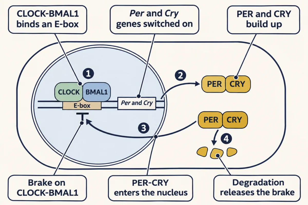
The delays, steep enough repression and the rate at which PER and CRY are broken down together set a period near 24 hours, not exactly 24. That is why the clock needs a daily reset.
How light keeps the clock on time
Light reaches the suprachiasmatic nucleus (SCN) through the retinohypothalamic tract, a tract being a bundle of axons within the central nervous system.
Many of the retinal ganglion cells that send axons to the SCN sense light themselves. They express melanopsin, a photopigment that signals through a different G-protein cascade from the rod’s cGMP pathway (see the senses and cGMP and the retina). Rods and cones help too. In the mouse SCN, light pulses strong enough to reset the clock rapidly induced Per1 transcripts (Shigeyoshi and colleagues, 1997), so light reaches into the gene loop itself.
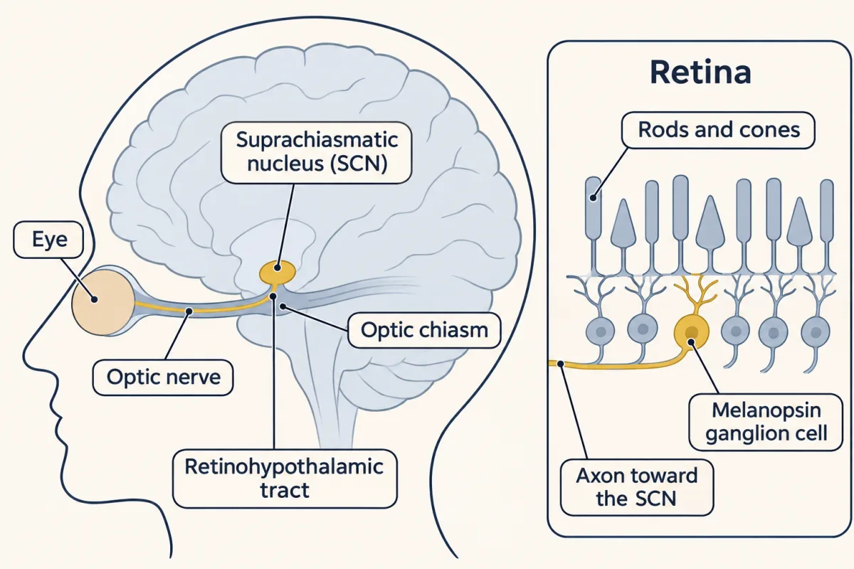
Which way light moves the clock
Whether light pushes the clock earlier or later depends on when it arrives.
- Light in your biological evening tells the clock that the day is not over yet, so the clock runs later. That is a phase delay.
- Light in your biological morning tells the clock that the day began early, so the clock runs earlier. That is a phase advance.
In healthy people under controlled conditions, bright light before the daily low point of core body temperature delayed the clock and light after it advanced the clock. That low point usually falls in the early morning, a couple of hours before habitual waking.
Predict. A typical sleeper goes to bed near midnight and wakes near 8 in the morning, so the temperature low point falls around 6. Bright light at 23:00, or at 07:00 on a morning they are already up: which way does each move the clock?
Check your prediction
Light at 23:00 comes before the temperature low point, in the biological evening, so it delays the clock. Light at 07:00 comes after it, in the biological morning, so it advances the clock.
Worked problem: free-running drift. Czeisler and colleagues (1999) measured a mean intrinsic period of 24.18 h in healthy young and older adults under controlled dim light. That is a study mean; individuals vary. Assume an idealized clock with exactly that period, no time cues, starting in phase with a 24.00-h day.
Step 1: excess per cycle. 24.18 − 24.00 = 0.18 h, which is 0.18 × 60 = 10.8 min per cycle.
Step 2: seven internal cycles. 7 × 0.18 h = 1.26 h, which is 75.6 min. The clock’s marker now falls 75.6 min later than on the 24-h day.
Step 3: check. Internal time is 7 × 24.18 = 169.26 h. External time is 7 × 24.00 = 168.00 h. The difference is 1.26 h, as before.
Step 4: what entrainment needs. To stay at 24.00 h, this clock needs an average net light-driven advance of 10.8 min per day. Each day the morning advance must outweigh the evening delay by that much.
- Excess per cycle (period − 24.00 h)
- —
- Net drift per day (excess − shift)
- —
- Marker offset at the cursor
- —
- Check: internal time against external time
- —
- To stay at 24.00 h this clock needs
- —
—
In this sketch: an idealized clock with a constant period, no time cues, starting in phase with a 24.00-h day; light is a fixed net shift per day, with no phase-response curve. The lesson names the timing of light but does not give it as a rule this sketch uses: in controlled human studies, bright light before the core-body-temperature minimum delayed the clock and light after it advanced it. 24.18 h is a study mean (Czeisler and colleagues, 1999, healthy young and older adults, controlled dim light); individuals vary.
Offset after n cycles = n × (period − 24.00 h); with a 24.18-h period that is 10.8 min per cycle, 75.6 min after 7 cycles (7 × 24.18 = 169.26 h against 7 × 24.00 = 168.00 h), and an average net advance of 10.8 min per day holds the marker at 24.00 h.
Without that daily light, this clock would drift later each day. Because the rule’s levels ride on the clock, the timing of sleep would drift with it.
Reading a night
Read the transcript of “Reading a Night”
Film transcript: Reading a Night
Sleep pressure is highest when you fall asleep, and it drains all night. What does a recording show? Sleep is read from brain waves, eye movements and chin muscle tone. The night is cut into 30-second windows. Slower, larger brain waves mean many cortical neurons are active, then silent, in step. That happens as NREM sleep deepens. In stage N3, the deepest, large slow waves take up at least a fifth of the window. In REM sleep, the brain waves can look much like waking. The eyes and the muscles tell them apart. So where in the night will most of the deepest sleep fall?
Early, when pressure is highest. REM sleep grows toward morning, with the clock and with time already asleep. How does the brain flip? In one model, sleep-active neurons and wake-promoting groups inhibit each other. Whichever side is ahead silences the other. So the brain flips quickly, instead of drifting. Once sleep wins, pressure must drain well below where sleep began before waking can win again. That memory is one way to picture the gap between the two levels. Orexin neurons are thought to hold the switch on the wake side, like a finger holding a light switch up. In narcolepsy type 1, these neurons are severely reduced, and waking becomes unstable. Muscle tone can also fail while the person stays conscious.
Keep this. Deep sleep mainly follows the pressure. REM grows with the clock and with time asleep. A switch with a memory may explain the gap that keeps sleep in one block.
From the film
- Sleep is scored in 30-second epochs from brain waves, eye movements and muscle tone, and most N3 falls in the first half of the night, when pressure is highest.
- In the model, REM grows toward morning, following both the clock and the time already slept.
- In one flip-flop model, a switch with a memory is one way to explain the gap between the two levels.
- Orexin neurons are thought to hold the switch steady in waking, and narcolepsy type 1 is associated with severe loss of them.
The rule says pressure is highest when you fall asleep and drains through the night. What does that look like in a recording?
Sleep is scored by polysomnography: scalp electroencephalogram (EEG), the electro-oculogram (EOG) for eye movements and the chin electromyogram (EMG) for muscle tone. The night is cut into 30-second epochs, and each epoch gets one stage.
EEG rhythms are named by frequency: beta at about 13 to 30 Hz (common in alert waking), alpha at 8 to 13 Hz (common in relaxed waking with eyes closed), theta at 4 to 7 Hz and delta below 4 Hz. Slower, larger waves mean that many cortical neurons are active and silent in step, which happens as NREM sleep deepens. In adults:
- N1: alpha gives way to theta.
- N2: sleep spindles, trains of 11 to 16 Hz waves lasting at least 0.5 s, and K-complexes, well-defined biphasic waves lasting at least 0.5 s.
- N3: slow waves of 0.5 to 2 Hz with peak-to-peak amplitude above 75 µV in frontal leads, filling at least 20% of the epoch. N3 merges what older scoring rules called stages 3 and 4.
- REM: an EEG that can look much like waking, rapid eye movements on the EOG, and the lowest muscle tone of the night on the EMG.
So the EOG and EMG separate REM from waking. Through a night, NREM and REM sleep alternate in several cycles.
Predict. While pressure drains, the clock heads toward its low point in the early morning. In which half of the night will most N3 fall? And most REM?
Check your prediction
Most N3 falls in the first half of the night, when pressure is highest. In the simple model, about 70% of the sleep-onset pressure is left after an hour and a half of sleep, and about a fifth after six and a half hours. REM grows toward morning. Deep sleep mainly follows the pressure; the rise of REM toward morning follows both the clock and the time already slept.
Worked problem: scoring a hypnogram. In this made-up recording, a subject is in bed for 960 epochs. The scorer counts 60 epochs of wake, 48 of N1, 432 of N2, 192 of N3 and 228 of REM.
Step 1: total time. 960 × 30 s = 28,800 s = 8.0 h. The counts add to 60 + 48 + 432 + 192 + 228 = 960.
Step 2: sleep. 960 − 60 = 900 epochs, or 900 × 30 s = 27,000 s = 7.5 h.
Step 3: sleep efficiency, sleep divided by time in bed: 900 / 960 = 0.9375, or 93.75%.
Step 4: stage shares of sleep. REM 228 / 900 = 25.3%. N3 192 / 900 = 21.3%. N2 432 / 900 = 48.0%. N1 48 / 900 = 5.3%. Rounded, they add to 99.9%; the unrounded shares add to 100%. These are practice numbers, not norms.
The switch: why sleep comes in one block
The rule’s two levels need a switch that flips to sleep at a high pressure and back only at a much lower one. How could the brain do that?
In the ventrolateral preoptic area (VLPO), sleep-active neurons release the inhibitory transmitter GABA and inhibit wake-promoting cell groups: histamine neurons of the tuberomammillary nucleus, noradrenaline neurons of the locus coeruleus, serotonin neurons of the raphe and cholinergic neurons of the brainstem (see transmitter systems). In one model, those groups inhibit the VLPO in return, a mutual inhibition called a flip-flop: whichever side is ahead silences the other, so transitions are quick and intermediate states are rare.
Mutual inhibition also gives the switch a memory, which engineers call hysteresis. Once the sleep side wins, it silences the wake side, which can then no longer push back. Pressure has to drain well below the level at which sleep began before waking can win again. That memory is one way to explain the gap between the rule’s two levels. It is a model, and its details have exceptions (see Going deeper).
Orexin holds the switch
A flip-flop switches quickly but can flip too easily, because near the balance point a small push from either side tips it. Neurons in the lateral hypothalamus that make the peptides orexin A and B (also called hypocretin 1 and 2) excite many of the wake-promoting groups, and are thought to hold the switch on the wake side, like a finger holding a light switch up. Remove the finger and the switch still works, but it flips at the slightest nudge: waking breaks into short bouts.
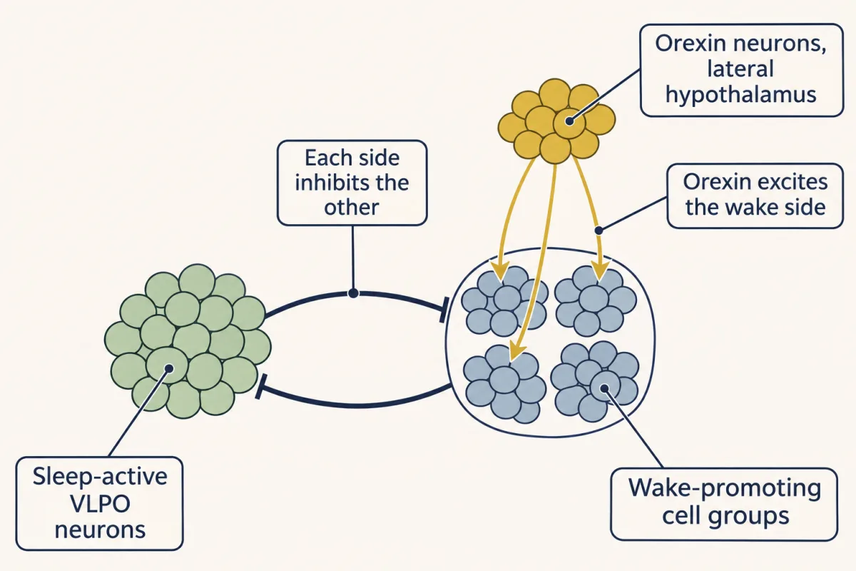
Three lines of evidence tie orexin to narcolepsy, and each comes from a different species.
- Dogs. In a familial dog model, Lin and colleagues (1999) found an autosomal recessive mutation in the hypocretin receptor 2 gene.
- Mice. Conditional loss of orexin neurons in adults fragmented waking and increased transitions between sleep and wake states (Tabuchi and colleagues, 2014).
- People. Thannickal and colleagues (2000) counted 85 to 95% fewer hypocretin neurons in four postmortem narcolepsy brains than in twelve controls, while neighbouring neurons making melanin-concentrating hormone were not reduced.
Most human patients do not carry the dog’s receptor mutation; narcolepsy type 1 is associated with severe loss of brain orexin. An autoimmune attack on these neurons is hypothesized, not established. Cataplexy, the sudden muscle weakness many patients have, occurs with consciousness preserved, so it is not falling asleep. It is thought to be rapid eye movement (REM) sleep’s loss of muscle tone breaking into waking.
Adamantidis and colleagues (2007) expressed channelrhodopsin-2 in hypocretin neurons of mice and delivered light through an optical fibre into the lateral hypothalamus of freely moving animals. Stimulation increased transitions from slow-wave and REM sleep to waking.
What the sleeping brain does with its hours, including replay of hippocampal activity, is the subject of memory consolidation.
Design problem
A colleague says the optogenetic result and the human cell counts prove orexin neurons are needed for waking. Design a mouse experiment that asks whether they are needed for waking itself or for stable waking: what you measure, your controls, and your prediction.
Saved in this browser only.
One way to do it
Driving the neurons shows that their activity is enough to wake a mouse under that stimulation: sufficiency, not necessity. The human series is correlational: far fewer cells stain for hypocretin in four patients, but the counts cannot say when or why that happened.
To test necessity, silence or ablate the neurons with an inhibitory opsin or a toxin expressed only in orexin neurons, and do it in adult mice so development cannot compensate. Include control mice that lack the opsin or toxin but receive the same light or the same induction, to control for heat, light, handling and the treatment itself. Record EEG and EMG continuously and have blind scorers assign stages in fixed epochs.
Measure total wake per 24 h, the mean duration of wake bouts, the number of transitions between states, and episodes of abrupt atonia that come straight out of sustained waking. If the neurons are needed for stable waking, you predict much shorter wake bouts and many more transitions with little loss of total wake, plus cataplexy-like episodes, scored as abrupt atonia lasting at least 10 s with a theta-dominated EEG after at least 40 s of waking, with video showing immobility. If they were needed for waking itself, total wake would fall sharply.
Two limits remain. Orexin neurons release other transmitters, so silencing the cells does not isolate the peptide; a receptor antagonist or peptide knockout tests that. And the result holds for these mice and this timing, not for human patients.
Going deeper
The two-process model in numbers
A simple version of the rule, in the style of the model of Daan, Beersma and Borbély (1984), uses illustrative values, not measurements. S rises toward 1 with a time constant of 18 h while awake and falls toward 0 with a time constant of 4 h while asleep. The upper level is 0.67 and the lower level 0.17, each plus the same daily cosine of amplitude 0.12, highest at about 17:00. Run with these values, the model:
- sleeps from about 00:20 to about 08:10, about 7.8 h, and stays locked to the 24-hour day;
- if both levels are replaced by one midway between them, flips back and forth at that line once pressure reaches it, because the waking and sleeping curves both point toward it all day; how short those episodes get is set by the simulation’s time step, not by biology, and other single lines can allow longer bouts;
- with the clock’s swing set to zero, alternates about 16.6 h awake and 5.5 h asleep, a cycle of about 22.1 h;
- if kept awake from 08:10 until midnight the next night, about 40 h, sleeps about 8.7 h instead of 7.8 h.
These outputs show the rule hangs together; they are not measurements of people.
Who showed each step of the clock loop
Gekakis and colleagues (1998) showed that CLOCK and BMAL1 bind E-boxes as a pair and activate Per1 in mouse cells. In reporter assays either CRY1 or CRY2 strongly inhibited CLOCK-BMAL1-driven transcription (Kume and colleagues, 1999). Kinases set part of the loop’s timing: casein kinase 1 phosphorylates PER, and the hamster tau mutation is an allele of casein kinase 1 epsilon (Lowrey and colleagues, 2000).
How the light-sensing ganglion cells were found
Berson, Dunn and Takao (2002) recorded from SCN-projecting retinal ganglion cells in isolated rat retina and found that they still depolarized to light with rod and cone input blocked. Those cells express melanopsin (Hattar and colleagues, 2002). Mice failed to entrain to light-dark cycles only when melanopsin and rod and cone phototransduction were all removed (Hattar and colleagues, 2003).
REM atonia and dreaming
In rodents, glutamatergic neurons of the sublaterodorsal nucleus are thought to produce rapid eye movement (REM) atonia by recruiting inhibitory neurons in the medulla and spinal cord. Dreams are not limited to REM: people woken from NREM sleep also report them (Siclari and colleagues, 2017).
Qualifications to the flip-flop model
The sleep-active ventrolateral preoptic area (VLPO) neurons release galanin as well as GABA. The flip-flop scheme also lists cholinergic neurons of the basal forebrain among the groups the VLPO inhibits. Each link in the scheme has to be tested on its own: a slice response to a transmitter does not show which neurons supply it.
In slice recordings, putative sleep-promoting VLPO cells were inhibited by noradrenaline, acetylcholine and mostly serotonin, not directly by histamine (Gallopin and colleagues, 2000). In mice, Chung and colleagues (2017) found that activating preoptic GABA neurons that project to the tuberomammillary nucleus increased NREM sleep, while activating preoptic GABA neurons in general increased waking. Projection target together with transmitter, not location alone, defines a sleep-promoting cell: glutamatergic preoptic neurons with the same target promoted waking.
Why an autoimmune cause is suspected
Narcolepsy type 1 is strongly associated with the immune-system gene variant HLA-DQB1*06:02, and T-cell findings point the same way. Neither shows that an immune attack destroys the orexin neurons, so the hypothesis remains unproven.
Orexin experiment details
In the Tabuchi experiment, dark-period wake time was significantly reduced from 3 to 10 weeks after induction, with recovery by 11 weeks (Tabuchi and colleagues, 2014). In the Adamantidis experiment, trains at 5 to 30 Hz shortened the latency to wake, while 1 Hz did not (Adamantidis and colleagues, 2007).
Your notes
Select any text in the lesson to highlight it or add a note to it. Your highlights and notes are saved in this browser and collected in your notebook.
This browser is not letting the site save anything (a private window or strict privacy settings), so highlights and notes will be lost when you leave the page.
How we measure it
Lesion and transplant studies
Destroy a region and see what is lost; then replace it with donor tissue and see what returns. A lesion tests necessity, but an electrolytic lesion also destroys passing fibres and every cell type in its path, and a transplant with a genetically marked donor shows which tissue sets a property such as period.
Bioluminescence reporters (PER2::LUCIFERASE)
Fuse a clock protein to firefly luciferase in a knock-in mouse, culture a tissue with luciferin and count the photons it emits over days. The glow rises and falls with the clock protein, so each tissue's rhythm can be followed in real time.
Polysomnography (EEG, EOG and EMG)
Record scalp EEG, eye movements (EOG) and chin muscle tone (EMG) through the night, then assign one stage to each 30-second epoch. EEG alone cannot tell REM sleep from waking; the muscle and eye channels can.
Optogenetics
Express a light-gated channel such as channelrhodopsin-2 in one genetically defined cell type and drive it through an optical fibre; silencing needs a different, inhibitory opsin. Driving cells tests sufficiency; only silencing or removing them tests necessity, and either holds only for the cells, timing and behaviour tested.
Practice questions on this lesson (3) → MCAT-style, original, each with an explanation
Where this lesson fits
Before thisReview first
- Autonomic Control and the HypothalamusThis course · Lesson 18
The suprachiasmatic nucleus sits in the hypothalamus, and you need that map of its groups and roles.
- The RetinaThis course · Lesson 10
Light reaches the clock through retinal ganglion cells, so you need the retina's cell types and light pathway from there.
- Transmitter Systems and DrugsThis course · Lesson 6
The sleep switch relies on GABA, histamine, noradrenaline and serotonin groups acting on each other, which that lesson introduces.
After thisStudy next
- Plasticity, Networks and ConsolidationThis course · Lesson 23
It takes up what the sleeping brain does with its hours, including replay of hippocampal activity.
Elsewhere on the siteFilms, simulations and method films in other courses
- From One Day to One YearFilm · Life Science
Introduces the circadian rhythm of about 24 hours that the clock genes here generate.
- A Blind Mouse That Knows It Is DayFilm · Sensory Systems
Shows the melanopsin cells that set the body clock to the day in mice lacking rods and cones.
Suggested by an AI model from the lessons themselves, then checked. How we use AI
Finish line
Test yourself
8 questions
Practice and MCAT-style, with explained answers.
Next · Lesson 22
Memory Systems
H.M. got better at mirror drawing over three days and did not remember ever having done it. What kind of memory improved?
Continue →