Cell Signaling · Unit 4: Receptors with enzymes attached
Cytokine Receptors and JAK–STAT
How does a signal reach a gene when the only relay between the receptor and the DNA is one kind of protein?
Pause when the reading reaches a film, and offer to play it. Saved on this device.
Watch the film now, or keep listening
Builds on:Phosphorylation: Kinases and PhosphatasesProtein Modules: SH2, SH3, PTB, PDZ and PHA Field Guide to ReceptorsWhy these?
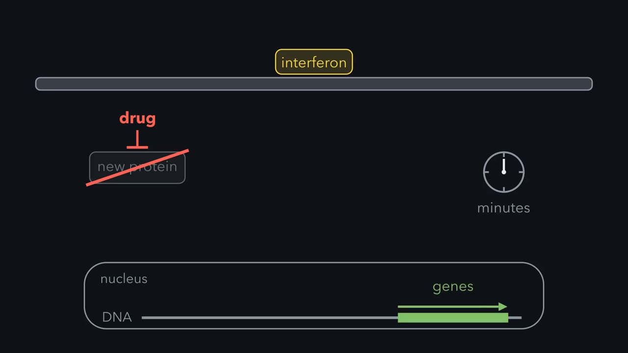 ▶ Watch One Protein From Receptor to Gene · 2:31
Read the transcript of “One Protein From Receptor to Gene”
Film transcript: One Protein From Receptor to Gene
Add interferon to human cells, and a set of genes switches on within minutes. Interferon is a protein that warns cells of a virus. The genes still switch on when a drug stops the cells from making any new protein. So every part of the path, from the cell surface to those genes, must already be in the cell, waiting. What is the path made of?
Interferon is a cytokine. A cytokine is a small secreted protein that tells other cells what to do. Each chain of this kind of cytokine receptor crosses the membrane once, and it has no enzyme at all. Instead, its inside tail holds a separate tyrosine kinase. This kinase is called a JAK, and it is there before any cytokine arrives. Binding does one job. The cytokine sets receptor chains, and so their JAKs, in position against each other. In the simplest case there are two of each. Now the two JAKs are close enough to phosphorylate each other. Each adds a phosphate to its partner, and both become fully active. A receptor with no enzyme of its own has borrowed one.
The active JAKs now phosphorylate tyrosines along the receptor tails. These phosphorylated tyrosines are docking sites. A protein called a STAT waits in the cytoplasm. It carries an SH2 domain, a module that binds a phosphate on a tyrosine. With that domain, the STAT docks on the receptor tail, next to the JAK. The JAK then adds one phosphate to the docked STAT, on a single tyrosine. Now the same SH2 domain has something new to hold. Many STATs already sit in loose pairs. Phosphorylation switches them into a pair held by this grip: each domain holds the partner's phosphorylated tyrosine. Two grips hold the pair, where one grip held the STAT to the receptor.
So the same domain is used twice. The pair enters the nucleus, binds a specific DNA sequence, and switches on transcription. Count the proteins between the receptor complex and the gene. There is one kind, the Stat, with no second messenger and no chain of kinases. Interferon adds one partner, I R F nine. The Stats are already waiting, so genes answer within minutes. So keep this rule: a Stat, with one phosphate, carries the signal from receptor to gene.
Add interferon, a protein that warns cells of a virus, to human cells and a particular set of genes switches on within minutes, even when a drug has stopped the cells from making any new protein. Every part of the path from the cell surface to those genes must therefore be in the cell already, waiting. The people working on it judged the second messengers of Unit 3 a poor fit: a rise in a second messenger is a fairly nonspecific event, and interferon’s action is highly specific. The answer was a receptor that borrows a kinase and a transcription factor that waits in the cytoplasm for one phosphate.
The idea: a receptor with no enzyme borrows one
A cytokine is a small secreted protein that tells other cells what to do, most often cells of the blood and the immune system. Interferons are cytokines. So are the interleukins, which carry messages between white blood cells. Several hormones use receptors of the same family: erythropoietin (EPO), which tells the bone marrow to make red blood cells, and also growth hormone, prolactin and leptin.
Each receptor chain crosses the membrane once. A receptor tyrosine kinase, such as the insulin receptor from the receptor field guide, is its own kinase. A cytokine receptor of this family (the class I and class II cytokine receptors) has no enzyme in it at all. A stretch of its inside tail, near the membrane, holds a separate tyrosine kinase called a JAK (Janus kinase), and the JAK is there before any cytokine arrives. Think of an antenna with no electronics of its own and a radio bolted to its base.
Binding does one job: it sets receptor chains, and so their JAKs, in the right position against each other. The simplest case is two chains and two JAKs; some receptors assemble more chains around the cytokine. One growth hormone molecule has two different faces, and each face grips one copy of the same receptor chain. For some receptors, the EPO receptor among them, crystal structures suggested the chains might be loosely paired beforehand, with binding re-setting their geometry. Tracking single receptor chains in living cells points the other way: the chains come together when the hormone binds. The question is still argued. Either way, two JAKs end up close enough to phosphorylate each other.
A JAK has two kinase-like domains side by side. One is a working kinase. The other, the pseudokinase domain, has the kinase fold but little or no catalytic activity, and it restrains the working one. The family is named for Janus, the Roman god with two faces. Mammals have four: JAK1, JAK2, JAK3 and TYK2.
The interferon puzzle: activating a factor without making more of it?
Read the transcript of “Two Labs, One Missing Kinase”
Film transcript: Two Labs, One Missing Kinase
A drug stops the cells making any new protein, and interferon still switches genes on. So the factor must already be in the cell. Two laboratories went looking, from opposite ends. James Darnell's laboratory started at the gene. It found a protein complex, ISGF3, that binds a short DNA sequence shared by interferon-responsive genes. In August 1992, Christian Schindler, Ke Shuai and colleagues reported that interferon puts phosphate on its proteins' tyrosines.
Staurosporine, a drug that blocks many kinases, stopped the activation. Some kinase had to act first. Which one, it could not say. George Stark and Ian Kerr's laboratory started with genetics. Sandra Pellegrini built human cells with a marker gene controlled by interferon-responsive DNA. Only cells deaf to interferon survived. She screened about 100 million mutagenized cells. Each mutant had lost a part of the pathway. She put DNA from normal cells into a mutant, and looked for the rare cell that recovered. In July 1992, her group in Paris reported the gene that rescued one mutant: TYK2. Two weeks apart, the results fit together. A JAK is required, and the DNA-binding proteins carry phosphotyrosine.
Darnell's laboratory named those proteins STATs.
A cell can raise a transcription factor’s activity by making more of it. The inhibitor experiment rules that out here, so the factor must exist already and be activated some other way. Two laboratories went looking, from opposite ends.
James Darnell’s laboratory at Rockefeller University started at the gene. Interferon-responsive genes share a short DNA sequence, and the laboratory identified a protein complex, ISGF3, that binds it. Its proteins wait in the cytoplasm and gain the ability to bind DNA within minutes of interferon reaching the cell. In August 1992 Christian Schindler, Ke Shuai and their colleagues reported that interferon causes proteins of the complex to be phosphorylated on tyrosine, and that staurosporine, a drug that inhibits many kinases, blocks the interferon-induced activation of the complex. Some kinase had to act first. The experiment could not say which.
George Stark and Ian Kerr’s laboratory in London started with genetics. Sandra Pellegrini built human cells in which an interferon-responsive stretch of DNA controlled a marker gene, so that in the right medium only cells deaf to interferon survived. She mutagenized the cells and screened about a hundred million of them. Each mutant line that came through had lost a part of the pathway. To find which part, you put DNA from normal cells into the mutant and look for the rare cell whose response returns, a test called complementation. Pellegrini did this in her own new laboratory at the Institut Pasteur in Paris. In July 1992, two weeks before the Darnell paper, her group reported the gene that rescued one mutant. It encoded TYK2.
The two results fit together: a Janus kinase (JAK) is required, and the DNA-binding proteins carry phosphotyrosine. Darnell’s laboratory named those proteins signal transducer and activator of transcriptions (STATs), signal transducers and activators of transcription.
The mechanism: one phosphate, and an SH2 domain used twice
Try this before reading on: design a signaling pathway that is activated by receptor oligomerization and tyrosine phosphorylation. (Oligomerization means several chains coming together.) The cell’s version has six steps.
- The cytokine sets two receptor chains in position.
- The two Janus kinases (JAKs) phosphorylate each other on their activation loops, the switch described in the phosphorylation lesson, and become fully active.
- The JAKs phosphorylate tyrosines along the receptor tails.
- A signal transducer and activator of transcription (STAT) from the cytoplasm docks on one of those phosphotyrosines by its SH2 domain, the phosphotyrosine-binding module from the lesson on protein interaction modules.
- The JAK phosphorylates the docked STAT on a single tyrosine near its C-terminal end (tyrosine 701 in STAT1).
- Two phosphorylated STATs pair, each SH2 domain holding the other’s phosphotyrosine. (Many STATs already sit in loose pairs before this, held together another way; phosphorylation switches them into this active form.) The active pair builds up in the nucleus, binds a specific DNA sequence and switches on transcription.
The SH2 domain is used twice. It first holds the STAT on the receptor, next to the JAK. Once the STAT carries its own phosphotyrosine, the same domain holds a partner STAT instead, and two grips hold the pair where one held the STAT to the receptor.
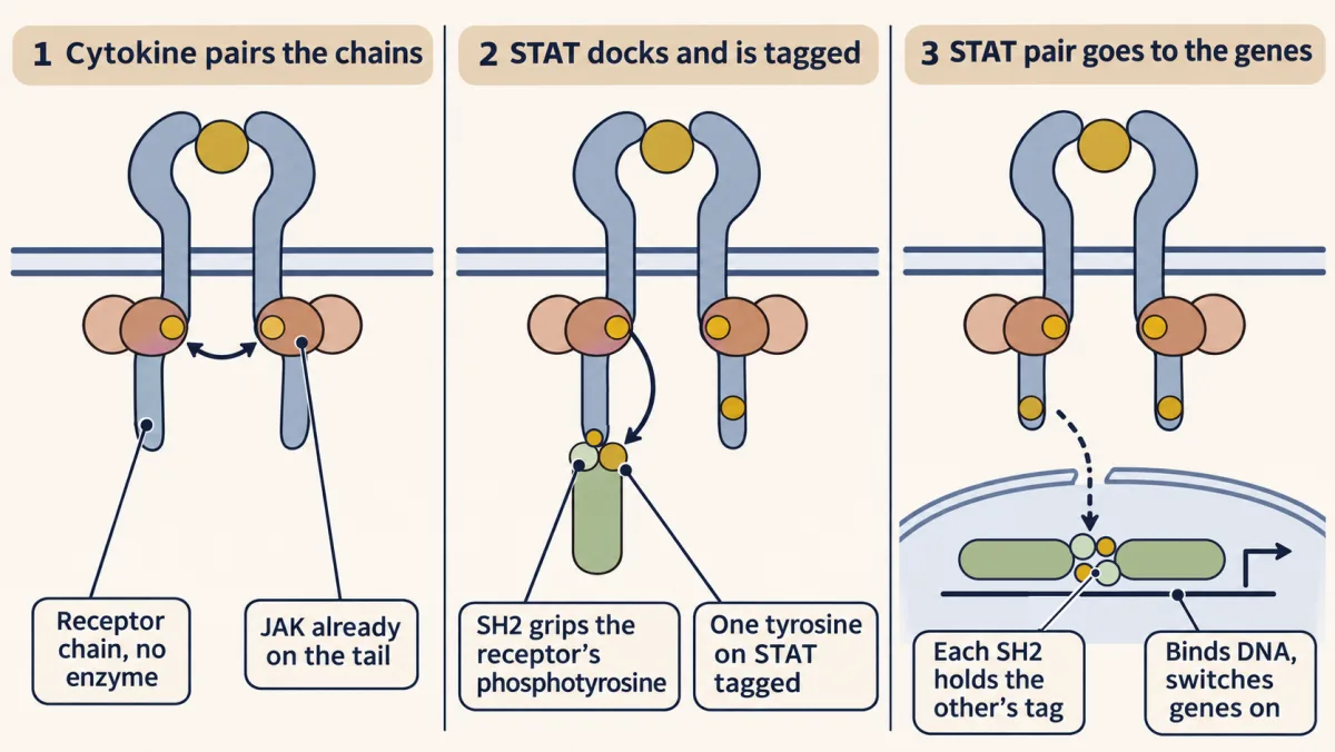
Count the kinds of protein that carry the message from the receptor complex to the gene: one, the STAT. (Type I interferons add a partner, IRF9, to make ISGF3.) With no second messenger and no chain of kinases, this is about the shortest route there is from the cell surface to a gene.
Design problem
Design the shortest possible path from a receptor at the cell surface to a gene in the nucleus. Say what each part must be able to do. Then compare your design with a STAT protein, domain by domain, and say what this short path gives up when you set it beside a three-kinase cascade.
Saved in this browser only.
One way to do it
The shortest path has one moving part: a protein that is modified at the receptor and then goes to the DNA itself. It must do four things: tell an active receptor from an idle one, take a mark that records the meeting, change its behaviour once marked so that marked copies build up in the nucleus and bind particular DNA sequences. (Unmarked STATs pass in and out of the nucleus too, but they do not bind the DNA in the same way.)
A STAT has a part for each job. The SH2 domain finds the active receptor, since only an active receptor carries phosphotyrosines. The single tyrosine near the C-terminus takes the mark. The SH2 domain, used again, reads that mark on a partner and makes the pair. A DNA-binding domain in the middle of the chain grips the DNA, and the C-terminal end recruits the machinery that starts transcription.
What does the short path give up? Some gain, and some places to adjust the signal. In a three-kinase cascade each active kinase can phosphorylate many copies of the next, so a few receptors can give a large output. The stacked steps, together with positive feedback, can also make an all-or-none switch, as in Ferrell’s frog oocytes, and each step is a point where another pathway can cut in. Here the only kinase is the JAK. It marks many STATs in turn, but no further kinase stage multiplies the signal again. The reward is a direct line from occupied receptors to genes.
One family, many jobs: four JAKs, seven STATs
Mammals have four Janus kinases (JAKs) and seven signal transducer and activator of transcriptions (STATs) (STAT1, 2, 3, 4, 5A, 5B and 6) to serve dozens of cytokines. The receptor does most of the sorting. Each signaling chain’s tail holds particular JAKs and carries particular docking tyrosines, and the amino acids around each phosphotyrosine decide which STAT’s SH2 domain fits.
Mice with one gene deleted, called knockout mice, show what each STAT is for:
- STAT1: no response to interferons; the mice succumb to infections that normal mice survive.
- STAT4: impaired response to interleukin-12.
- STAT6: impaired response to interleukin-4.
- STAT5A and STAT5B: failed responses to prolactin and growth hormone; the two partly cover for each other.
- STAT3: the embryo dies early, so its jobs were worked out one tissue at a time.
You might expect the JAKs to cover for each other as well, since the four are similar in sequence. Two results say that on at least two receptors they do not.
James Ihle’s laboratory had shown in 1993 that JAK2 is the kinase on the erythropoietin (EPO) receptor’s tail. In 1998 it reported mouse embryos lacking JAK2. They were anaemic and died at about day 12.5 of gestation, and their fetal livers, where a mouse embryo makes its adult-type red cells, held no red-cell precursors that could be grown in culture. Klaus Pfeffer’s laboratory reported the same result in the same issue. The genes for JAK1, JAK3 and TYK2 were untouched, and none of those kinases took over.
The human case is severe combined immunodeficiency (SCID), in which a child is born without working T cells, and so with almost no adaptive immune defence. In 1993 Warren Leonard’s laboratory reported that boys with the X-linked form carry mutations in the gamma chain of the interleukin-2 receptor. The disease is far worse than losing interleukin-2 alone, because the receptors for interleukins 4, 7, 9, 15 and 21 use the same chain, now called the common gamma chain. Then a patient studied with Rebecca Buckley of Duke University had the disease with a normal gamma chain. In 1995 John O’Shea’s and Leonard’s groups found that patient’s mutations in JAK3, the kinase that sits on the gamma chain, one in each copy of the gene. A group in Italy working with O’Shea had reported JAK3 mutations in other SCID patients about two months before.
Switching it off, and what happens when a JAK is stuck on
Two kinds of brake do most of the work of ending the signal. Tyrosine phosphatases remove the phosphates from the Janus kinases (JAKs), the receptor tails and the signal transducer and activator of transcriptions (STATs). A STAT that loses its phosphate in the nucleus returns to the cytoplasm, ready for reuse.
The pathway also makes its own brake. Among the genes that STATs switch on are those for the SOCS proteins, short for suppressors of cytokine signaling. A SOCS protein has an SH2 domain, so it goes to the phosphotyrosines on the active receptor and JAK. Some SOCS proteins block the JAK’s catalytic site directly. All of them carry a region that recruits a ubiquitin ligase; for several, including SOCS1 and SOCS3, this can mark the receptor complex for the proteasome. This is negative feedback as you met it in the signaling circuits lesson: more signal makes more brake, after a delay.
In 2005 several research groups reported the same mutation in most patients with polycythaemia vera, a disease in which the bone marrow makes too many red blood cells: valine 617 of JAK2 changed to phenylalanine. Residue 617 lies in the pseudokinase domain. The change weakens that domain’s restraint and helps two JAK2 molecules hold together in the active pair, so JAK2 signals from the erythropoietin (EPO) receptor with little or no EPO present.
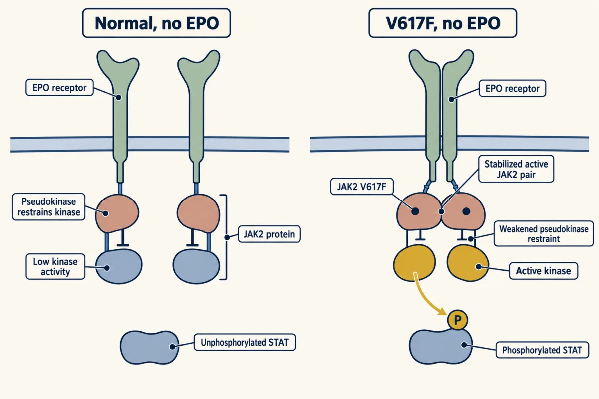
Missing JAK2 leaves a mouse embryo with no adult-type red cells, and overactive JAK2 gives people too many, so JAKs became drug targets. Ruxolitinib, which inhibits JAK1 and JAK2, was approved in the United States in 2011 for myelofibrosis, a bone-marrow disease related to polycythaemia vera. Tofacitinib followed in 2012 for rheumatoid arthritis. Its logic came from the SCID patients: a person without JAK3 has an immune defect and little else, so blocking JAK3 should quiet an overactive immune system and spare other tissues. In practice tofacitinib blocks JAK1 as strongly as JAK3, and JAK2 more weakly, and much of its effect comes through JAK1. Like imatinib, these drugs sit in the kinase’s ATP pocket. The four JAK pockets are alike, so selectivity between them is hard to win, and anaemia from partly blocked JAK2 is a known side effect of several. One way round this is to aim at the pseudokinase domain instead. Deucravacitinib binds the pseudokinase of TYK2 and holds it in its restraining state, which blocks TYK2 while sparing the other JAKs. It is now approved for psoriasis.
Your notes
Select any text in the lesson to highlight it or add a note to it. Your highlights and notes are saved in this browser and collected in your notebook.
This browser is not letting the site save anything (a private window or strict privacy settings), so highlights and notes will be lost when you leave the page.
How we measure it
Mutant screen in cultured cells, with complementation
Treat a large population of cultured cells with a mutagen and select the rare ones that have lost a response, here the response to interferon. Fusing two mutant lines and checking whether the response returns tells you whether they lack the same gene, and putting normal DNA back into a mutant until the response returns identifies the gene. This is how TYK2 was tied to interferon signaling.
Gel-shift assay (electrophoretic mobility shift)
Mix a short labelled piece of DNA with a cell extract and run it through a gel. DNA with a protein bound to it moves more slowly, so a shifted band reports a DNA-binding factor and how much of it is active. It is how an interferon-activated DNA-binding complex can be seen appearing within minutes.
Gene knockout in mice
Disrupt both copies of one chosen gene in a mouse and see what fails. Because every other gene is intact, the result tests whether related proteins can cover for the missing one. Mouse embryos without Jak2 make no adult-type red cells.
Anti-phosphotyrosine detection
Separate a cell's proteins by size and probe them with an antibody that binds only phosphotyrosine. Comparing cells before and after a cytokine shows which proteins gain a tyrosine phosphate and how quickly.
Method films · the methods library
- Binding assays: immunoprecipitation, pull-down and gel shift 3:43
- Complementation (cis-trans) test 2:48
- Cryo-electron microscopy 4:06
- Forward genetic screens 4:07
- Gel electrophoresis, SDS-PAGE and western blot 4:01
- Gene targeting: knockout and knock-in mice 4:13
- Pharmacological blockade and inhibitors 4:10
- Reporter gene assays 2:42
- X-ray crystallography 4:06
Practice questions on this lesson (3) → MCAT-style, original, each with an explanation
Where this lesson fits
Before thisReview first
- Phosphorylation: Kinases and PhosphatasesThis course · Lesson 6
JAK activation-loop trans-phosphorylation and phosphatase removal of phosphates rely on the kinase and phosphatase switch described there.
- Protein Modules: SH2, SH3, PTB, PDZ and PHThis course · Lesson 9
STAT docking on the receptor and STAT pairing both use the SH2 domain reading a phosphotyrosine, as taught there.
- A Field Guide to ReceptorsThis course · Lesson 4
The lesson sets enzyme-less cytokine receptors against receptors that carry their own kinase, a split drawn in that field guide.
After thisStudy next
- The Shape of a Signal: Size, Duration, Frequency and PlaceThis course · Lesson 35
It asks how a cell reads signal duration and size, which STAT signaling and its SOCS feedback illustrate.
- Oncogenes: When the Switch SticksThis course · Lesson 41
Stuck-on JAK2 V617F in polycythaemia vera is an oncogene-style gain-of-function switch taken further there.
- Drugs Against a PartThis course · Lesson 42
It takes up how kinase inhibitors for the ATP pocket achieve selectivity, as with JAK inhibitors and TYK2.
Elsewhere on the siteFilms, simulations and method films in other courses
- RNAi and antisense knockdownMethod film
Knocking down one JAK or STAT with siRNA is a way to test what each part does.
Suggested by an AI model from the lessons themselves, then checked. How we use AI
Finish line
Test yourself
8 questions
Practice and MCAT-style, with explained answers.
Next · Lesson 23
TGF-β and Smads
Why does the protein that tells healthy cells to stop dividing end up helping late tumours?
Continue →