Physiology · Unit 6: Endocrine systems
Thyroid Hormone: Tissues Decide What T4 Becomes
Rewritten on : new text, figures and flashcards. Your notes and highlights stay, but the text around them may have moved.
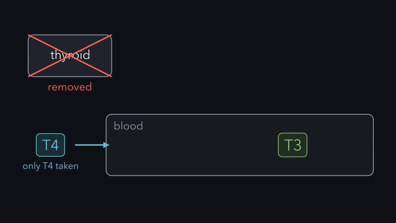
A person with no thyroid who takes only T4 still has T3 in the blood. Where does it come from?
Pause when the reading reaches a film, and offer to play it. Saved on this device.
Watch the film now, or keep listening
Builds on:Endocrine Axes and How to Test ThemBody Water and Its CompartmentsWhy these?
 ▶ Watch Tissues Decide What T4 Becomes · 2:26
Read the transcript of “Tissues Decide What T4 Becomes”
Film transcript: Tissues Decide What T4 Becomes
A person has had their thyroid removed. They take only thyroxine, called T4. Yet their blood still holds the more active hormone, T3. Where does it come from? If the gland were the only source, there should be none. The thyroid secretes mostly T4. T4 binds the thyroid hormone receptor much more weakly than T3 does. So T4 is best treated as a prohormone. Inside cells, T3 binds receptors that regulate transcription. That is the form that acts. Tissues contain three enzymes called deiodinases. Each removes one iodine. D1 and D2 remove an iodine from the outer ring of T4. That makes T3, and it switches the hormone on. D3 removes an iodine from the inner ring instead, turning T4 into reverse T3, which is inactive. D1 can do this too.
The ring that loses the iodine decides: on or off. In 1970, Braverman, Ingbar and Sterling tested this. They studied 13 patients treated with T4. All but two were athyreotic, meaning without a functioning thyroid, or hypothyroid. They gave labelled T4, and separated the iodine compounds of serum by chromatography. Labelled T3 appeared. A specially purified lot of tracer held less than 0.1 percent T3, and gave the same result. In two more patients, T4 went into a vein, bypassing the gut. Labelled T3 still appeared. The result ruled in conversion outside the thyroid, in humans. The study did not identify the enzyme or the organ. It did not measure how much T3 is made this way.
Later work is commonly summarized as roughly 80 percent, in healthy people with enough iodine. That is an approximate estimate. Hormone does not simply diffuse into cells. It enters through transporters such as MCT8. Inside the cell, D2 often makes T3 from T4. So the T3 inside a cell need not match the T3 in plasma. In mice lacking D2, body temperature fell in the cold, because brown fat made less heat. Yet plasma T3 was normal. Keep the rule. The thyroid sends mostly T4, and the ring that loses its iodine decides: on or off.
A person whose thyroid has been removed, and who takes only thyroxine (T4), still has the more active hormone, triiodothyronine (T3), in the blood. Where does it come from?
From the rest of the body. The thyroid secretes mostly T4, and tissues remove one iodine atom to make T3, so tissues, as well as the gland, decide how much active hormone each cell gets. T3 then acts on nuclear receptors: it is needed for the growth of the brain and skeleton, and in adults it raises metabolic rate and cardiac output. The first look at the axis is in endocrine system; paired hormone levels and assays are in endocrine axes and testing.
The one idea to carry through this lesson. Thyroid hormone runs around one loop with four stations. The gland makes it, mostly as T4. The blood carries it, almost all bound to protein. Each tissue converts it, switching T4 on to T3 or off. The pituitary senses it and sets thyroid-stimulating hormone (TSH), the hormone that drives the gland. With the loop intact and at steady state, the loop holds the free hormone in the blood steady. Binding proteins change how much hormone is stored (the total), not how much is free (unbound), and each tissue then adjusts its own dose of T3, from the free T4 it converts and the free T3 it takes up. To read any thyroid test, ask which station failed, then let the loop predict the rest.
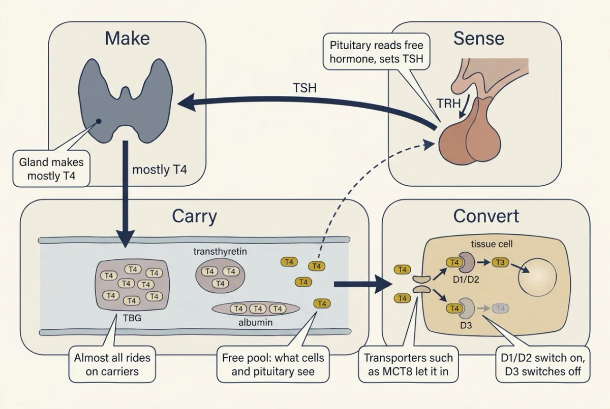
The rule has four limits, each met later in the lesson:
- It holds at steady state. After a change, the loop takes weeks to settle.
- It assumes a working sensor. When the pituitary or hypothalamus is the fault, TSH cannot report the problem, so a normal TSH can mislead.
- It needs an intact loop. The person in the opening question, with no thyroid and a fixed dose of T4, has an open loop: free T4 is set by the absorbed dose and clearance, not by TSH.
- Blood tests report the supply, not the tissue. Where conversion or entry into cells is the fault, blood values and the T3 inside cells can disagree. That boundary is part of the rule.
The gland sends mostly T4; tissues decide how much becomes T3
The film at the top of the page tells this section in short. Its closing rule, “the ring that loses its iodine decides: on or off”, is the convert station of the loop, and it applies to T4.
Start with the two hormones. T4 and T3 are the same two linked rings, except that T4 carries four iodine atoms and T3 carries three. T3 binds the thyroid hormone receptor much more tightly than T4 does, so T3 is the form that acts. T4 is best treated as a prohormone: a supply that tissues can switch on. This section follows two stations of the loop, carry and convert.
Carry: almost all of it rides on proteins
Thyroid hormones dissolve poorly in water, so blood carries them on three proteins: thyroxine-binding globulin (TBG), which holds most of the T4, transthyretin and albumin. In human blood about 99.97 percent of T4 is bound, so only about 3 molecules in 10,000 are free. T3 is held less tightly: about 3 in 1,000 are free (99.70 percent bound). The free fraction, the unbound part, is what transporters carry into cells and what the pituitary senses. The bound hormone is a store.
Predict. In the simplest picture, the cells that break thyroid hormone down can take up only free molecules. T4 is about ten times less free than T3. Which should last longer in the blood?
Check your prediction
T4. Most of it is parked on carriers, so the clearing cells reach it only a little at a time, as molecules come off. In people with normal thyroid function (euthyroid), the half-life of T4 is about 6.2 days, against about 1 day for T3. Binding is one reason for the difference. The long half-life is also why one tablet of T4 a day gives steady blood levels.
Convert: which ring loses an iodine decides on or off
Each tissue changes T4 with three deiodinases, enzymes that each remove one iodine atom. T4 has an outer ring and an inner ring, each carrying two iodines.
- D1 and D2 remove an iodine from the outer ring and make T3. This switches the hormone on. D2 often works inside the very cell that will use the T3.
- D3 removes an iodine from the inner ring. It turns T4 into reverse T3, which is inactive, and T3 into 3,3’-diiodothyronine (3,3’-T2). This switches the hormone off.
For T4, the ring decides: outer ring, on; inner ring, off. (D1 can also take iodine from the inner ring; Going deeper, “D1 works on both rings”.)
Predict. One tissue raises its D3 and lowers its D2. Plasma T4 and T3 stay normal. What happens to the T3 reaching that tissue’s nuclei?
Check your prediction
It falls. Less T4 is switched on inside those cells, and more of both T4 and T3 is switched off. A blood test may not see this, because one tissue’s change is small against the whole body’s supply.
Getting in: transporters
T4 and T3 do not simply slip through cell membranes. They enter, and leave the gland, through transporters such as MCT8. Dumitrescu, Refetoff and colleagues (American Journal of Human Genetics, 2004) reported what happens when that door is faulty: in two unrelated families whose affected boys had global developmental delay, they found mutations in MCT8, a gene on the X chromosome. In the syndrome, T3 is high, T4 is low or low-normal and thyroid-stimulating hormone (TSH) is normal or mildly raised, yet the developing brain, which depends on MCT8 to take the hormone in, behaves as if starved of it. This is the rule’s fourth limit, supply versus tissue.
The evidence: T3 is made outside the gland
The opening question was tested directly in people. If the gland were the only source of T3, a person without a functioning thyroid (athyreotic) given only T4 should have none. Lewis Braverman, Sidney Ingbar and Kenneth Sterling (Journal of Clinical Investigation, 1970) gave radiolabelled T4 to 13 patients treated with T4, all but two athyreotic or hypothyroid, and found labelled T3 in their serum (the scientist card on this page gives the method). Two controls ruled out dull explanations. A specially purified lot of labelled T4, with less than 0.1 percent contaminating T3, gave the same result, so the T3 had not simply been injected. And in two more patients T4 given into a vein still produced labelled T3, so conversion was not confined to the gut during absorption.
The result ruled in conversion outside the thyroid, in humans. It did not identify the enzyme or organ, or measure how much T3 is made this way; later work is commonly summarized as roughly 80 percent in healthy people with enough iodine, an approximate estimate.
So the T3 inside a cell need not match the T3 in plasma; the design problem at the end tests this in mice.
What T3 does: development first, then heat and the heart
Thyroid hormone is first of all a development hormone. In humans, too little around birth impairs growth of the skeleton and the brain, and many countries screen newborns for congenital hypothyroidism. MCT8 deficiency, above, shows the same need from the other side.
T3 acts mainly through thyroid hormone receptors, nuclear receptors that regulate transcription, so its main effects build over hours to days. “Mainly” matters: in mice whose receptors could not bind DNA (Hönes and colleagues, PNAS, 2017), heart rate and body-temperature regulation were preserved, while skeletal development (TRα) and feedback (TRβ) were not.
In adults the best-known action is on basal metabolic rate, energy use by an awake, resting, fasting person in the thermoneutral zone (thermoregulation). How does more hormone become more heat? Part of the extra heat is thought to come from making less ATP per fuel burned (mitochondrial uncoupling, oxidative phosphorylation), and part from other ATP-using processes that turn ATP into heat: pumping by the sodium-potassium pump, calcium cycling by SERCA with sarcolipin, and futile substrate cycles. Which matters most is still argued.
Thyroid hormone raises heart rate and contractile force and lowers systemic vascular resistance (SVR), the resistance of the whole systemic circulation to flow, so cardiac output rises (cardiac output and venous return; resistance, compliance and local flow). Since mean arterial pressure is roughly cardiac output times SVR, mean pressure may change little.
Sense: one loop holds free hormone steady
Thyrotropin-releasing hormone (TRH) from the hypothalamus reaches the anterior pituitary through portal vessels (hypothalamus and autonomic control) and stimulates thyrotropes to release thyroid-stimulating hormone (TSH). TSH acts through a G protein-coupled receptor on follicular cells (G proteins) to raise synthesis and release, and over weeks it enlarges the gland. T3 inhibits TRH and TSH release. Much of the T3 acting in the pituitary is thought to be made there from T4 by D2, so the pituitary reads free T4 as well as free T3. This helps explain why TSH tracks free T4 so closely, and TSH and free T4 are the pair usually measured first.
TSH changes steeply with small changes in free hormone, so it is usually the most sensitive single readout of a problem in the gland itself.
Worked problem: a rise in binding protein
In this made-up example, an adult at steady state has a total plasma T4 of 80 nmol per L, 99.97 percent protein-bound. Estrogen, for example from an oral contraceptive, raises thyroxine-binding globulin (TBG); suppose TBG doubles. Assume intact feedback, unchanged carrier affinities, and TBG carrying 75 percent of bound T4 (a textbook approximation), transthyretin and albumin the other 25 percent.
Predict before reading the steps: once things settle, what has happened to total T4, to free T4 and to TSH?
- Free T4 before. 80 nmol per L × 0.0003 = 0.024 nmol per L = 24 pmol per L.
- The transient. The extra TBG takes up hormone, so free T4 dips, and TSH rises.
- Why free T4 comes back. At steady state the gland’s output must again match the clearance of free hormone, and TSH keeps pushing output up until free T4 is back where the pituitary holds it. The extra hormone parks on the new TBG.
- The new steady state. With free T4 back at 24 pmol per L, each carrier is as saturated as before, so bound hormone scales with carrier concentration. TBG-bound: 0.75 × 80 = 60 nmol per L, doubled to 120. Other carriers: 0.25 × 80 = 20 nmol per L, unchanged. Total = 120 + 20 = 140 nmol per L.
- Answer. Total T4 rises 75 percent in this made-up example (140 / 80 = 1.75); free T4 and TSH end where they started.
- How fast. Over weeks. TBG itself rises over weeks after estrogen starts, and how fast the total catches up depends on how hard the loop pushes.
free T4 = total T4 × free fraction
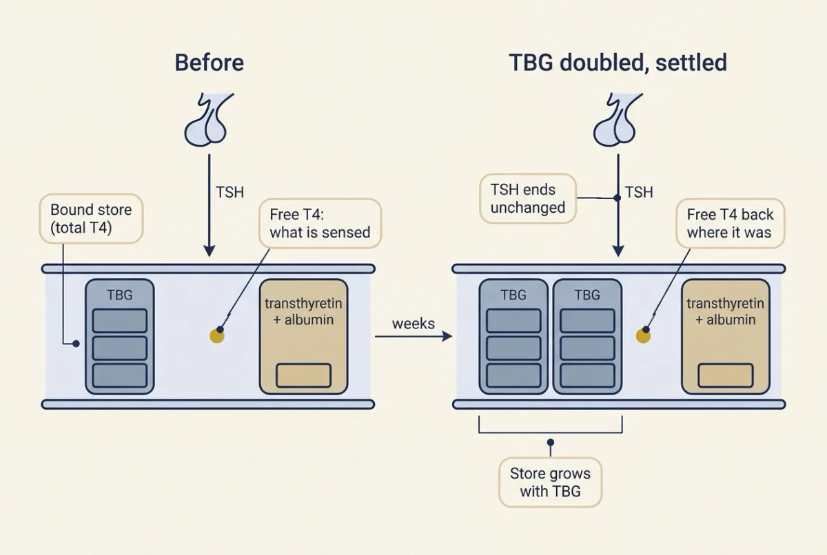
Measured in real women starting estrogen, the rule holds when the loop is intact and fails when it is open (Going deeper, “Estrogen with an open loop”).
Back to the opening patient: an open loop
Suppose the fixed daily T4 dose of the person in the opening question changes. If binding and clearance stay the same, treat the patient’s T4 as one well-mixed pool with first-order elimination and the euthyroid half-life of 6.2 days. Each half-life closes half the remaining gap, so after n half-lives T4 is 1 − 2−n of the way to its new steady level: 50 percent at 6.2 days, 75 percent at 12.4, 93.75 percent at n = 4 (24.8 days) and 96.875 percent, about 97, at n = 5 (31.0 days).
fraction of new steady state = 1 − 2−t / half−life
The same half-life sets the fall when input stops: with no new input, 100 nmol of T4 falls to 25 nmol in 12.4 days, two half-lives. T4-driven changes take about a month to settle, so an early measurement can mislead.
Make: how the follicle builds hormone, and what the scan sees
The last station is the gland itself. The thyroid is a gland of follicles: hollow balls of epithelial cells around a lumen of colloid, a protein gel made mostly of thyroglobulin. Each follicular cell faces blood on its basolateral side and colloid on its apical side. Synthesis runs from one face to the other.
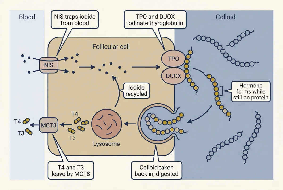
- Trapping. The sodium-iodide symporter (NIS) in the basolateral membrane moves iodide into the cell against its gradient, driven by the inward sodium gradient that the sodium-potassium pump maintains: secondary active transport (membrane transport). In the classic measurement on rat NIS in frog oocytes (Eskandari and colleagues, 1997), two sodium ions move in with each iodide, one net positive charge per cycle. NIS only traps iodide; it makes no hormone.
- Iodination. Iodide crosses into the colloid (pendrin is thought to be one route). At the apical surface, thyroperoxidase (TPO) uses hydrogen peroxide from dual oxidase (DUOX) to oxidize iodide and attach it to tyrosines of thyroglobulin, making monoiodotyrosine (MIT) and diiodotyrosine (DIT) residues. Fixing iodine onto protein is organification.
- Coupling. Still on thyroglobulin, the iodinated ring of a donor residue is joined onto an acceptor DIT. DIT plus DIT gives T4; MIT plus DIT gives T3. The hormone stays stored in the colloid as part of the protein. Coupling happens mainly at particular exposed, flexible, nearby tyrosine pairs, placed there by structure, mutagenesis and hormone-production assays (Coscia and colleagues, Nature, 2020; Adaixo and colleagues, Nature Communications, 2022).
- Release. Under thyroid-stimulating hormone (TSH) stimulation the cell takes colloid back in by endocytosis, the vesicles fuse with lysosomes, proteases digest thyroglobulin, and T4 and T3 leave through basolateral transporters such as MCT8.
- Recycling. Iodotyrosine dehalogenase strips iodide from the freed MIT and DIT for reuse.
Iodine is the raw material, so when it is short, it limits the hormone supply: TSH can rise and T4 can fall, while T3 can be preserved (Going deeper, “An iodine-poor population with more T3”). The gland’s parafollicular C cells secrete calcitonin, taken up in calcium, phosphate and bone.
Locating a fault: read the pair, then the scan
Now use the whole loop. Name the station that failed, and the loop predicts the tests.
Predict. Two adults both have a low free T4. In one, the gland itself is failing. In the other, the pituitary’s TSH-making cells (thyrotropes) have been damaged. What should thyroid-stimulating hormone (TSH) do in each?
Check your prediction
With a failing gland, TSH is high: the pituitary senses low hormone and responds. With damaged thyrotropes, TSH is low or inappropriately normal, because the cells that should respond are gone. This is the rule’s second limit, a broken sensor.
So read the pair: a TSH not as high as a low free T4 demands suggests a problem at the pituitary or hypothalamus.
| Lesion (untreated, in humans; overt disease, with free T4 abnormal, unless noted) | Station at fault | TSH | Free T4 | T3 |
|---|---|---|---|---|
| Graves disease: antibodies switch on the TSH receptor | Make, driven without TSH | Suppressed | High | High, sometimes rising before free T4 |
| Toxic adenoma: one nodule makes hormone on its own | Make, in one nodule | Suppressed | High | High |
| Thyroiditis (early, thyrotoxic phase): damaged follicles leak stored hormone | Make, store leaking | Suppressed | High | High or high-normal; raised less than T4 |
| Primary hypothyroidism: the gland itself fails | Make, failing | High | Low | Can stay normal; may fall when severe |
| Secondary hypothyroidism (pituitary): too little TSH | Sense | Low or inappropriately normal | Low | Often low or low-normal; not used to define it |
| Compensated iodine deficiency: too little raw material | Make, short of iodine | Normal or high | Normal or low | Normal or high |
Three exceptions. In subclinical disease TSH is abnormal while free T4 and T3 are still normal. Some thyrotoxic patients have a raised T3 with a normal free T4. And in secondary hypothyroidism TSH can even be mildly raised but less active, especially in hypothalamic disease. A rise in thyroxine-binding globulin (TBG) is not a lesion, so it is not in the table: at steady state TSH, free T4 and free T3 stay normal while total T4 and total T3 rise (worked problem above).
The radioiodine scan asks what the gland is doing
Radioiodine uptake asks a different question: give a tracer dose of radioactive iodine, commonly iodine-123, and measure hours later what fraction the gland has trapped; a scan shows where. Uptake reports how much tracer the gland still holds at the time of measurement, which depends on trapping by NIS, organification, the stable iodide supply and tracer loss. No single numerical normal range applies everywhere; it depends on tracer, timing, laboratory and local iodine intake. The patterns are what count:
- Graves disease: usually high uptake, even across both lobes; in a minority of cases it is normal or uneven.
- Toxic adenoma: one bright nodule, the rest faint because TSH is suppressed.
- Thyroiditis: low or absent uptake. Low uptake is not specific to it: thyroid hormone taken as a drug, and hormone made outside the gland, do the same.
- Iodine deficiency: high uptake, with little stable iodide competing with the tracer.
- A recent large iodine load, such as contrast dye: lower uptake, partly because stable iodide dilutes the tracer.
Same blood, different scan: Graves disease and thyroiditis
Predict. Graves disease and early thyroiditis both give a high free T4 and a suppressed TSH. Which one should show high uptake on the scan?
Check your prediction
Graves disease, whose gland is making new hormone from new iodide and so traps tracer.
In Graves disease, autoantibodies bind and activate the TSH receptor; Faust and colleagues (Nature, 2022) solved structures of the receptor with such an antibody, showing how it can mimic the hormone. The loop predicts the rest. Free T4 and T3 rise. TSH falls to suppressed levels, yet stimulation continues because it never depended on TSH. The gland enlarges diffusely and uptake is usually high and diffuse. Downstream, thyrotoxicosis (excess thyroid hormone) exaggerates the actions of T3 above: higher metabolic rate and heat, faster heart, higher cardiac output, lower systemic vascular resistance (SVR).
In the early thyrotoxic phase of thyroiditis, hormone is again high and TSH suppressed, but uptake is low: damaged follicles trap little iodide, and the excess hormone is stored hormone leaking out, not new synthesis. The scan is read with the hormone pattern, never alone.
Try the loop below: pick a lesion and read the arrows, then double TBG on a normal axis.
In this sketch: the arrows are directions only, taken from the lesson’s table for untreated people and its MCT8 paragraph, and carry no numbers. The only numbers are the lesson’s made-up worked problem (an adult at steady state with a total T4 of 80 nmol per L, 99.97 percent protein-bound, TBG carrying 75 percent of bound T4, unchanged carrier affinities). The dark scan panel, its brightness shades, the equal gland sizes and the outlined block are drawing choices made up for this sketch, and the scan shows a pattern, not a measurement.
The rule to keep: ask which station failed, and read TSH and free T4 as a pair. Low hormone with high TSH points to the gland; low hormone with TSH not high suggests the pituitary or hypothalamus; high hormone with suppressed TSH is thyrotoxicosis, and the uptake scan, read with the hormone pattern and never alone, tells high synthesis from leakage. In the worked problem, total T4 rose with the binding protein; free T4 is the level feedback defends.
Design problem
de Jesus and colleagues (Journal of Clinical Investigation, 2001) studied mice lacking type 2 deiodinase (Dio2-knockout). In the cold they became hypothermic because brown adipose tissue thermogenesis was impaired, yet plasma T3 was normal. A single injection of T3 reversed the hypothyroid-like abnormalities measured in their brown fat cells. What does this show about where active hormone is made, and what controls would you add? (Brown fat in adult humans is in skin and human heat.)
Saved in this browser only.
One way to do it
Plasma T3 was normal, yet the tissue behaved as if it lacked thyroid hormone. So brown fat relies on T3 made inside its own cells from T4 by D2, and plasma T3 does not fully report thyroid status in that tissue.
The T3 rescue supports a lack of T3 in the tissue rather than some function of D2 unrelated to making T3. A rescue confined to brown fat would test it more directly.
Controls: wild-type littermates in the same cold; both genotypes housed at thermoneutrality; plasma T4 and TSH as well as T3; T3 content of brown fat itself; a check that sympathetic drive to brown fat arrived normally, since cold-induced thermogenesis depends on noradrenaline; and a rescue dose too small to make the whole animal hyperthyroid.
Limits: this is mouse brown fat in the cold, not an adult human at rest, and a whole-body knockout lacks D2 everywhere, so not every phenotype belongs to brown fat.
Going deeper
The main line above does not depend on these details.
How T3 becomes heat: the muscle NMR study
The uncoupling idea in the main text draws on a study in human muscle. Lebon and colleagues in Gerald Shulman’s laboratory (Journal of Clinical Investigation, 2001) studied seven healthy adults before and after three days of T3. Carbon-13 nuclear magnetic resonance (NMR) spectroscopy estimated tricarboxylic acid (TCA) cycle flux in skeletal muscle, phosphorus-31 saturation-transfer NMR estimated phosphate-to-ATP flux as a measure of ATP synthesis, and indirect calorimetry measured whole-body energy expenditure. If T3 acted only by raising ATP demand, TCA flux and ATP synthesis should rise together. Instead TCA flux rose by about 70 percent while estimated ATP synthesis did not increase. Energy expenditure rose by about 15 percent and heart rate by about 30 percent. Taken at face value, ATP made per turn of the cycle fell to about 0.6 of baseline (1.0 / 1.7 = 0.59). The authors’ reading is mitochondrial uncoupling: more fuel oxidized per ATP made, the difference released as heat. The study does not identify the leaking protein or apply beyond muscle, and the fluxes are model-based estimates in seven people over three days.
Vascular resistance in hyperthyroid patients
The main text’s lower systemic vascular resistance (SVR) with little change in mean pressure has been measured in patients. In one 2024 study (Suonsyrjä and colleagues), SVR was 18 percent lower in 20 still-hyperthyroid patients, measured a median of six days after starting antithyroid medication, than in 60 matched euthyroid controls. Aortic pressures were similar, radial pulse pressure was about 5 mmHg higher and finger blood pressure was lower.
D1 works on both rings
D1 also takes iodine from the inner ring, so it makes some reverse T3 as well, and it is the main enzyme that clears reverse T3. When D1 removes an outer-ring iodine from reverse T3, the product is inactive 3,3’-T2. That outer-ring step is clearance, not activation, which is why the lesson states the ring rule for T4.
An iodine-poor population with more T3
In 285 people from Nomane, an iodine-deficient region of New Guinea, Chopra and colleagues (1975) found a higher mean serum T3 than in an American comparison group, alongside a lower mean T4 and a raised mean thyroid-stimulating hormone (TSH). The population had goitre (an enlarged gland), a sign of long TSH drive, so it shows iodine shortage further along than the mildest, compensated cases in the table.
Estrogen with an open loop
Arafah (New England Journal of Medicine, 2001) followed postmenopausal women starting estrogen therapy. In women with normal thyroid function, total T4 and thyroxine-binding globulin (TBG) rose while free T4 and TSH did not change, as the worked problem predicts. In women with hypothyroidism taking T4, total T4 and TBG rose in the same way, but free T4 fell and TSH rose: their supply was a fixed dose of T4, so the rise in TSH could not raise it. The study concluded that estrogen may increase the need for T4. So the loop, not binding chemistry alone, is what reliably defends the free level.
Your notes
Select any text in the lesson to highlight it or add a note to it. Your highlights and notes are saved in this browser and collected in your notebook.
This browser is not letting the site save anything (a private window or strict privacy settings), so highlights and notes will be lost when you leave the page.
How we measure it
Isotope tracing with chromatographic separation
Give a hormone labelled with a radioactive atom, then separate the related compounds in serum by chromatography and measure the label in each. Label that appears in a different compound shows a conversion, provided the tracer was pure and the route is controlled.
Functional expression cloning in frog oocytes
Make RNA from a library of complementary DNA, inject it into Xenopus oocytes, and measure a function such as iodide uptake above the background of uninjected eggs. Pools that confer the function are narrowed to a single clone, which identifies the gene by what it does.
Radioiodine uptake and thyroid scintigraphy
Give a small tracer dose of radioactive iodine and measure, hours later, the fraction the gland has taken up; a scan shows where in the gland it went. Uptake reports how much tracer the gland retains, which depends on trapping, organification and iodide supply, not how much hormone is in the blood.
13C and 31P NMR spectroscopy with indirect calorimetry
Nuclear magnetic resonance of carbon-13 from a labelled substrate estimates tricarboxylic acid cycle flux in muscle, and phosphorus-31 saturation-transfer NMR estimates phosphate-to-ATP flux, which is taken as an estimate of ATP synthesis rather than a direct measure of it. Indirect calorimetry converts oxygen consumption and carbon dioxide output into whole-body energy expenditure.
Practice questions on this lesson (4) → MCAT-style, original, each with an explanation
Where this lesson fits
Before thisReview first
- Endocrine Axes and How to Test ThemThis course · Lesson 2
You need its paired hormone levels and feedback testing here to read TSH with free T4.
- Body Water and Its CompartmentsThis course · Lesson 1
Free versus bound hormone in plasma builds on how substances distribute between body compartments.
After thisStudy next
- The Adrenal Cortex and MedullaThis course · Lesson 20
Another hormone axis where tissue enzymes and binding proteins shape what reaches the cell, with a similar feedback loop.
- Exercise: The Whole Body at WorkThis course · Lesson 30
Thyroid hormone's effects on metabolic rate and cardiac output matter when the whole body works hard.
Suggested by an AI model from the lessons themselves, then checked. How we use AI
Finish line
Test yourself
9 questions
Practice and MCAT-style, with explained answers.
Next · Lesson 20
The Adrenal Cortex and Medulla
The kidney's aldosterone receptor binds cortisol just as tightly, and plasma carries far more cortisol. So why does aldosterone run sodium transport?
Continue →