Neuroscience · Unit 5: Homeostasis and affect
Emotion and Stress
Rewritten on : new text, figures, flashcards and film. Your notes and highlights stay, but the text around them may have moved.
A rat freezes to a tone that once came before a shock. Damage one region of its hypothalamus and it still freezes, but its learned rise in blood pressure is impaired.
Pause when the reading reaches a film, and offer to play it. Saved on this device.
Watch the film now, or keep listening
Builds on:Autonomic Control and the HypothalamusSynaptic PlasticityFeeding, Motivation and RewardWhy these?
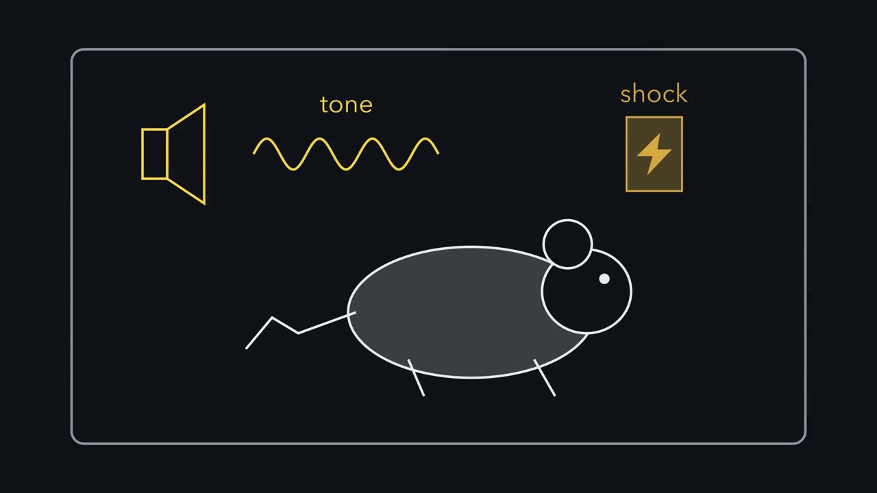 ▶ Watch One Signal, Three Routes · 2:15
Read the transcript of “One Signal, Three Routes”
Film transcript: One Signal, Three Routes
A rat hears a tone that once came just before a mild shock. What does its brain do with that tone? Two things happen at once. The rat stops moving, and its blood pressure rises. Neither one is the feeling of fear. But both can be measured, and that lets us trace them in the brain. First, is the response learned? One group of rats hears tones that end in a shock. A second group gets the same tones and the same shocks, but kept apart in time. Later, the paired rats freeze far more to the tone alone. That difference is our estimate of what was learned. Where is it learned? The tone travels to the amygdala, a cluster of nuclei deep in the brain. The lateral amygdala is the input stage.
The central amygdala sends the signal out. Three routes leave it. One runs to the periaqueductal gray, in the midbrain. It drives freezing. A second runs to the lateral hypothalamus. It drives the rise in blood pressure. A third, mostly indirect, reaches a hormone chain: hypothalamus, pituitary, adrenal. In people, its hormone is cortisol. Two of these routes are fast. Freezing and the pressure rise appear while the tone is still playing. The hormone route is slow. The adrenal must make its hormone, so levels peak tens of minutes later. Feedback then turns further release down. So here is the rule. A learned threat cue reaches the amygdala, then leaves by separate routes.
In this rat task, damaging neurons in one target impaired mainly its own response. Damage to the amygdala can impair both. This map is for learned responses to a cue outside the body, measured in rats. It does not say what the rat feels. Later chapters show where the map stops. The amygdala is needed to learn the threat. Separate routes carry out the response, two fast and one slow.
From the film
- Paired minus unpaired freezing estimates what a rat learned about the tone.
- A learned threat cue reaches the amygdala, then leaves by separate routes: one for freezing, one for the pressure rise, and a mostly indirect one to the hormone response.
- Freezing and the pressure rise appear while the tone is still playing, but the hormone response peaks tens of minutes later.
- This map describes learned responses to an outside cue in rats; it does not say what the rat feels.
Play a rat a tone that has repeatedly ended in a mild footshock, and two things happen when it hears the tone alone: it stops moving, and its arterial pressure rises.
Neither response is the feeling of fear. Both can be measured, and with them you can ask which parts of the brain learn that a tone predicts harm and which turn that knowledge into a reaction.
One signal, several routes, two speeds
Three brain structures and one hormone chain carry the story. The amygdala, deep in the brain, is needed for the rat to learn that the tone predicts harm. It sends that signal on to the periaqueductal gray in the midbrain and the lateral hypothalamus, which helps drive the sympathetic nervous system, and, mostly indirectly, to a hormone chain, the hypothalamus–pituitary–adrenal (HPA) axis, whose end product is cortisol in humans (corticosterone in rats).
The one idea to carry through this lesson. In a rat that has learned that a tone predicts shock, the tone reaches the amygdala, and the learned responses leave by separable routes. Freezing depends on the caudal periaqueductal gray and the blood-pressure rise on the lateral hypothalamus; both appear while the tone is still playing. Amygdala circuits also recruit, mostly indirectly, a hormone response through the HPA axis on a slower clock: the hormone peaks tens of minutes after the stress begins, and negative feedback then limits further release. Damage the neurons of one output target and mainly its response is impaired; damage the shared amygdala node and both learned responses should be impaired. These are separable contributions within a larger network, not three independent channels.
The rule covers learned responses to a cue from outside the body, measured in rats, and it does not say what the rat feels. “Where the rule stops” marks its other edges.
Learning what predicts harm
In Pavlovian threat conditioning, an initially innocuous conditioned stimulus (CS), such as a tone, is paired with an aversive unconditioned stimulus (US), such as a brief footshock. The US produces an unlearned reaction. After pairing, the CS alone produces a conditioned response (CR): in rats, freezing (a defensive behaviour in which the animal holds still except for breathing) and autonomic changes (made by the involuntary, autonomic nervous system) such as a rise in arterial pressure.
A rat that freezes to a tone after shocks might simply be jumpy. The test of association is the unpaired control: a second group hears the same tones and receives the same shocks, but the tones and shocks are kept apart in time.
Predict. Both groups heard the same tones and felt the same shocks. If the paired rats freeze much more to the tone than the unpaired rats, what is left to explain the difference?
Check your prediction
Only the pairing: in one group the tone predicted the shock, and in the other it did not. So paired minus unpaired is the standard estimate of learning about the tone, the subtraction the worked problem uses.
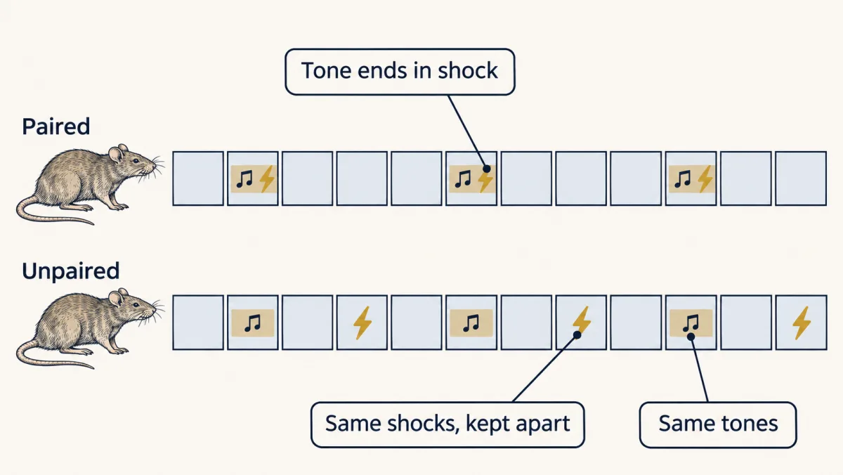
It is an estimate, not a perfectly neutral baseline: an explicitly unpaired tone can come to signal safety, which is why some studies use a truly random schedule instead.
The map: one node, several routes
Picture a diagram drawn from left to right. The tone travels from the ear through brainstem auditory relays to the auditory thalamus, and from there reaches the amygdala by two parallel routes: directly from the thalamus, and indirectly through auditory cortex. The amygdala is a cluster of nuclei (a nucleus here is a compact group of neuron cell bodies in the brain). It lies deep in the medial temporal lobe in humans, and in the corresponding part of the ventral forebrain in the rat.
Inside it are two stages. The lateral amygdala is the input stage: the tone arrives here. The central amygdala is the output stage: it sends the signal on to the hypothalamus and brainstem. Three routes leave it.
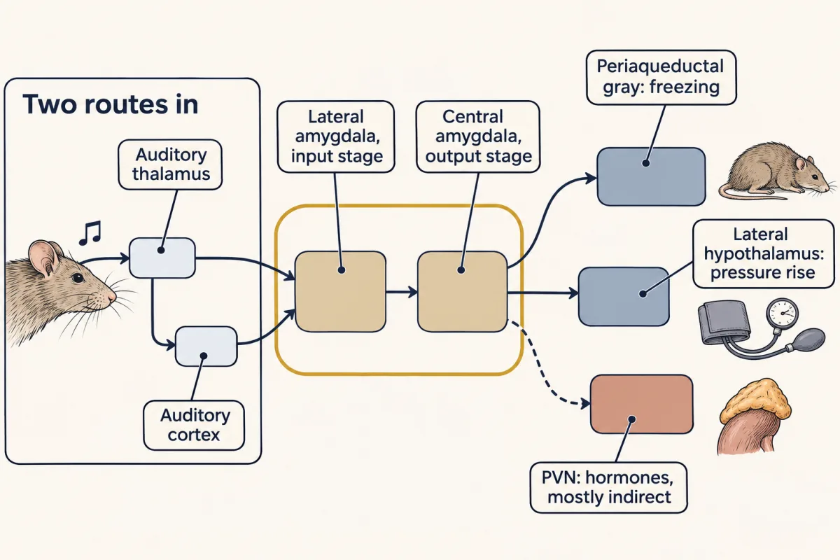
| Route out of the central amygdala to | Response it drives | Clock |
|---|---|---|
| Periaqueductal gray, in the midbrain | Freezing | While the tone is playing |
| Lateral hypothalamus | Blood-pressure rise | While the tone is playing |
| Paraventricular nucleus of the hypothalamus, mostly indirectly through relay structures | Glucocorticoid release through the HPA axis: corticosterone in rats, cortisol in humans | Rises after a lag; peaks tens of minutes after stress begins |
This is the simplest working map. The two-stage picture is a simplification: the basal amygdala, inhibitory circuits within the central amygdala and plasticity at more than one site also contribute. The damage that matters in the studies below is bilateral: both amygdalae.
If the lateral amygdala stores the tone-shock association, its response to the tone should change with pairing. Rogan, Stäubli and LeDoux (1997) recorded tone-evoked field potentials, the summed response of many neurons near an electrode, in the rat lateral amygdala. After pairing, the tone-evoked response grew larger, as a synapse’s response does after long-term potentiation (LTP), a lasting increase in synaptic strength after strong activity (see synaptic plasticity). The change was absent in rats given the tones and shocks unpaired. An LTP-like change that tracks the contingency (whether the tone predicts the shock) fits plasticity as part of the learning; it does not show that this one change is all of it.
The key experiment: cutting one route at a time
Read the transcript of “Cutting One Route at a Time”
Film transcript: Cutting One Route at a Time
Freezing and the pressure rise arrive together. Do they travel one route out of the amygdala, or two? In 1988, Joseph LeDoux and his colleagues asked exactly that, in rats. First they damaged one target of the central amygdala. Two weeks later they trained the rats, and the next day played the tone alone. In the same rats, they measured both responses: freezing, and the rise in blood pressure. Picture two bars for each group. Intact rats that had the pairing show two tall bars. Intact rats without the pairing show two short bars. That short pair is our baseline. For these invented numbers, we assume the damage leaves the baseline alone, and that each score is the baseline plus a learned part.
So the excess over the baseline estimates the learned response. Suppose both responses need the same route through these lateral hypothalamus neurons. What should damage there do to freezing? If both responses needed those neurons, both would fall. They do not. The pressure rise falls almost to the baseline. Freezing stays almost at the top. So freezing does not need the lateral hypothalamus, but the pressure rise does. Now damage the caudal, or rear, periaqueductal gray instead. What do you expect this time?
The reverse. Freezing drops almost to the baseline, and the pressure rise survives. Each kind of damage impairs one response and spares the other. That pattern is called a double dissociation. It means different targets below one amygdala node contribute to different responses. In the worked problem, damage to the central amygdala itself leaves neither learned response above the baseline, as a shared source predicts. One detail matters. The caudal midbrain damage used ibotenic acid, a toxin that kills cell bodies but spares passing axons. So the freezing loss points to neurons in the periaqueductal gray, not to cut fibres. The same toxin in the lateral hypothalamus impaired the pressure rise while sparing freezing.
This experiment finds the routes out of the amygdala. It says nothing yet about a feeling.
From the film
- In rats, damage to lateral hypothalamus neurons impaired mainly the pressure rise, and damage to the caudal periaqueductal gray impaired mainly freezing.
- Each lesion impairing one response and sparing the other is a double dissociation, so different targets below the amygdala contribute to different responses.
- Damage to the shared central amygdala node should impair both learned responses.
- This experiment finds the routes out of the amygdala, but it says nothing about a feeling.
LeDoux, Iwata, Cicchetti and Reis (1988) asked whether one route below the central amygdala carried both freezing and the pressure rise, or whether each response had its own. The design: damage (make a lesion in) one candidate target of the central amygdala, let the rats recover for two weeks, condition them, and the next day play the tone alone and measure both responses in the same animals.
Predict. Suppose freezing and the pressure rise both need a common route through the lateral-hypothalamic neurons being lesioned. What should the lesion do to freezing? What if those neurons are needed for the pressure rise but not for freezing?
Check your prediction
If both responses need those neurons, both should be impaired. If only the pressure rise needs them, pressure conditioning should be impaired while freezing is spared. The experiment distinguishes these two specific hypotheses.
The result was the second. Lateral-hypothalamic lesions impaired the conditioned pressure increase and spared freezing. Lesions of the caudal periaqueductal gray (PAG; also called the central gray, the sheath of neurons around the midbrain’s fluid canal; caudal means toward the rear) disrupted freezing and spared the pressure response. The hypothalamus’s sympathetic outputs are covered in the hypothalamus and the autonomic nervous system.
How the lesions were made matters. An electrolytic lesion passes current and destroys everything near the tip, including fibres of passage, axons that only travel through the region, so a deficit after it might come from cut axons going elsewhere. Ibotenic acid, an excitotoxin, kills local neuron cell bodies and spares those fibres. The caudal PAG lesions used it, so the freezing deficit points to PAG neurons rather than to cut fibres. Fibre damage is a real worry in the lateral hypothalamus, which large fibre tracts run through; ibotenic-acid lesions there also impaired the pressure response and spared freezing, so that deficit too points to local neurons and not only to passing axons.
Worked problem. These numbers are invented, laid out in the design of the 1988 study; they are not its data. Rats were tested with the tone alone after conditioning. Freezing is the percentage of a 60-s tone spent frozen; pressure is the rise in mean arterial pressure in mmHg.
| Group | Freezing (%) | Pressure rise (mmHg) |
|---|---|---|
| Intact, paired | 60 | +20 |
| Intact, unpaired | 8 | +3 |
| Lateral hypothalamic lesion, paired | 57 | +4 |
| Caudal PAG lesion, paired | 9 | +19 |
| Central amygdala lesion, paired | 6 | +3 |
Assume a lesion would not change how an unpaired rat responds, so the intact unpaired score is the baseline for every group, and that the measures add linearly: each score is the baseline plus a learned part.
Step 1: the learned response. Subtract unpaired from paired in intact rats.
learned freezing = 60 − 8 = 52 percentage points; learned pressure = 20 − 3 = 17 mmHg
Step 2: each lesion as a share of what was learned. Subtract the baseline from each lesion group and divide by the learned response.
hypothalamus: freezing (57 − 8)/52 = 49/52 = 94%; pressure (4 − 3)/17 = 1/17 = 6%
PAG: freezing (9 − 8)/52 = 1/52 = 2%; pressure (19 − 3)/17 = 16/17 = 94%
central amygdala: freezing (6 − 8)/52 = −4%, slightly below baseline, read as no learned freezing left; pressure (3 − 3)/17 = 0%
Step 3: read it. Each output lesion selectively impairs one response and spares the other: a double dissociation, evidence for separable contributions below one amygdala node. The amygdala lesion leaves neither response above baseline, as a shared source predicts.
- Freezing
- —
- Pressure rise
- —
—
—
In this sketch: invented numbers laid out in the design of LeDoux and colleagues (1988), not its data; rats; the unpaired baseline is assumed unchanged by lesions and the measures are assumed to add linearly. No lesion combinations are offered because the lesson gives none. LH = lateral hypothalamus, PAG = caudal periaqueductal gray, CeA = central amygdala. “Spared” and “impaired” here mean at least or under half of the learned response, a sketch convention.
Each output lesion removes one response and keeps the other, a double dissociation below one amygdala node; the unpaired group is the subtraction that isolates learning about the tone; the result localizes outputs of a defensive response and says nothing about a feeling.
The slow route: the HPA loop
Read the transcript of “The Slow Route, and Where the Map Stops”
Film transcript: The Slow Route, and Where the Map Stops
Freezing begins while the tone still plays. So why does the hormone peak only tens of minutes later? Because it runs through three relays: hypothalamus, pituitary, adrenal. Neurons in the hypothalamus release CRH into tiny vessels that lead to the pituitary. The pituitary answers by releasing ACTH into the general circulation. ACTH reaches the adrenal cortex, which makes cortisol, or corticosterone in rats. The pituitary answers within minutes. But the adrenal must make its hormone, so levels peak tens of minutes later. As the hormone rises, it feeds back and turns down further release. A low baseline carries on. This thermostat has two speeds.
A fast arm acts over seconds to minutes. A slow arm works by changing which genes are read, over hours to days. Even with no threat at all, the hormone rises and falls. It has a daily rhythm, and pulses about once an hour. Is each hourly pulse a command from the hypothalamus? Walker and colleagues tested this in rats. They chose a time of day when the rat's own CRH is low. So new pulses were unlikely to be the rat's own commands. Then they dripped in CRH at a perfectly steady rate. If every pulse is a command, what should you see?
Commands alone would give a steady, raised level. Instead, the hormone still rose and fell, about once an hour. So in this preparation, pulsed CRH was not needed. Given a steady infusion, the pituitary and adrenal loop can make its own rhythm. Now, where does our rule stop? In rats, learning about a place also needed the hippocampus. A patient with selective damage to both amygdalae could still say which stimulus went with which. The learned bodily response was missing. And one breath of carbon dioxide made three women with that damage panic. That is three people, from one rare disease. Rat circuits can find necessary steps for a measured response.
They cannot alone say what is felt. The amygdala is needed to learn an outside threat. Neural routes respond fast; the hormone route is slower, because the adrenal must make its hormone.
From the film
- The hormone route runs through the hypothalamus, pituitary and adrenal, so levels peak tens of minutes after the stress begins.
- As the hormone rises, it feeds back to turn down further release, with a fast arm over seconds to minutes and a slow arm over hours to days.
- Hormone levels also rise and fall with a daily rhythm and pulses about once an hour, and in one rat preparation a steady CRH infusion still gave hourly pulses.
- The map stops at feeling: rat circuits can find necessary steps for a measured response, but cannot alone say what is felt.
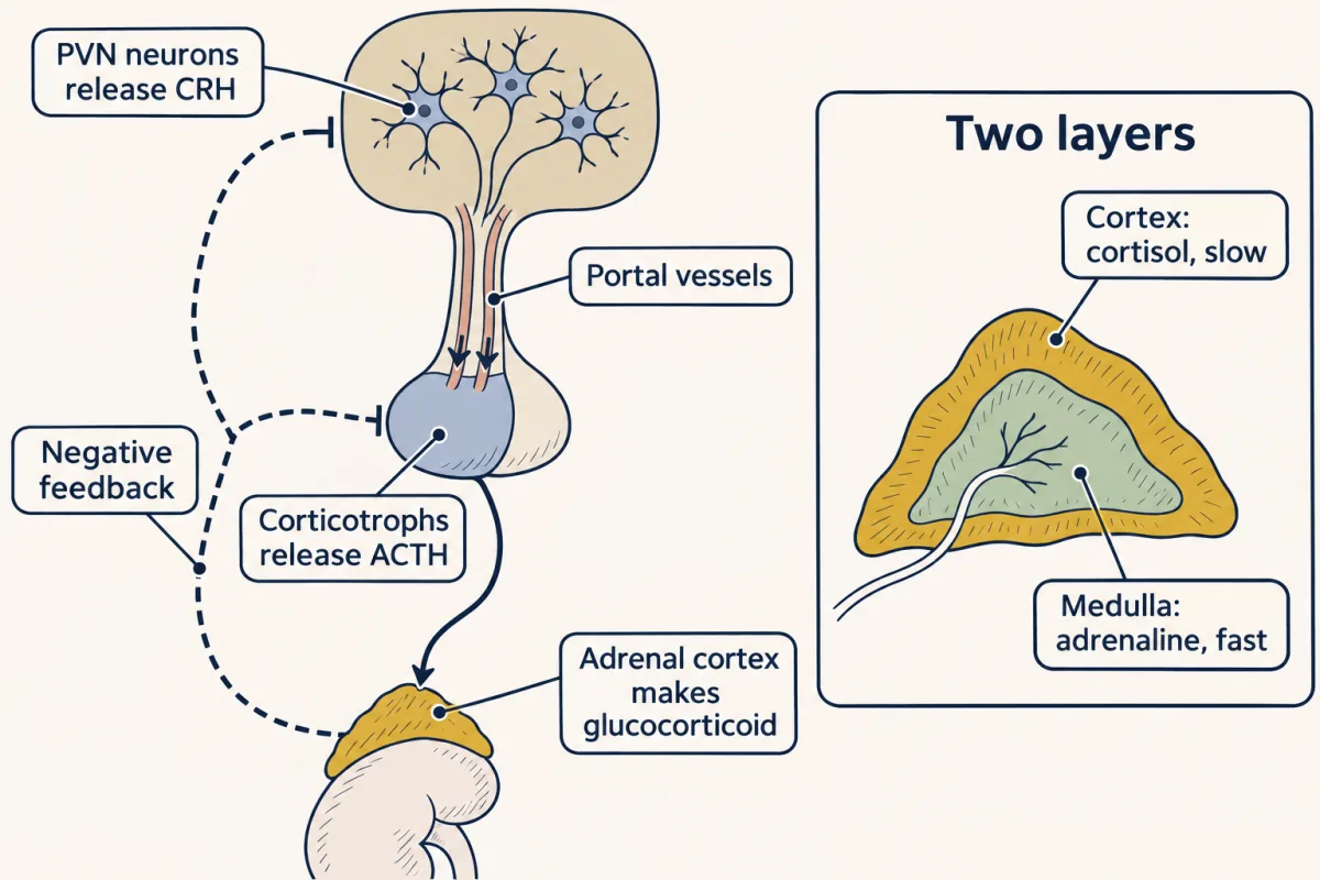
Freezing and the pressure rise arrive while the tone is still playing. The third output, the hypothalamus–pituitary–adrenal (HPA) axis, is slower. Picture a chain of three hormone relays, drawn as boxes from the top of the page to the bottom.
- Hypothalamus. Neurons in the paraventricular nucleus (PVN) of the hypothalamus make corticotropin-releasing hormone (CRH), a major activator of the axis. They release it into tiny blood vessels, the hypophyseal portal vessels, which carry it a short distance down the pituitary stalk to the gland. Because it travels this private route, CRH reaches the pituitary without being diluted in the whole bloodstream.
- Pituitary. In the anterior pituitary, cells called corticotrophs respond to CRH by releasing ACTH (adrenocorticotropic hormone) into the general circulation.
- Adrenal cortex. Circulating ACTH reaches the adrenal cortex, the outer layer of the adrenal gland, and stimulates it to make and secrete a glucocorticoid: cortisol in humans, corticosterone in rats and mice.
ACTH can rise within minutes. The adrenal cortex, however, does not store glucocorticoid: it makes the steroid when ACTH arrives. So glucocorticoid rises after a lag and typically peaks tens of minutes after the stress begins. That synthesis, not the trip through the blood, sets the slow clock.
How does a learned tone get into this chain? Anticipated threats, such as a cue that predicts harm, act through limbic structures (forebrain regions involved in emotion and memory), including the amygdala. They work largely by releasing the PVN from tonic inhibition: rather than pushing the PVN, they mostly lift a brake that normally holds it back. Physical challenges take a different route, through brainstem relays. So the amygdala is one way into the slow route, not the only one.
Keep the adrenal’s two layers apart. Adrenaline comes from the inner medulla, driven within seconds by sympathetic nerves, so it acts on the fast clock; it is not a fourth route in this map. Cortisol comes from the outer cortex, driven by a blood-borne hormone, ACTH; it is the slow arm.
A thermostat with two speeds
Glucocorticoids also limit their own secretion by negative feedback: they act on brain regions and the anterior pituitary to reduce further CRH and ACTH output, much as a thermostat turns the furnace down as the room warms. Feedback limits the response; it does not stop secretion altogether, and the level also falls as the drive ends and the hormone is cleared.
Rodent studies distinguish two speeds for this thermostat.
- A fast arm acts over seconds to minutes. It is nongenomic: it does not wait for genes to be switched on.
- A delayed arm acts over hours to days. It is genomic: glucocorticoid and mineralocorticoid receptors bind the hormone and can regulate transcription after moving to the nucleus (see nuclear receptors).
Predict. Give a rat a drug that blocks new gene transcription but leaves the receptors’ fast actions intact, then stress it. Does corticosterone still turn ACTH down within minutes?
Check your prediction
Yes. The fast arm does not need transcription, so it still works. Over hours, the feedback that depends on new gene products would be weaker. A drug that blocks the receptor itself is a different test, because the fast arm may use the same receptor.
A rhythm with no threat at all
Cortisol is not released only during stress. Under basal conditions glucocorticoids are secreted in a circadian rhythm, highest around the start of the active phase (around waking in humans, around dark onset in nocturnal rats), and in an ultradian rhythm of roughly hourly pulses.
Is each hourly pulse a command from the hypothalamus? Walker and colleagues (2012) tested this in conscious male rats by adding a constant intravenous CRH infusion, 0.5 µg/h for 6 h, sampling blood every 5 min. They worked at the circadian trough, when the rat’s own CRH drive is expected to be low and corticosterone is not pulsatile. The controlled input was the infusion; the rat’s own hypothalamic secretion was not clamped, so pulses appearing there were unlikely, not certain, to be its own CRH bursts.
Predict. If every pulse is a command from the hypothalamus, what should a steady, unchanging CRH drip produce in corticosterone? If the pituitary and adrenal oscillate on their own, what then?
Check your prediction
Commands only: a steady drip gives a steady, raised level. A pituitary-adrenal oscillator: the level keeps rising and falling even though the input is flat.
Picture the record: a flat line at the trough, then, under the steady drip, peaks and troughs about an hour apart. Corticosterone still oscillated, at a mean 0.89 pulses per hour. So pulsatile CRH was not required in this preparation, which points to a pituitary-adrenal loop that, given a steady drive, can make its own rhythm.
Turn the frequency into a period, treating the output as an ideal oscillator:
period = 1/f = 1/(0.89 per h) = 1.124 h = 67.4 min; cycles in 6 h = 0.89 × 6 = 5.34
That is model arithmetic, not a count of observed peaks, and a rat rate, not a human constant.
Where the rule stops
The rule has five edges:
- what the rat, or a person, feels (the last section);
- learning about a place, which also needs the hippocampus;
- knowing the pairing as a fact, which a patient with selective damage to both amygdalae can still do;
- a threat signalled from inside the body, such as one breath of CO2;
- the hypothalamus–pituitary–adrenal (HPA) axis without a learned cue: its resting rhythm, above, and physical challenges, which reach it through the brainstem.
Learning about a place: the hippocampus as well
Phillips and LeDoux (1992) conditioned rats to a tone in a distinctive chamber, so that a conditioned rat came to freeze in the chamber itself as well as to the tone. Amygdala lesions disrupted conditioning to both the tone and the chamber; hippocampal lesions disrupted conditioning to the chamber (the context) while sparing conditioning to the tone. In this task the hippocampus was needed for context. That does not mean the hippocampus never takes part in cue learning.
Knowing the pairing: patients
Patients test what rats cannot report. Bechara and colleagues (1995) studied people with naturally occurring bilateral damage. Each did a conditioning task, and afterwards each was asked what had been paired with what. So each patient gives two measures: the learned autonomic response, and the stated facts.
Predict. One patient has selective damage to both amygdalae. Will that patient show the conditioned autonomic response? Will that patient be able to say which stimulus went with which?
Check your prediction
By the rule, the learned bodily response needs the amygdala, so it should be missing. The rule says nothing about stating facts. That is what happened: the patient with selective amygdala damage did not acquire conditioned autonomic responses, yet learned the declarative facts (facts one can state) about which stimulus went with which.
A patient with selective hippocampal damage showed the reverse, acquiring the conditioning but not the facts, and a patient with damage to both acquired neither. Three people are a double dissociation, not a population estimate; the kinds of memory involved return in memory systems.
A threat from inside the body: one breath of CO2
Patient SM has bilateral amygdala damage from Urbach-Wiethe disease, a rare genetic condition. Feinstein and colleagues (2011) found that she reported at most minimal fear in the external-threat situations they tested, while her capacity for other basic emotions was not abolished.
Then Feinstein and colleagues (2013) changed the threat. Three women with the same bilateral damage, SM among them, inhaled a single breath of 35% CO2. All three panicked, against 3 of 12 neurologically intact comparison women on the first inhalation. Fear was rated on a 0 to 100 visual analogue scale. Their anticipatory autonomic responses before the challenge were deficient. Three patients, from one rare disease: enough to show that fear can occur without the amygdala, not to say how often (the arithmetic is in Going deeper).
Two simpler rules are tempting, and these edges refute both: “The amygdala is the fear centre” (the CO2 result) and “Cutting the amygdala removes every stress response” (the brainstem route into the HPA axis and its resting rhythm). The rule claims less: in rats, the amygdala is a necessary node for the learned freezing and pressure responses to an outside cue.
Design problem
You are reviewing the CO2 result above. Interpret it, and design the follow-up you would want before generalizing it.
Saved in this browser only.
One way to do it
Interpretation: the amygdala is not needed for every fear. A threat signalled from inside the body (rising blood CO2) may engage circuits outside the amygdala, for instance brainstem regions that sense CO2; that is a hypothesis this study does not test.
Limits: three patients, two of them (AM and BG) monozygotic twin sisters, whose damage is thought to have arisen during development, give no population panic rate and may have compensated over years. The physiological records came from only two patients (heart rate) and one (skin conductance), so the autonomic conclusions rest on fewer people than the ratings.
Follow-up: keep an air inhalation as a sham, blind participants and raters to which gas is given, and randomize order. Record autonomic measures and fear ratings separately for every participant, so the reported feeling and the body’s response can dissociate. Add patients with adult-onset bilateral amygdala damage to address compensation, and image the brainstem during the challenge to test the alternative-circuit hypothesis.
What a freezing rat can tell us
A rat cannot tell you how it feels. Freezing and a pressure rise are defensive responses, and a single defensive measure is not an unambiguous measure of fear. Researchers disagree on what follows. Fanselow and Pennington (2018) argue that fear is best treated as an integrated autonomic, behavioural and cognitive-emotional response, so animal circuits model fear itself. LeDoux has argued for separating defensive survival circuits from the conscious feeling of fear, which on that view needs other circuitry. The disagreement is about what the measurements license.
The rat work can locate necessary steps for a measured response under defined conditions. It cannot alone say what the animal experiences, or that a human feeling uses the same route. Cain (2023) points to differences in tasks and outcome measures across species as a limit on translation. The human cases show why both kinds of evidence are needed: SM’s minimal fear to external threats and her panic to CO2 came from one person tested two ways, so the difference points to the kind of threat rather than the patient, although the tests also differed in procedure and date.
Going deeper
Valence in the mouse amygdala
In mice, Kim and colleagues (2016) identified genetically distinct basolateral-amygdala excitatory populations linked to behaviours of opposite valence. Testing such circuits by driving and silencing defined projections is the subject of designing a circuit experiment.
The receptors behind each relay
Corticotropin-releasing hormone (CRH) and adrenocorticotropic hormone (ACTH) act through G-protein-coupled receptors; the machinery is in G proteins and their receptors and cAMP and PKA. Glucocorticoids act through nuclear receptors, the delayed arm of the feedback above.
The constant-infusion experiment in detail
The rats were male Sprague-Dawley rats. Analysed animal by animal, the pulse frequency under 0.5 µg/h CRH ranged from 0.79 to 0.93 per hour. With samples every 5 min over the first 25 min of infusion, ACTH was significantly above basal by 10 min, whereas corticosterone first differed significantly at 20 min: the pituitary leads and the adrenal follows, a small lag the feedback model predicts. These significance times do not establish exact physiological onset times.
Does the pattern of cortisol matter?
Timing can matter for the brain. In 15 healthy men, Kalafatakis and colleagues (2018) found that, with their own cortisol production suppressed, the same daily replacement dose given in different patterns changed fMRI responses to emotional faces, although momentary mood did not differ between the two subcutaneous patterns.
How strong is the CO2 result?
Three of 3 patients panicked, against 3 of 12 comparison women, and the paper reports p < 0.05 by Fisher’s exact test. Recomputed on those counts, the test gives p ≈ 0.044 (one- and two-sided agree here). If one more comparison woman had panicked (4 of 12), it would give p ≈ 0.077; that sensitivity check is this lesson’s calculation, not the paper’s. Both treat participants as independent. Two patients are monozygotic twins, a possible within-family dependence these tests do not adjust for; the size of any distortion is unknown, and their shared background also limits independent replication. The two values show how far a result resting on three people can move with one person.
Your notes
Select any text in the lesson to highlight it or add a note to it. Your highlights and notes are saved in this browser and collected in your notebook.
This browser is not letting the site save anything (a private window or strict privacy settings), so highlights and notes will be lost when you leave the page.
How we measure it
Pavlovian threat conditioning with an unpaired control
A neutral tone (the conditioned stimulus) is paired with a mild footshock (the unconditioned stimulus), and the response to the tone alone is tested later. A control group gets the same tones and shocks, not paired, so a difference between the groups points to the association rather than exposure to shock or tone. An explicitly unpaired tone can itself become a safety signal, so the comparison is an estimate rather than a pure measure.
Electrolytic and excitotoxic lesions
An electrolytic lesion destroys everything near the electrode tip, including axons passing through. An excitotoxin such as ibotenic acid kills local neuron cell bodies and spares fibres of passage, so a deficit after it points to the neurons in that region rather than to axons passing through.
Evoked field-potential recording
An extracellular electrode records the summed response of many neurons to a stimulus, here the auditory tone in the rat lateral amygdala. Tracking its size before and after training shows whether learning changed the response.
Serial blood sampling and spectral analysis
In conscious rats, blood is drawn every few minutes through a catheter and hormones are measured by immunoassay. A discrete Fourier transform of the detrended series gives the dominant pulse frequency.
Practice questions on this lesson (3) → MCAT-style, original, each with an explanation
Where this lesson fits
Before thisReview first
- Autonomic Control and the HypothalamusThis course · Lesson 18
You need its hypothalamic sympathetic outputs and autonomic control to follow the pressure-rise route here.
- Synaptic PlasticityThis course · Lesson 8
The lateral amygdala change after pairing is an LTP-like strengthening that this lesson takes as understood.
- Feeding, Motivation and RewardThis course · Lesson 19
It uses hypothalamic circuit logic and prediction learning that this lesson builds on for cue-outcome learning.
After thisStudy next
- Memory SystemsThis course · Lesson 22
It takes up the facts-versus-conditioning split seen in patients with amygdala or hippocampal damage.
- Plasticity, Networks and ConsolidationThis course · Lesson 23
It takes the amygdala and hippocampus learning of fear further, including tagged cells that drive freezing.
- Designing a Causal Circuit ExperimentThis course · Lesson 28
It extends the logic of cutting routes to causal tests, with controls for what a manipulation really does.
Elsewhere on the siteFilms, simulations and method films in other courses
- One sample, many answersSimulation · Physiology
Reading cortisol and its feedback pair shows how the HPA axis is tested in practice.
- An Enzyme Guards the ReceptorFilm · Physiology
It shows how the adrenal makes cortisol and how a loop controls it, the hormone end of this stress route.
- When the Body Pushes BackFilm · Biology
It shows how a body pushes back against a change through feedback, the idea behind the HPA fast and slow feedback.
Suggested by an AI model from the lessons themselves, then checked. How we use AI
Finish line
Test yourself
8 questions
Practice and MCAT-style, with explained answers.
Next · Lesson 21
Circadian Rhythms and Sleep
Stay up all night and you feel worst near dawn, then better by the afternoon, still without sleep. How can more time awake make you less sleepy?
Continue →