Physiology · Unit 9: Reproduction
Pregnancy, Birth and Lactation
Rewritten on : new text, figures and flashcards. Your notes and highlights stay, but the text around them may have moved.
In the minutes after birth, a newborn's lungs go from taking a small share of the heart's output to carrying nearly all that the right ventricle pumps. What has to change for blood to make that turn?
Pause when the reading reaches a film, and offer to play it. Saved on this device.
Watch the film now, or keep listening
Builds on:Cardiac Output and Venous ReturnThe Ovarian Cycle and FertilityWhy these?
 ▶ Watch The Lungs Take Over From the Placenta · 2:56
Read the transcript of “The Lungs Take Over From the Placenta”
Film transcript: The Lungs Take Over From the Placenta
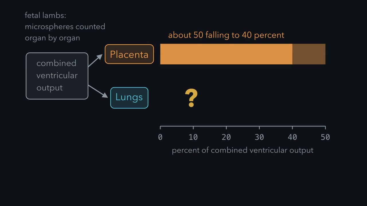
A fetus does not use its lungs to exchange gas, so where does its blood go? Researchers injected radioactive microspheres into fetal lambs and counted them organ by organ. The placenta took about half of the output in young lambs and about 40 percent near term. The lungs took a small share that rose with gestation. Those are sheep numbers from an instrumented preparation, and human proportions are inferred from imaging. Three shunts route fetal blood past the liver and the lungs; the fetal lungs are fluid filled, with high resistance. The ductus venosus carries a variable share of the umbilical vein blood past the liver, toward the inferior vena cava.
The foramen ovale, an opening in the atrial septum, passes a stream from the right atrium to the left. Most of the blood leaving the pulmonary artery crosses the ductus arteriosus into the aorta. Umbilical arteries carry blood back to the placenta, a low resistance bed in parallel with the fetal body. At birth, air fills the lungs and pulmonary vascular resistance falls. Pulmonary blood flow rises, and pulmonary venous return raises pressure in the left atrium. Once left atrial pressure exceeds right atrial pressure, the flap of septum primum is pressed against the septum. The foramen ovale then stops carrying a right to left stream. Losing the placenta removes a low-resistance circuit, so systemic vascular resistance and ventricular afterload rise.
Oxygen constricts the ductus arteriosus, shown directly on fresh human ductal rings studied outside the body. These endpoints do not arrive together. In 50 healthy term neonates followed by ultrasound, the ductus arteriosus had closed in 94 percent before day 3. Only 12 percent had a closed ductus venosus by then, and Doppler found foramen ovale flow in every infant. Instrumented preterm lambs, six in each group, were clamped before ventilation or ventilated before clamping. Clamping first dropped mean right ventricular output by about 66 percent. The group means fell from 114.6 to 38 .8 milliliters per minute per kilogram. Heart rate fell about 40 percent too.
Ventilating first gave stable flows and pressures. The reading is that, before aeration, umbilical venous return supplies much of the filling of the left ventricle. This is a mechanism in preterm lambs, with no human outcome. The study cannot separate a transfusion of blood volume from preload replacement. Aeration lets pulmonary venous return take over the filling, so the order decides whether preload falls as afterload rises.
In the minutes after birth, a newborn’s lungs go from taking a small share of the heart’s output to carrying nearly all that the right ventricle pumps.
Before birth the lungs are full of fluid, so the fetal heart sends most of its blood around them. Once the shunts close, everything the right ventricle pumps goes through them. What has to change for blood to make that turn?
The lesson builds the answer in order: the placenta, the mother who supplies it, the fetal circulation built around it, the swap at birth, then labour and milk. The ovarian cycle, fertilization and implantation come first, in ovarian cycle and fertility and animal reproduction.
The circulation runs on one rule, which needs one term. Beds in parallel are fed side by side from the same arteries and drain into the same veins, so they share one driving pressure.
The one idea to carry through this lesson. Blood divides among beds in parallel in proportion to how easily each one lets it through, and where two chambers or vessels connect, blood crosses from the higher-pressure side to the lower. In the fetus the placenta is the easy bed and the fluid-filled lungs the hard one, so the open shunts let blood go to the placenta and past the lungs. Birth swaps the two beds while the shunts are still open: the pressures across them reverse, the foramen ovale’s flap is pushed shut, and flow through the ductus arteriosus turns around until chemistry closes it. Once the shunts close, the lungs sit in series with the body and carry the whole output of the right ventricle, whatever their resistance.
The mother needs only the whole-circuit version, MAP = CO × systemic vascular resistance (SVR).
The rule does not explain why the mother’s vessels relax, what closes the ductus arteriosus, how the placenta moves solutes, or labour and milk. Those get their own short rules: progesterone holds the uterus quiet until its block is lifted, and prolactin fills the alveolus, oxytocin empties it.
The placenta: the fetus’s lung, gut and kidney
The placenta breathes, feeds and excretes for the fetus, without letting the two bloods mix. In the human placenta, fetal villi hang in a pool of maternal blood, the intervillous space. A villus is a branched tree of fetal capillaries in a core of connective tissue, covered by the syncytiotrophoblast, a continuous multinucleated layer with no gaps between cells. Maternal blood bathes the outside of the villus; fetal blood runs inside it. The two lie close together and normally do not mix, so everything that moves between mother and fetus crosses the syncytiotrophoblast and the fetal capillary wall.
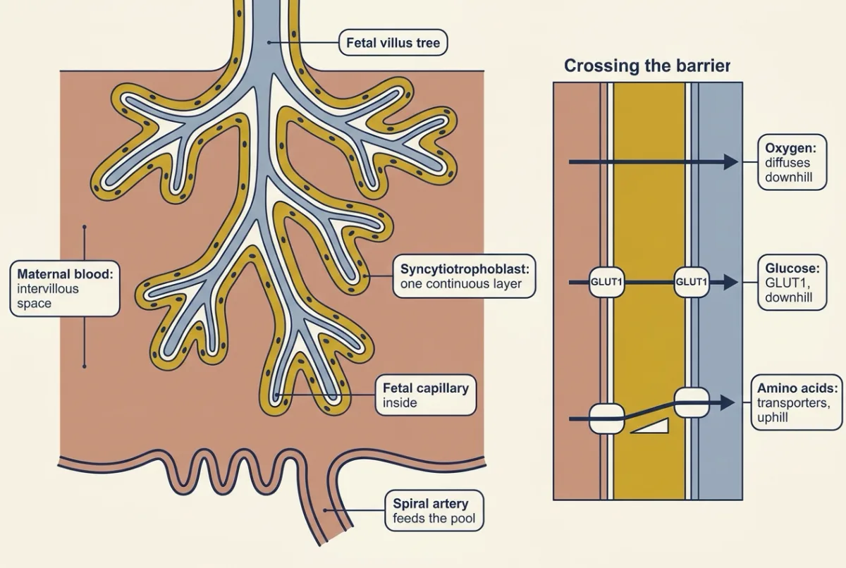
How each solute crosses depends on which way its gradient runs.
- Oxygen diffuses down its partial pressure gradient. On the far side, fetal hemoglobin holds oxygen more tightly at a given PO2 than adult hemoglobin does (oxygen transport).
- Glucose moves down a maternal-to-fetal concentration gradient by facilitated diffusion, mainly through the transporter GLUT1.
- Amino acids still move from mother to fetus, but against their gradient: several are more concentrated in fetal than maternal plasma. Transport proteins in both syncytiotrophoblast membranes carry them, two steps in series, first the membrane facing maternal blood, then the one facing the fetal capillary. Transfer also depends on blood flow on both sides and on the concentrations in each compartment.
So oxygen and glucose go downhill; several amino acids are carried uphill by transporters. The intervillous pool is not as still as diagrams draw it, either (Going deeper, “A placenta that squeezes”).
The placenta is also an endocrine organ
The placenta’s hormones each solve a problem of the pregnancy.
Keeping progesterone flowing. Early in pregnancy, human chorionic gonadotropin (hCG) from the trophoblast, the placenta’s outer cell lineage, supports the maternal corpus luteum, so progesterone production continues past the point where the luteum would otherwise regress (animal reproduction covers that handover). The placenta then becomes the main source, making progesterone from cholesterol precursors. Progesterone acts through its nuclear receptor (nuclear receptors) and promotes quiescence of the myometrium, the smooth muscle of the uterus wall (smooth muscle).
Making estrogens with partners. The placenta lacks the enzyme that turns progesterone-type steroids into androgens (17α-hydroxylase/17,20-lyase), so it cannot make the androgen precursors that estrogens are built from. It borrows them. It converts DHEAS, a weak androgen precursor from maternal and fetal adrenal glands, to estradiol, and fetal 16-hydroxylated DHEAS to estriol, as gestation advances. The arrangement is called the maternal-fetal-placental unit because no one tissue holds the whole route (adrenal glands).
Saving glucose for the fetus. In the second half of pregnancy the mother becomes insulin resistant, an adaptation that spares glucose for the fetus. Her tissues take up glucose less readily, so more stays in her blood for the fetus, and her insulin secretion rises to compensate, with an expansion of beta-cell mass that is large in rodents and more modest in women. Two hormones of the syncytiotrophoblast are the likely drivers. Human placental lactogen (hPL), structurally close to growth hormone and prolactin, antagonizes insulin action and, in mice, promotes expansion of maternal beta cells. Placental growth hormone replaces pituitary growth hormone in maternal blood from mid-gestation and is thought to be a major driver of the insulin resistance. If insulin secretion does not rise enough to match the resistance, blood glucose rises: that is gestational diabetes (insulin, glucagon and glucose). hCG, progesterone, estrogens and hPL are the four to know, and placental growth hormone the fifth to recognise; the placenta may make many more (Going deeper, “How many hormones?”).
The mother re-sets her circulation, breathing and kidneys
To supply a second body, the mother pumps more blood, breathes more and filters more. One fact looks odd: in healthy human pregnancy, cardiac output rises by around 40 percent, yet mean arterial pressure changes only slightly.
Predict. A made-up healthy woman at rest has a mean arterial pressure of 90 mmHg. In pregnancy her cardiac output rises 40 percent and her systemic vascular resistance falls 28 percent. Roughly where does her pressure end up? The card below gives the working.
In words Mean arterial pressure equals cardiac output times systemic vascular resistance, with right atrial pressure taken as zero.
- MAP“M A P”
- mean arterial pressuremmHg
- CO“C O”
- cardiac output, the flow out of the left ventricleL/min
- SVR“S V R”
- systemic vascular resistance, the resistance of all the body's beds in parallelmmHg·min/L
Sanity check Pressure changes by relative output times relative resistance: here 1.40 × 0.72, about 1.01, so pressure barely moves. Raise output alone and pressure rises in proportion.
Try one number
Before pregnancy, with cardiac output 5.0 L per min: SVR = 90 / 5.0 = 18.0 mmHg min per L.
In pregnancy: CO = 5.0 × 1.40 = 7.0 L per min, and SVR = 18.0 × 0.72 = 12.96 mmHg min per L.
MAP = 7.0 × 12.96 = 90.7, about 91 mmHg.
The reported fall in resistance is 25 to 30 percent. At the edges of that range, a 25 percent fall gives 94.5 mmHg and a 30 percent fall gives 88.2 mmHg.
Need a refresher? rates and “per” · units and prefixes
So a pressure measured at the arm hides both changes (blood pressure regulation). This is a first approximation: measured pressure does dip a little in the first and second trimesters and returns to nonpregnant levels in the third.
What lowers the resistance? Not mainly the placenta. Resistance falls in the first weeks, before the uteroplacental bed is large, through vasodilation across the mother’s body: nitric oxide and hormonal signals relax the vessels (relaxin’s role is shown best in animals). Even at term, the placental bed is thought to account for only part of the fall; vasodilation in other beds, such as the kidney and skin, accounts for the rest.
Blood volume. Plasma volume expands proportionally more than red-cell mass, so hemoglobin concentration falls by dilution rather than because red cells were lost (blood cells and hemostasis). Does that dilution cost oxygen delivery? Hematocrit is the fraction of blood volume taken up by red cells.
- Before: plasma 2.5 L and red cells 1.5 L give blood volume 4.0 L, hematocrit 1.5 / 4.0 = 37.5 percent.
- In pregnancy: plasma rises 44 percent to 3.6 L and red cells 20 percent to 1.8 L. Blood volume is 5.4 L, hematocrit 1.8 / 5.4 = 33.3 percent.
- Oxygen content: with hemoglobin per litre of red cells and arterial saturation unchanged, and dissolved oxygen ignored, content scales with hematocrit: 33.3 / 37.5 = 0.889.
- Oxygen delivery: relative output times relative content, 1.40 × 0.889 = 1.24.
Delivery is about 24 percent higher although content is about 11 percent lower.
- Pressure: output × resistance
- Blood volume: plasma + red cells
- Hematocrit: red cells ÷ blood volume
- Relative oxygen delivery
In this sketch (all of this is made up for the sketch unless the lesson gives it): the baselines are the lesson’s made-up round numbers for one healthy adult woman at rest, with cardiac output 5.0 L per min, mean arterial pressure 90 mmHg, resistance 18.0 mmHg min per L (90 ÷ 5.0), plasma 2.5 L and red cells 1.5 L (hematocrit 37.5 percent); right atrial pressure is taken as 0 mmHg; hemoglobin per litre of red cells is unchanged and dissolved oxygen is ignored, so oxygen content scales with hematocrit; the slider ranges, and any setting other than the lesson’s worked example, are choices made for the sketch. The lesson reports a cardiac output rise of around 40 percent and a fall in resistance of approximately 25 to 30 percent, and only supposes the plasma and red-cell rises. Real values are group values that vary with gestational age and method, and measured pressure dips a little in the first and second trimesters and returns to nonpregnant levels in the third, so this is a first approximation.
Rule to keep: pressure follows the product of output and resistance, so a 40 percent rise in output against a 25 to 30 percent fall in resistance nearly cancel, while hematocrit follows the share of blood volume that is red cells, so oxygen delivery can rise even though oxygen content per litre of blood falls. Resistance falls because the body's beds sit in parallel and many of them widen at once, mostly by vasodilation across the mother's body, such as in the kidney and skin, rather than in the placental bed alone; the same parallel-bed rule later steers the fetal circulation.
Breathing. Minute ventilation rises early, by up to about 48 percent in the first trimester, and stays raised. It rises through larger tidal volumes at an essentially unchanged respiratory rate. Progesterone raises the sensitivity of the respiratory centre to carbon dioxide (control of breathing), so arterial PCO2 settles at a plateau around 32 mmHg. Functional residual capacity falls as the diaphragm is displaced upward.
How much more breathing does that plateau take? In the steady state, arterial PCO2 is proportional to CO2 production divided by alveolar ventilation. Start at 40 mmHg and suppose CO2 production rises 20 percent. For PCO2 to settle at 32 mmHg, alveolar ventilation must rise by the factor 1.20 × 40 / 32 = 1.50, that is 50 percent. Check: 40 × 1.20 / 1.50 = 32 mmHg. (That is alveolar ventilation, not the measured minute ventilation; Going deeper, “Minute and alveolar ventilation”.) The payoff is for the fetus: a lower maternal PCO2 steepens the gradient that carries fetal CO2 across the placenta.
Kidneys. Glomerular filtration rate is already up to 40 to 50 percent higher in the first trimester. In pooled data from uncomplicated pregnancies, serum creatinine was lowest at 15 to 21 weeks, 23.2 percent below nonpregnant values (glomerular filtration).
Each of these numbers is a group value from healthy pregnancies that varies with gestational age and method.
Where fetal blood goes
The film at the top of the page, “The Lungs Take Over From the Placenta”, covers this section and the next.
Apart from the one-way flap of the foramen ovale, the fetal circulation has no valves to steer with. It steers mainly by resistance, helped by streaming in the right atrium that aims the best-oxygenated stream at the foramen ovale.
Picture the fetal arteries feeding three beds in parallel: the body, the placenta and the lungs. The blood divides among them in proportion to how easily each one lets it through. The placenta is a wide, low-resistance bed. The fluid-filled lungs, whose small arteries are held constricted by low oxygen and compressed by lung liquid, are a high-resistance bed. In fetal lambs about half of the combined output of both ventricles goes to the placenta when the fetus is young and about 40 percent near term, and only a small share goes to the lungs, a share that rises with gestation (Going deeper, “Counting where the blood goes”). Human fetuses send their lungs a larger share, but the pattern is the same.
Three short cuts make that split work. Follow a red cell back from the placenta.
- Ductus venosus. Oxygenated blood returns in the umbilical vein. A variable share of it runs through the ductus venosus, past the liver, toward the inferior vena cava; the rest perfuses the liver.
- Foramen ovale. In the right atrium, the well-oxygenated stream from the ductus venosus and inferior vena cava is aimed at the foramen ovale, an opening in the wall between the atria, and much of it crosses into the left atrium. So relatively well-oxygenated blood reaches the left ventricle. A flap on the left side of the opening lets blood through only from right to left, like a door that opens one way. (The film calls this flap the septum primum.)
- Ductus arteriosus. Blood that reaches the right ventricle is pumped into the pulmonary artery. There it meets the high resistance of the lungs, and most of it takes a short vessel, the ductus arteriosus, into the aorta. The ductus is held open mainly by prostaglandin E2, much of it made by the placenta and some by the ductal wall itself, with nitric oxide also contributing.
Umbilical arteries then carry blood back to the placenta, the low-resistance bed in parallel with the fetal body.
Predict. The first branches of the aorta feed the heart muscle and the head, and the ductus arteriosus joins the aorta after those branches. Which blood does the brain mostly get?
Check your prediction
The better-oxygenated stream that crossed the foramen ovale and was pumped out by the left ventricle. The less-oxygenated blood from the right ventricle enters the aorta through the ductus, downstream of the head and neck branches, and heads to the lower body and back to the placenta. The layout gives the best blood the fetus has to the heart and brain.
At birth the lungs and the placenta trade places
Two resistances change within minutes.
- The lungs open. The first breaths fill the lungs with air, the air and the rise in oxygen relax the small pulmonary arteries, and pulmonary vascular resistance falls several-fold. Pulmonary blood flow rises.
- The placenta goes. Losing the placental bed removes a low-resistance circuit, so systemic vascular resistance and ventricular afterload, the pressure the ventricles pump against, rise.
The lungs are now the easy path and the body the harder one. Apply the rule.
Predict. For the first hours the ductus arteriosus is still open. Which way does blood flow through it now?
Check your prediction
Mostly the other way, from the aorta into the pulmonary artery. On the first day flow is often both ways (in half of healthy term babies in one Doppler study), while pulmonary pressure is still close to systemic. As pulmonary resistance falls below systemic, aortic pressure exceeds pulmonary artery pressure, and the open ductus carries blood from higher pressure to lower. Pulmonary resistance has to fall below the systemic side, not merely fall, and losing the placenta helps by raising that side.
Now the atria. Far more blood flows through the lungs and returns to the left atrium, so left atrial pressure rises. The umbilical return that used to help fill the right atrium has stopped, so right atrial pressure falls. Once left atrial pressure exceeds right, the one-way flap is pressed against the opening and the foramen ovale stops carrying a right-to-left stream. A pressure reversal is all it takes to stop the right-to-left flow through this shunt.
A pressure reversal does not close the ductus arteriosus; it only turns its flow around. The ductus closes itself. Its smooth muscle constricts as the blood in it becomes well oxygenated (smooth muscle), and the prostaglandin E2 that held it open falls, because the placenta that made much of it is gone and the newly perfused lungs break it down. The film gives oxygen alone; the fall in prostaglandin E2 is the other half (Going deeper, “Closing the ductus”). The ductus venosus, with no umbilical flow left to carry, closes too, more slowly.
| Fetus | After the first breaths | |
|---|---|---|
| Easiest bed (shunts still open) | placenta | lungs |
| Pulmonary vs systemic resistance | pulmonary higher | pulmonary lower |
| Ductus arteriosus | pulmonary artery → aorta | aorta → pulmonary artery (often both ways on day 1), then constricts |
| Atrial pressures | right higher | left higher |
| Foramen ovale | open, right to left | flap pressed shut |
The closures run on different clocks. In healthy term babies the ductus arteriosus mostly stops carrying flow before day 3. The ductus venosus stops by day 18. The foramen ovale’s flap is pressed shut but not yet sealed, so Doppler still finds some flow across it, as the film says. After the first hours most of that is a small left-to-right leak past the flap’s edge rather than the fetal right-to-left stream, though brief right-to-left flow can occur early or when the baby cries (the study in Going deeper, “The closure tallies”, recorded flow, not its direction). Stopping flow is not the same as fusing: the tissues fuse into ligament or scar later still, and in about a quarter of adults the foramen ovale never fully seals.
The order of the two changes matters. If the cord is clamped before the lungs are aerated, the likely reading is that the heart loses the umbilical venous return that supplied much of its filling (its preload) before pulmonary venous return can take over; the experiment cannot separate this from a lost transfusion of blood volume. In preterm lambs, right ventricular output fell by about two-thirds. Aerate first and output largely holds. These are preterm lambs, with no human outcome (Going deeper, “Clamp first or breathe first”, which also gives the film’s numbers).
Predict. In persistent pulmonary hypertension of the newborn, pulmonary vascular resistance fails to fall after birth. Run the rule backwards: what happens at the foramen ovale and the ductus arteriosus?
Check your prediction
Both shunts keep working the fetal way. Pulmonary blood flow stays low, so pulmonary venous return and left atrial pressure stay low. Right atrial pressure can remain the higher of the two, so the foramen ovale flap is not held shut and flow continues right to left. Pulmonary artery pressure can stay at or above aortic pressure, so the ductus arteriosus keeps carrying blood toward the aorta. Systemic arterial PO2 stays low, removing one stimulus that constricts the duct.
Labour: lifting progesterone’s block
The short rule: progesterone holds the uterus quiet until its block is lifted. Labour needs that block lifted and the muscle made ready.
How the block is lifted differs between species. In humans circulating progesterone stays elevated until birth, so the withdrawal that releases the myometrium is thought to be functional or local rather than systemic, and no single human trigger is established. Sheep, where parturition is better understood, differ: a rise in fetal adrenal cortisol near term shifts the placenta’s steroid output away from progesterone and toward estrogens and prostaglandins, so progesterone itself falls.
Near term several signals make the myometrium ready and drive it.
- Estrogens are thought to prepare the muscle. Near term the myometrium gains oxytocin receptors and gap junctions, which link its cells so they contract together.
- Prostaglandins contract the myometrium and soften the cervical stroma.
- Oxytocin drives a positive-feedback loop. Stretch of the cervix and vagina can release oxytocin, oxytocin increases uterine contraction, and stronger contraction increases stretch (tissues and homeostasis sets out that vocabulary).
Oxytocin helps, but in mice, at least, labour does not depend on it: mice lacking oxytocin delivered without obvious deficits (Nishimori and colleagues, 1996).
Milk: prolactin fills, oxytocin empties
Milk has two hormones with two jobs, and students reverse them.
Prolactin, from the anterior pituitary, supports secretion of milk by the alveolar cells of the breast (endocrine axes and testing covers its dopamine control). Oxytocin, released from the posterior pituitary by magnocellular hypothalamic neurons (hypothalamus and autonomic control), contracts the myoepithelial cells wrapped around each alveolus and ejects milk already stored. Suckling is the main afferent limb for both, and infant cues such as crying can also trigger oxytocin release.
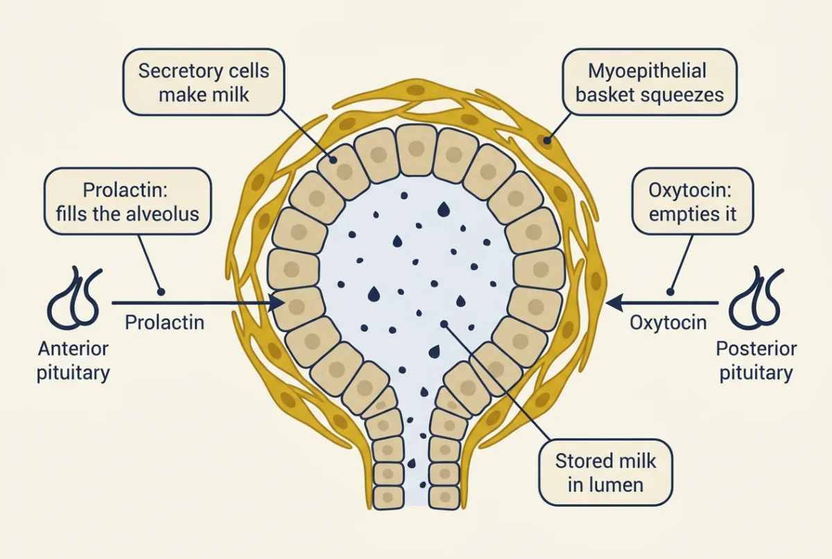
The same mice fit “oxytocin empties”: they fail to nurse their pups, because the milk is made but not ejected.
Predict. Prolactin rises steadily through pregnancy and is high by late pregnancy. Why is there no full milk production until after birth?
Check your prediction
Progesterone appears to hold it back. While the placenta is in place, progesterone restrains the switch to full milk production. After birth the placenta is gone, and the fall in progesterone, with prolactin maintained, permits the switch, two to three days after birth. Physiologists call this switch secretory activation. The same hormone that keeps the uterus quiet keeps the breast waiting.
The lesson in one paragraph. The mother’s pressure barely moves because her resistance falls to about 0.72 of its old value, close to the reciprocal of the 1.4-fold rise in her output. While the shunts are open, the fetus favours the easier parallel bed, the placenta, over its lungs. At birth the two trade places: the foramen ovale’s flap is pressed shut and ductal flow turns mainly aorta to pulmonary artery before oxygen and falling prostaglandin E2 close the duct, leaving the lungs in series with the body. Then progesterone’s block lifts for labour, prolactin fills the alveolus and oxytocin empties it.
Design problem
In the preterm-lamb experiment, aerating the lungs before clamping the cord largely preserved right ventricular output (a fall of about 22 percent against about 66 percent). Two mechanisms could explain that: pulmonary venous return supplies replacement preload, or the placenta transfuses blood volume into the lamb during the extra interval. Using instrumented lambs, design the measurements and groups that would separate them, and say what each explanation predicts.
One way to do it
Record continuously: pulmonary arterial, ductal and carotid flow, arterial and central venous pressure, and umbilical flow before clamping. Add a blood-volume measure, such as dilution of a tracer that stays in the vascular space, plus serial hematocrit.
Groups, gestation-matched and randomised: clamp then ventilate; ventilate then clamp; ventilate first with the umbilical vein occluded for the same interval, so no placental blood can return (if the arteries stay patent, fetal blood can drain into the placenta, so read the volume measure with that in mind); and clamp first, then give a measured transfusion before ventilation. No single group settles it; the comparison across groups does.
Predictions. If replacement preload does the work, the ventilate-first and vein-occluded groups both show early pulmonary flow and largely preserved output, and transfusion without aeration preserves output only while it runs. If transfusion does the work, the vein-occluded group loses much of the benefit, the intact ventilate-first group shows a measurable rise in blood volume, and transfusion alone preserves output without aeration.
What would ruin it: unmatched gestational age, unequal aeration, and hematocrit alone as a volume measure, since fluid shifts change it with no transfusion. The limit stands: preterm lambs, a mechanism, not a practice.
Going deeper
The main line above does not depend on these details.
Counting where the blood goes
The classic measurements of the fetal layout were made in fetal lambs. Rudolph and Heymann, working in the 1960s, catheterized fetal lambs inside the uterus and injected radioactively labelled microspheres sized to lodge in the first capillary bed they reached, then counted radioactivity organ by organ; the count in an organ is proportional to the share of cardiac output it received at that moment. The placenta took a large share, the lungs only a small one, and substantial streams crossed the foramen ovale and the ductus arteriosus; the main text gives the shares. Those are sheep numbers from an instrumented preparation. Human proportions differ; the film says they are inferred from imaging. Doppler measurements put the lung share at about 13 percent of combined output at 20 weeks, rising to about 25 percent by 30 weeks (Rasanen and colleagues, 1996). Sources: Rudolph and Heymann, Circulation Research 21 (1967) 163-184; Rudolph and Heymann, Circulation Research 26 (1970) 289-299.
A placenta that squeezes
Magnetic resonance imaging of human pregnancies in utero (Dellschaft and colleagues, 2020) found net blood speeds in some placental regions of around 0.04 cm per second in the healthy group (between faster jets of inflowing blood), and dynamic scans showed local contractions of the placental bed expelling maternal blood from the intervillous space. A net velocity from imaging is not volumetric perfusion, and no benefit to the fetus was shown.
The evidence on hPL
hPL antagonizes insulin action: in an early study in adult volunteers, not a pregnancy cohort, twelve-hour infusions of hPL impaired glucose tolerance in seven of eight people tested. In mice, prolactin and placental lactogen act through the prolactin receptor on maternal beta cells, and deleting that receptor from beta cells left beta-cell mass late in pregnancy (gestational day 16.5) about half that of controls, though it still rose.
How many hormones?
The four-hormone picture is an abridgement. Single-cell RNA sequencing of first- and second-trimester human placental cells (Liu and colleagues, 2018) distinguished fourteen cell subtypes and detected expression of 102 polypeptide-hormone genes. A transcript is not a secreted hormone, so read that number as a map of what the organ might make.
Closing the ductus
Prostaglandin E2 holds the fetal ductus arteriosus open predominantly by acting on EP4 receptors, evidence from mouse studies. It circulates high in the fetus partly because little blood passes through the lungs that clear it, evidence largely from fetal lambs. Oxygen constricts the ductus arteriosus, shown directly on fresh human ductal rings studied ex vivo (Michelakis and colleagues, 2002). The 26 ducts came from infants about nine days old, and the experiment shows how isolated tissue responds to oxygen rather than the order of events in a newborn. The prostaglandin half of closure has a clinical mirror: drugs that block prostaglandin synthesis, such as indomethacin and ibuprofen, are used to close a ductus that stays open, and prostaglandin E1 is used to keep one open.
The closure tallies
Fifty healthy term neonates were examined repeatedly by ultrasound over their first 18 days (Fugelseth and colleagues, 1997). The ductus arteriosus had closed in 94 percent before day 3; on day 1 its flow was bidirectional in 26 of the 50. The ductus venosus had closed in only 12 percent by then, and in all of them before day 18. Doppler detected flow across the foramen ovale in every infant. Fusion of the foramen ovale is never completed in a sizeable minority: an autopsy study of 965 normal hearts found it still probe-patent in about 27 percent (Hagen and colleagues, Mayo Clinic Proceedings, 1984).
Clamp first or breathe first
These are the film’s numbers (Bhatt and colleagues, 2013). Preterm lambs were delivered by caesarean section at 126 plus or minus 1 days of gestation, with flow probes on the pulmonary artery, the carotid artery and the ductus arteriosus. Two orders of events were compared in six lambs each: clamping the cord before ventilation, or starting ventilation before clamping.
Clamping first dropped mean right ventricular output from 114.6 plus or minus 14.4 to 38.8 plus or minus 9.7 mL per min per kg (group means plus or minus SEM) before ventilation began, a fall of about 66 percent. Heart rate was also down by about 40 percent, so the fall in output is not purely a stroke-volume effect, and afterload rose at the same moment. Ventilating first gave stable flows and pressures across the transition. The design problem above asks how to separate preload replacement from a transfusion of blood volume.
Minute and alveolar ventilation
Alveolar ventilation is not minute ventilation, since part of each breath ventilates dead space. With dead space roughly fixed, deeper breaths raise alveolar ventilation proportionally more than minute ventilation. The 20 percent rise in CO2 production in the worked example is a made-up round number, so the worked 50 percent (alveolar) is illustrative; it and the measured 48 percent (minute) describe different quantities and are not meant to match.
Your notes
Select any text in the lesson to highlight it or add a note to it. Your highlights and notes are saved in this browser and collected in your notebook.
This browser is not letting the site save anything (a private window or strict privacy settings), so highlights and notes will be lost when you leave the page.
How we measure it
Radioactively labelled microspheres
Inject particles sized to lodge in the first capillary bed they meet, each batch carrying a different label, then count radioactivity in each organ afterwards. The count in an organ is proportional to the share of the cardiac output it received at the moment of injection, which let the flow through the fetal shunts be measured in the intact fetus inside the uterus.
Implanted flow probes and arterial catheters
Place ultrasonic flow probes around named vessels and catheters in arteries before the event you want to study, then record flow and pressure continuously through it. In lambs this resolves the minute-by-minute changes around the first breaths, which single measurements before and after would miss.
Serial Doppler ultrasound of the newborn
Return to the same infants on successive days and look for flow through each shunt with pulsed and colour Doppler. Because it is repeated and non-invasive, it separates the dates on which different shunts stop carrying flow, which a single examination cannot.
Velocity-sensitive and dynamic placental MRI
Image the placenta in the uterus with sequences sensitive to the motion of blood, and repeat them over time. This gives regional net blood speed and shows changes in placental shape and intervillous content, with the limit that a net velocity is not the same quantity as volumetric perfusion.
Practice questions on this lesson (5) → MCAT-style, original, each with an explanation
Where this lesson fits
Before thisReview first
- Cardiac Output and Venous ReturnThis course · Lesson 7
Maternal output rising 40 percent and the preload lost when the cord is clamped both rest on venous return.
- The Ovarian Cycle and FertilityThis course · Lesson 28
The corpus luteum and progesterone that hCG rescues in early pregnancy come from this lesson.
After thisStudy next
- Exercise: The Whole Body at WorkThis course · Lesson 30
It uses the same chain of output, oxygen delivery and ventilation to ask which link limits the whole body.
Elsewhere on the siteFilms, simulations and method films in other courses
- Why Blood Needs an S-Shaped CarrierFilm · Organismal Biology
Fetal oxygen supply relies on hemoglobin carrying oxygen and releasing it where tissues need it.
- Radioactive and stable isotope tracersMethod film
The placenta lesson's transfer of oxygen, glucose and amino acids is measured by following labelled molecules across it.
Suggested by an AI model from the lessons themselves, then checked. How we use AI
Finish line
Test yourself
12 questions
Practice and MCAT-style, with explained answers.
Next · Lesson 30
Exercise: The Whole Body at Work
At maximal exercise a healthy young adult takes up ten or more times the oxygen used at rest. Which link in the chain sets the ceiling?
Continue →