Organismal Biology · Unit 5: Defense, movement and reproduction
Animal Reproduction
Rewritten on : new text, flashcards and film. Your notes and highlights stay, but the text around them may have moved.
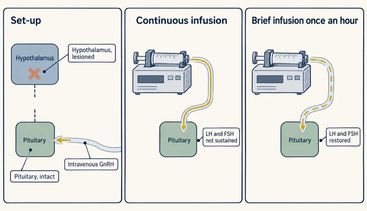
A monkey's pituitary, cut off from the brain's drive, was given its missing hormone either as a steady drip or once an hour, the drip giving at least as much. Only one schedule kept it working. Why would timing matter?
Pause when the reading reaches a film, and offer to play it. Saved on this device.
Watch the film now, or keep listening
Builds on:Hormones and the Endocrine SystemTissues, Organs and HomeostasisMeiosis and VariationWhy these?
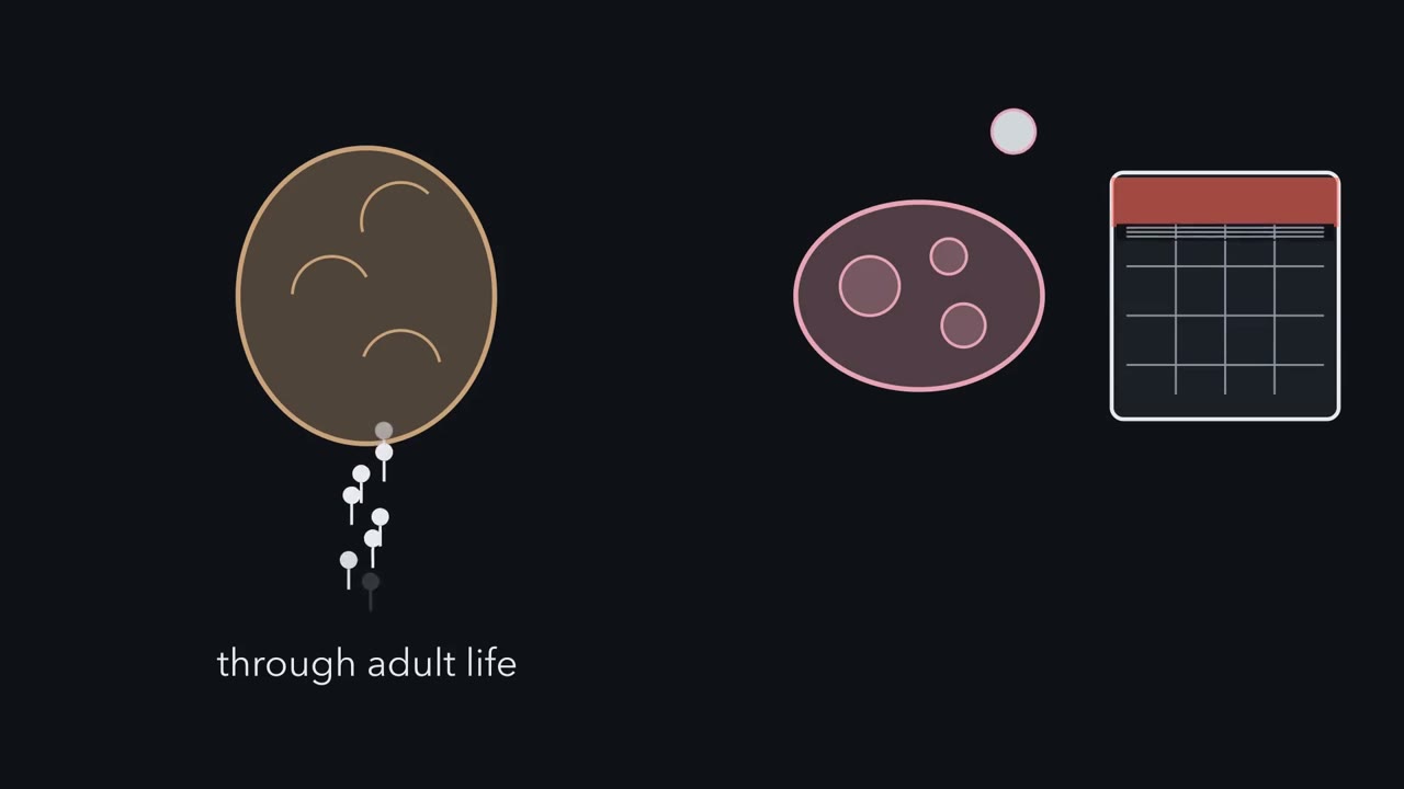 ▶ Watch A Loop That Keeps Time · 3:21
Read the transcript of “A Loop That Keeps Time”
Film transcript: A Loop That Keeps Time
A man's testes keep making sperm through adult life. A woman's ovaries usually release one egg per cycle. What keeps the gonads on schedule? The control sits at three levels, stacked top to bottom. The hypothalamus is at the base of the brain. The pituitary gland hangs just below it. Then come the gonads, the testes or ovaries. Neurons in the hypothalamus release a hormone called GnRH, gonadotropin-releasing hormone. Portal vessels carry it down to the pituitary. And it arrives in pulses, not a steady stream. The pituitary answers with two hormones of its own, LH and FSH. They travel in the blood to the gonads. Because they act on gonads, they are called gonadotropins.
Follow them into a testis. LH reaches Leydig cells, which sit between the sperm-making tubules. They make testosterone. FSH reaches Sertoli cells, which line the tubules and support the developing sperm. Now the loop closes. Testosterone travels back up and lowers the output of the hypothalamus and the pituitary. When a product turns down its own pathway, that is negative feedback. Think of it as a brake. With the brake on, LH and FSH fall. When testosterone drops, the brake eases, and LH and FSH climb back up. So here is a test of the loop.
Remove the testes. What happens to LH and FSH in the blood? Do they go up, or down? Both go up. With the testes gone, their testosterone is gone, so the main brake is off. The pituitary keeps driving, and LH and FSH climb. FSH has a second brake of its own. Sertoli cells release a hormone called inhibin B, which lowers FSH. Remove the testes, and that brake goes too. The ovary runs on the same loop. There the brake comes from the follicle, the sac of cells around a growing egg. Its hormone, estradiol, brakes the loop through most of the cycle's first half. But when estradiol stays high for long enough, the brake becomes an accelerator. After ovulation, progesterone brakes instead.
The lesson shows how. One thing is still odd. GnRH reaches the pituitary in bursts, not a stream. Is the burst just how it travels, or is it the message? So the brain drives the gonads in pulses, through the pituitary. And the gonads' hormones come back up as a brake, almost all the time.
From the film
- The hypothalamus releases GnRH in pulses, and the pituitary answers with two hormones, LH and FSH, that act on the gonads.
- In the testis, LH reaches Leydig cells, which make testosterone, and FSH reaches Sertoli cells, which support the developing sperm.
- Testosterone brakes the hypothalamus and pituitary, and inhibin B from Sertoli cells brakes FSH, so removing the testes makes LH and FSH rise.
- In the ovary, estradiol from the growing follicle brakes the loop through most of the cycle's first half; when it stays high for long enough, it becomes an accelerator. After ovulation, progesterone brakes.
A rhesus monkey whose hypothalamus can no longer drive her pituitary gets the missing hormone, gonadotropin-releasing hormone (GnRH), by pump, as a steady drip or an hourly burst, the drip giving at least as much. Only one schedule works. The first film builds her loop; the second runs the test.
The one idea. A mammal’s gonads (testes or ovaries) run on a loop that the brain times. The hypothalamus sends gonadotropin-releasing hormone (GnRH) to the pituitary in pulses, and the pituitary sends luteinizing hormone (LH) and follicle-stimulating hormone (FSH) to the gonads. The gonads’ hormones come back as a brake, except when estradiol from a growing ovarian follicle stays high for long enough: then it becomes an accelerator and sets off the LH surge that triggers ovulation. Whether the pulses themselves carry the message is what the second film tests.
Scope: the brake is simplified for a first course. In the testis, testosterone brakes the hypothalamus and pituitary, and inhibin B brakes FSH; in the ovary, estradiol brakes through most of the follicular phase and progesterone after ovulation. The timing result comes from rhesus monkeys.
Making sperm and eggs
Gametes are haploid sex cells, cells with one set of chromosomes. In fertilization two gametes, usually from two parents, join to give a diploid zygote with two sets, which is sexual reproduction. Meiosis, the division that halves the chromosome number and reshuffles the parents’ alleles (versions of a gene), makes gametes; see chromosomes and meiosis.
Spermatogenesis happens in the seminiferous tubules, coiled tubes inside each testis. Germ cells mature as they move from the tubule wall toward its centre:
- Spermatogonia (diploid germ cells, 46 chromosomes in humans, among them the stem cells of the line) divide by mitosis at the wall, renewing themselves through adult life and feeding cells into meiosis.
- A primary spermatocyte (46 chromosomes) completes meiosis I, which separates the paired chromosomes, to give two secondary spermatocytes, 23 chromosomes each, each still made of two sister chromatids: copies made by DNA replication, though crossing over in meiosis I may have made them no longer identical.
- Meiosis II separates the chromatids, giving four spermatids, each with 23 single chromosomes.
- Spermiogenesis remodels each spermatid into a sperm with a head and a tail.
The first film’s Leydig and Sertoli cells sit beside this path, and Sertoli support relies on testosterone’s help. Sperm then mature in the epididymis, a coiled duct on the testis. Spermatogenesis in the testis takes roughly 64 to 74 days by different estimates (Going deeper).
Oogenesis in human females starts before birth:
- In the fetal ovary, oogonia divide by mitosis and become primary oocytes (46 chromosomes), which enter meiosis I and arrest in prophase I.
- Each waits, for years or decades, in a follicle, the oocyte plus the supporting cells around it.
- After puberty, usually one follicle per cycle matures. Its oocyte finishes meiosis I shortly before ovulation, set going by the luteinizing hormone (LH) surge. The division is unequal: nearly all the cytoplasm goes to the secondary oocyte, and the other chromosome set leaves in a small polar body.
- The secondary oocyte, the egg of everyday speech and of the first film, is released arrested in metaphase II. It finishes meiosis II only if fertilized, giving the egg proper and a second polar body.
Adult human ovaries are not known to make new oocytes, though negative evidence cannot prove absence (see the anecdote on egg stem cells). The testis renews its stock and makes four gametes per meiosis; the ovary spends a stock laid down before birth, one gamete per meiosis.
Worked problem. The inputs are invented for practice. (a) Suppose 120 human primary spermatocytes complete both meiotic divisions. Each gives 4 spermatids: 120 x 4 = 480 spermatids. (b) Suppose a hypothetical 75 percent survive maturation into sperm: 480 x 0.75 = 360 sperm, so 480 - 360 = 120 are lost. This counts cells in the testis, not a semen sample.
Predict. Now suppose 120 human primary oocytes each completed both divisions. How many eggs would result, and where does the rest of the chromosome material go?
Check your prediction
120 eggs, one per oocyte. The other chromosome sets leave in the polar bodies, two or three per oocyte (the first polar body may or may not divide again). This is idealized: in humans meiosis II finishes only on fertilization. Both sexes halve 46 chromosomes to 23.
In this sketch: invented inputs; adult human; schematic cells, not to scale. The sperm count is of cells in the testis, not a semen sample. The oocyte count is idealized, because in humans meiosis II finishes only on fertilization.
Rule to keep: the halving from 46 to 23 is the same in both sexes; the yield per meiosis is four against one.
Gametes with a chromosome too many or too few are covered in chromosome changes.
After puberty, LH and follicle-stimulating hormone (FSH), released as gonadotropin-releasing hormone (GnRH) reaches the pituitary in bursts, keep spermatogenesis going and drive each cycle’s final follicle growth and ovulation. Is the burst itself the message? The second film follows the 1978 experiment that tested it.
Read the transcript of “Pulses, Not Amount”
Film transcript: Pulses, Not Amount
Same hormone, same monkey. Could the timing of a signal matter as much as its amount? The first film showed GnRH reaching the pituitary in pulses. This film asks whether the pulse is the message. Ernst Knobil's laboratory worked with female rhesus monkeys. Damage to the hypothalamus, called a lesion, had stopped their LH and FSH. But the pituitary itself was still intact. The design is simple. Remove the animal's own source of a hormone. Then give it back from outside on different schedules, with the steady one giving at least as much in total. It also needed a clean supply of the hormone. GnRH had just been isolated. Pure synthetic GnRH made this kind of experiment possible.
So the team could give GnRH through a vein. They read the pituitary's answer in blood samples. With no drive from above, the LH record started flat. First, brief infusions, once every hour. The pump gives a short burst, then stops, then bursts again. Hourly pulses restored LH and FSH secretion, and kept it going. Now switch the pump to a steady stream. Set it high enough that each day it delivers at least as much GnRH as the pulses. Will LH stay up, rise higher, or fall away?
It falls away. The steady supply did not sustain LH and FSH. If the pituitary counted only the total, the steady drip would have done at least as well. It did not. Then the team went back to hourly pulses. The authors reported that secretion came back. So the pituitary had not been damaged. It could still answer, when the timing was right. So the pituitary reads the timing of GnRH, not only its total. Hourly pulses keep it working. A steady supply, even a larger one, did not. This experiment has limits. It shows the rule in these monkeys. It does not show where the pulses come from, or what interval between pulses is normal in people. Nor does it show why a steady supply fails.
From the film
- Knobil's laboratory gave GnRH by pump to rhesus monkeys whose hypothalamus could no longer drive the pituitary.
- Brief infusions once an hour restored LH and FSH; a steady infusion, at rates giving at least as much GnRH each day, did not sustain them.
- Returning to hourly pulses brought secretion back, so the pituitary itself was still able to respond.
- In these monkeys the pituitary reads the timing of GnRH, not only its total; where the pulses come from, and why a steady supply fails, are separate questions.
The axis and its pulses
What the second film left out:
- The doses. The steady drip ran at one of four rates; at the two highest it delivered as much gonadotropin-releasing hormone (GnRH) as the pulses or more, so too little hormone cannot explain the failure.
- Where the pulses come from. In mice, kisspeptin neurons in the hypothalamus drive them (Going deeper); in primates that is unsettled. Nor did the experiment show what pulse interval is normal in humans.
- Why a steady supply fails. The usual answer is desensitization: under constant GnRH, the pituitary cells that make luteinizing hormone (LH) and follicle-stimulating hormone (FSH) turn their response down. The experiment did not identify the mechanism (Going deeper).
The whole loop is the hypothalamic-pituitary-gonadal axis (portal route: hypothalamus and autonomic control). LH and FSH, the gonadotropins, act through receptors of the family in G-protein-coupled receptors, and the first film’s brakes are negative feedback (the endocrine system).
The cycle and pregnancy
In human females the axis runs two linked cycles: the ovarian cycle (the follicles) and the uterine cycle (the uterine lining, or endometrium). The one idea again: the gonads’ hormones brake the loop, except when estradiol stays high long enough.
In the follicular phase, follicle-stimulating hormone (FSH) recruits a group of follicles and usually one becomes dominant. Luteinizing hormone (LH) drives theca cells to make androgens, and FSH-supported granulosa cells convert them to estradiol. Rising estradiol rebuilds the endometrium after menstruation (the proliferative phase) and, for most of this phase, brakes the axis.
When estradiol stays high late in the phase, its effect switches to positive feedback, in which a signal boosts its own trigger. LH release rises steeply in the LH surge, which sets the oocyte finishing meiosis I and triggers ovulation, the release of the secondary oocyte. Here the timing that matters is how long estradiol stays high, a different signal from the gonadotropin-releasing hormone (GnRH) pulses in the monkeys.
After ovulation, in the luteal phase, the emptied follicle becomes the corpus luteum, secreting progesterone and estradiol. Progesterone converts the thickened endometrium into a lining that can receive an embryo, the secretory phase, and it brakes LH and FSH again. Estradiol and progesterone act mainly through receptors inside the cell (nuclear receptors). If no embryo implants, the corpus luteum regresses, progesterone falls, and the lining breaks down in menstruation, which begins the next cycle.
| One time axis | Follicular phase | Around ovulation | Luteal phase |
|---|---|---|---|
| Ovary | One follicle becomes dominant | LH surge; oocyte released | Corpus luteum, regresses without an embryo |
| Feedback | Estradiol brakes | Sustained high estradiol accelerates | Progesterone brakes |
| Uterus | Menstruation, then proliferative phase | Lining thick | Secretory phase, until progesterone falls |
The names pair up: the follicular phase spans menstruation and the proliferative phase, and the luteal phase is the secretory phase.
The textbook 28-day cycle with day-14 ovulation is a drawing convention: in a large, self-selected app-based study (Bull and colleagues), cycles averaged near 29 days, luteal phases near 12, and lengths varied widely (numbers in the anecdote).
If an embryo implants, its outer cell layer (the trophoblast, which forms much of the placenta) secretes human chorionic gonadotropin (hCG). Acting through the LH receptor, hCG keeps the corpus luteum making progesterone, so the lining is not shed. From about weeks 7 to 9 of gestation, the placenta itself becomes the main source of progesterone. Placental exchange of oxygen and antibodies is in oxygen transport and adaptive immunity.
Predict. Suppose the corpus luteum were lost in week 5 of one pregnancy and in week 12 of another. Which pregnancy loses its main progesterone source, and why?
Check your prediction
The week-5 pregnancy. Before about weeks 7 to 9, progesterone still comes mainly from the corpus luteum, kept alive by hCG, so losing it removes the main source of the hormone that holds the lining. By week 12 the placenta has become the main source.
The same job across animals
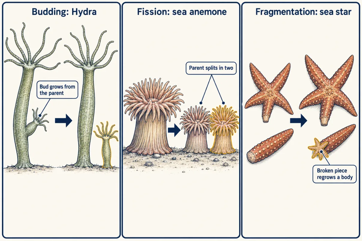
Asexual reproduction makes offspring from one parent without fertilization, so the young carry that parent’s genes. In budding a new individual grows out of the parent, as in Hydra; in fission the parent splits in two, as in some sea anemones and flatworms; in fragmentation a piece breaks off and regrows a whole body, as in some sea stars. In parthenogenesis an egg develops without being fertilized.
Sex carries what John Maynard Smith called the twofold cost of males. Suppose, in a made-up population, every female leaves two surviving young. An asexual female’s two are both daughters, and each will bear young; a sexual female’s are on average one daughter and one son, and the son bears no young himself. Counting only females and starting from one of each, asexual females go 1, 2, 4, 8 over three generations, while each sexual female leaves one daughter, so sexual females stay at 1.
Predict. Carry the tally one more generation, all else equal. What share of the females is asexual?
Check your prediction
16 asexual females to 1 sexual, so 16 of 17, about 94 percent.
Sex usually also means finding a mate, a cost asexual reproduction avoids; it offers new combinations of alleles. Why sex persists despite its costs is still argued; see life histories and selection in ecological context.
Some animals use both: water fleas (Daphnia) and many aphids make generations of females by parthenogenesis, then males and sexual eggs, often as the season turns. Hermaphrodites make both eggs and sperm. Earthworms make both at the same time, and two worms usually exchange sperm. Some fish change sex: clownfish start as males and the largest can become female, while many wrasses change from female to male. How sex is determined is in sex linkage and beyond.
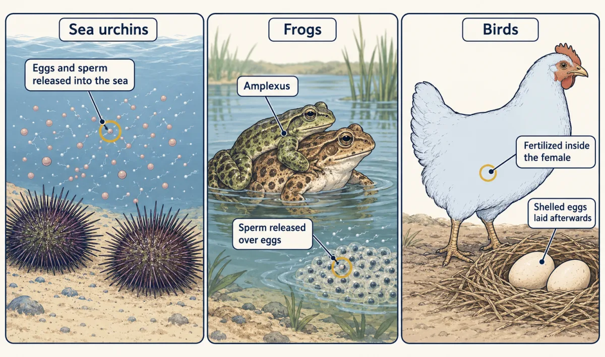
External fertilization happens outside the female’s body, usually in water. Sea urchins broadcast-spawn, releasing eggs and sperm into the sea, where many gametes never meet and eggs and larvae are exposed to predators. In most frogs, the male clasps the female (amplexus) and releases sperm over her eggs as she lays them.
Internal fertilization happens inside the female’s body. Sperm arrive close to the eggs, and on land the gametes are kept from drying out. Birds fertilize internally and then lay shelled eggs. Most mammals are viviparous (the young develop inside the mother and are born live), yet monotremes such as the platypus lay eggs, and some sharks and lizards bear live young.
Most mammals have estrous cycles instead of menstrual cycles: the endometrium is generally reabsorbed rather than shed, and females are usually receptive mainly around ovulation. Menstruation is known in humans, apes and Old World monkeys, and a few other mammals, such as some bats and the spiny mouse. The embryo’s next steps are in animal development, and the plant counterpart is plant reproduction.
Design problem
Lutes and colleagues (Nature, 2010) stained oocyte nuclei with DAPI, a fluorescent dye that binds DNA: chromosomes of the parthenogenetic Aspidoscelis tesselata had about 2.2 times the volume of those of the sexual A. gularis, which the authors read as a doubling of DNA. In body cells (nucleated red blood cells), DNA content differed by less than 1 percent. In another parthenogen, A. neomexicana, FISH probes (fluorescent in situ hybridization: fluorescent probes that bind particular chromosomes) showed that the partners paired in meiosis were sister chromosomes (copies of the same chromosome), not the two different homologues (the maternal and paternal versions) a sexual animal pairs. The oocytes also appeared to show the structures of meiotic pairing and crossing over. How can these all-female lizards keep meiosis and still pass on the mother’s genetic makeup, and what does each measurement rule out?
Saved in this browser only.
One way to do it
The doubled DNA suggests the chromosomes double once more before meiosis, so the oocytes that complete it carry a doubled set. Whether the doubling comes from extra DNA replication or from fusion of two germ cells is not settled, and a 2016 follow-up in A. neomexicana found that most oocytes enter meiosis without it and stall; only the rare doubled ones go on. The body-cell measurement rules out a simply bigger genome, so the doubling belongs to the germ line.
In a sexual animal crossing over mixes the paired maternal and paternal versions. If sister copies pair instead, crossing over swaps segments between identical copies and changes nothing, and the two divisions bring the doubled set back to the ordinary diploid number. The egg keeps both of the mother’s different versions of each chromosome (her heterozygosity) while meiosis and crossing over still happen.
Limits: the evidence is from these Aspidoscelis species, sister pairing was shown only for chromosomes the probes could tell apart, and other parthenogens may differ.
Going deeper
Why a steady supply fails
In desensitization, sustained gonadotropin-releasing hormone (GnRH) triggers adaptive changes in the gonadotrophs (the pituitary cells that make luteinizing hormone (LH) and follicle-stimulating hormone (FSH)), in their signalling beyond the receptor as well as in the receptors themselves. Mammalian GnRH receptors lack the C-terminal tail that drives rapid desensitization in many related receptors, so this turning-down is slower. A response that merely saturated would not fall below the pulsed response at the same or a higher dose, so saturation alone is ruled out. Belchetz, Plant, Nakai, Keogh and Knobil described the failure as desensitization or down-regulation but did not test which mechanism was at work.
The rest of the sperm route
From the epididymis, sperm pass on through the vas deferens, a duct that carries them onward; the seminal vesicles and prostate add fluid to make semen.
How long does spermatogenesis take?
Heller and Clermont estimated that spermatogenesis in the testis takes about 64 days in men, not counting the time in the epididymis. Textbooks often give about 74 days, and the true value is still uncertain.
Kisspeptin neurons in mice
In mice, Clarkson and colleagues (2017) recorded kisspeptin neurons in the arcuate nucleus of the hypothalamus; brief light-driven activation of those neurons evoked LH pulses, and silencing them suppressed the pulses.
Your notes
Select any text in the lesson to highlight it or add a note to it. Your highlights and notes are saved in this browser and collected in your notebook.
This browser is not letting the site save anything (a private window or strict privacy settings), so highlights and notes will be lost when you leave the page.
How we measure it
Hormone replacement with a changed time pattern
Remove an animal's own source of a hormone, then supply it again from outside on different schedules, such as continuous versus pulsed, with the continuous schedule giving at least as much in total. If the target responds only to pulses, it is reading the pattern of the signal and not only its quantity.
Thymidine pulse-chase radioautography
Give a short exposure to radioactively labelled thymidine, which only cells copying DNA take up, then take tissue samples at later times and expose sections to photographic emulsion. How far the labelled cells have advanced through recognizable stages gives the duration of a developmental process.
Measuring DNA content and identifying chromosome partners
Stain DNA with a fluorescent dye and measure chromosome volume in oocyte nuclei and DNA content in body cells, then use fluorescent probes that bind particular chromosomes (fluorescent in situ hybridization, FISH) to see which chromosomes pair in meiosis. Comparing germ cells with body cells separates a change confined to the germ line from a genome-size difference that would also show up in body cells.
Practice questions on this lesson (3) → MCAT-style, original, each with an explanation
Where this lesson fits
Before thisReview first
- Hormones and the Endocrine SystemThis course · Lesson 8
You need hormone feedback, brakes and releasing hormones from there to follow the GnRH, LH and FSH loop here.
- Tissues, Organs and HomeostasisThis course · Lesson 6
Negative feedback loops are set up there, and this lesson uses them for the gonad brake.
- Meiosis and VariationBiology · High school
Counting gametes from each meiosis here relies on the meiosis steps taught there.
After thisStudy next
- Animal DevelopmentThis course · Lesson 21
It picks up at fertilization and follows how the zygote divides and cells take their fates.
- Double Fertilization: How a Flower Makes a SeedThis course · Lesson 26
It shows how plants do the same job of making gametes and joining them, for comparison with animal fertilization.
Elsewhere on the siteFilms, simulations and method films in other courses
- Copies, Halves, and PairsFilm · Life Science
It contrasts one-parent copying with two-parent sex cells, the asexual and sexual comparison made here.
- How One Set Becomes TwoFilm · Biology
It covers how a cell copies its chromosomes before dividing, which sets up the divisions in making gametes.
- Set Up: Mealworms to BeetlesFilm · Biology Together
It shows one animal's life cycle from egg to adult, the offspring side of animal reproduction.
Suggested by an AI model from the lessons themselves, then checked. How we use AI
Finish line
Test yourself
8 questions
Practice and MCAT-style, with explained answers.
Next · Lesson 21
Animal Development
A frog egg divides into thousands of cells without growing at all. How does each cell come to its fate?
Continue →