Cell Signaling · Unit 8: Evolution, disease and tools
How We Know: Detecting a Signal
Under review Parts of this lesson are still being checked.
- One citation detail, a gel-shift result attributed to a 2003 BATF paper, is being checked against the original figure.
The text stays up while the check is done. If you spot a problem, report it. All lessons under review.
How do you measure one molecule among a trillion, and then show which kinase did the work?
Pause when the reading reaches a film, and offer to play it. Saved on this device.
Watch the film now, or keep listening
Builds on:Binding: Affinity, Occupancy and Dose–ResponsePhosphorylation: Kinases and PhosphatasesFinding a Signal: NGF, EGF and the BioassayWhy these?
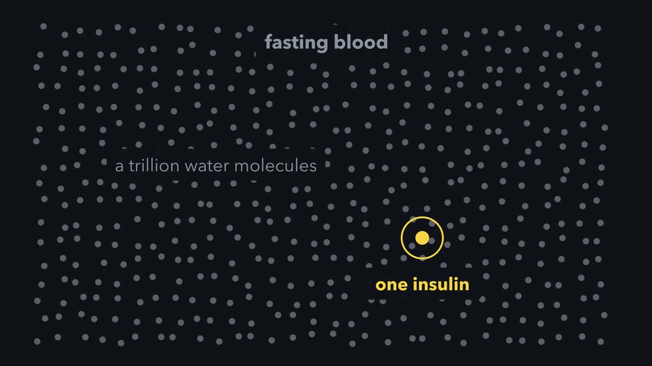 ▶ Watch More Hormone, Less Label · 2:19
Read the transcript of “More Hormone, Less Label”
Film transcript: More Hormone, Less Label
Fasting blood holds insulin at a few tens of picomolar. That is about one insulin molecule for every trillion molecules of water. Every pathway in this course rests on measuring something that scarce. How can anyone do it? Rosalyn Yalow and Solomon Berson found a way, and it began as an unexpected by-product of a different experiment. They were testing whether people with adult-onset diabetes destroy insulin too fast. So they tagged insulin with radioactive iodine, injected it, and followed the radioactivity as it left the blood. In people already treated with insulin, it left more slowly, because something in their blood was holding it. They argued that this something was an antibody.
Immunologists of the day doubted that a protein as small as insulin could raise antibodies. Then they used it as a scale. Take a tube. Put in a fixed, small amount of antibody. Add a fixed, small amount of radioactive insulin. Every tube gets the same amounts. The label is radioactive, so a counter can find it. The antibody has only a few binding sites, and the labelled insulin fills them. Now add the blood sample. Its own insulin carries no label, so it competes with the labelled insulin for the same few sites. A little sample insulin displaces a few labelled molecules, and a lot displaces many. The more insulin the patient had, the less radioactivity ends up on the antibody.
Now repeat the test with known amounts of insulin. Each tube holds the same antibody and the same labelled insulin. Those results build a curve. Separate the bound label from the free label, count each, and read the patient's concentration from the curve. This is the radioimmunoassay. Its range depends on the antibody and tracer. An antibody can tell cortisol from corticosterone, two steroids that differ by one hydroxyl group. So the assay is only as selective as its antibody. Bioassays measured what a hormone does. This counts what the antibody recognises. Keep this rule: more hormone in the sample means less label on the antibody.
Fasting blood holds insulin at a few tens of picomolar: about one insulin molecule for every trillion molecules of water, with the blood protein albumin outnumbering it roughly ten million to one. Every pathway in this course rests on someone measuring a thing that scarce, or picking one modified protein out of the thousands in a cell. This lesson is about how that is done and how much each kind of result is allowed to prove. The main point is the last one: each method answers one kind of question, and a claim about cause earns belief only when two methods that fail in different ways agree.
Measuring almost nothing: let the hormone compete with a labelled copy
The radioimmunoassay measures a hormone by competition. Put a fixed, small amount of antibody in a tube with a fixed, small amount of radioactive insulin, and add the blood sample. The sample’s own insulin carries no label, and it competes with the labelled insulin for the same few binding sites. The more insulin the patient had, the less radioactivity ends up on the antibody. Separate bound from free, count each, and read the concentration from a curve built with known amounts.
Rosalyn Yalow and Solomon Berson built this radioimmunoassay from an antibody they found by accident. Depending on the antibody and the tracer, it can measure a hormone from nanomolar down to a fraction of a picomolar, and the binding obeys the rules in binding and dose-response. Its selectivity is the antibody’s: Yalow pointed out in her Nobel lecture that an antibody can tell cortisol from corticosterone, two steroids that differ by one hydroxyl group. The bioassays in finding a signal measured what a hormone does. This one counts how much hormone the antibody recognises, which is usually, but not always, the same as how much active hormone is there.
The antibody came from an unexpected by-product of a different experiment. In the mid-1950s, at the Veterans Administration hospital in the Bronx, Yalow and Berson found that people already treated with insulin carried a serum globulin that held injected radioactive insulin. The full story is under Going deeper.
Seeing one protein among thousands: sort by size, then ask an antibody
Read the transcript of “Sort by Size, Then Ask an Antibody”
Film transcript: Sort by Size, Then Ask an Antibody
A cell extract holds thousands of different proteins. How can you follow just one? First, sort by size. This step is called SDS-PAGE. Heat the extract in SDS, a detergent. It unfolds each protein and coats it with negative charge, roughly in proportion to its length. Pull the mixture through a gel with an electric field. Small proteins travel farther than large ones. Next, find. This step is the western blot. Move the sorted proteins from the gel onto a membrane. Then soak the membrane in an antibody against your protein. A second antibody carries an enzyme that makes light or colour. It marks where the first one stuck. You see a band at one size.
One antibody is raised against a short piece of a protein with a phosphate on one residue. It is chosen so that it binds only when the phosphate is present. A blot with it reads one site, from cells, minutes after a signal arrives. Blot the same samples with an antibody to the whole protein as well. Otherwise you cannot tell more phosphate from more protein. An antibody has limits. It answers the question you built it for, so it cannot find a site nobody suspected. It reports that a band got darker, not what fraction of the molecules carry the phosphate. The antibody can bind the wrong protein. So a careful blot includes a sample in which the protein or the phosphate is missing. And a blot averages millions of cells.
A cell extract holds thousands of different proteins. Try this: describe three methods that follow one of them.
SDS-PAGE sorts. Heat the extract in SDS, a detergent that unfolds each protein and coats it with negative charge roughly in proportion to its length. Pull the mixture through a polyacrylamide gel with an electric field, and small proteins travel farther than large ones.
The western blot finds. Move the sorted proteins from the gel onto a membrane, then soak the membrane in an antibody against your protein. A second antibody, which carries an enzyme that makes light or colour, marks where the first one stuck. You see a band at one size.
Immunoprecipitation fishes. Here the extract is not heated. An antibody fixed to beads pulls its protein out of the mixture, and partners bound to that protein come out with it. Blot the catch for a suspected partner and you learn that the two were bound together in the extract, most likely as one complex. A tube with an unrelated antibody is the control that shows the partner did not stick to the beads on its own.
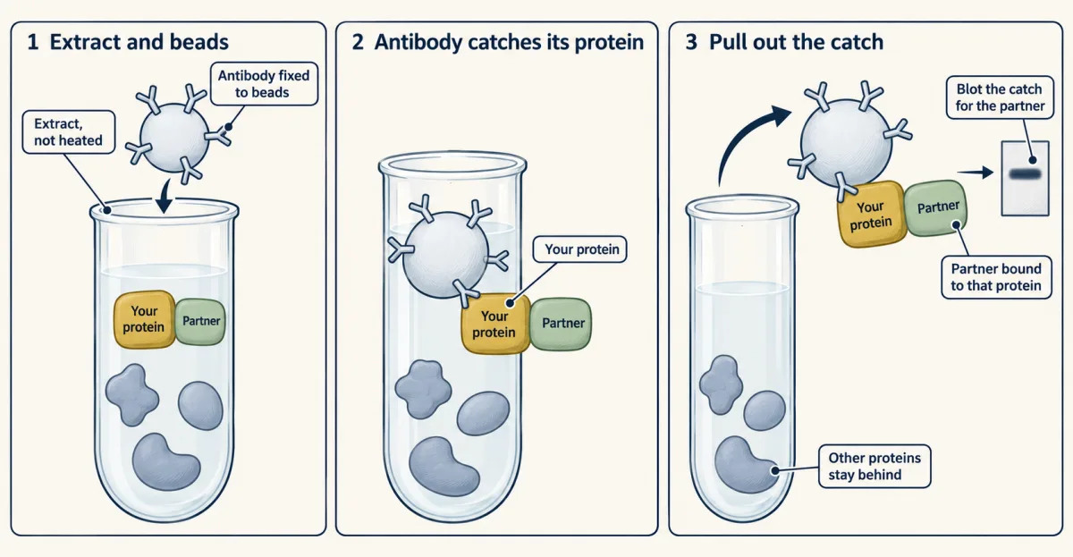
Among what he calls “other ways to detect phosphorylation events” is the tool behind much of this course’s evidence: the phosphospecific antibody. It is raised against a short piece of a protein with a phosphate on one residue, and chosen so that it binds only when the phosphate is present. A blot with it reads one site, from cells, minutes after a signal arrives. Blot the same samples with an antibody to the whole protein as well, or you cannot tell more phosphate from more protein.
An antibody has limits. It answers the question you built it for, so it cannot find a site nobody suspected. It reports that a band got darker, not what fraction of the molecules carry the phosphate. It can bind the wrong protein, which is why a careful blot includes a sample in which the protein or the phosphate is missing. And a blot averages millions of cells; watching one living cell takes the reporters of the next lesson.
”How do we determine where the protein is modified?” Weigh the pieces
Prediction tools such as Scansite search a sequence for the patterns that known kinases prefer, and databases such as UniProt list sites that have been reported. They are a start. A predicted site is a guess until someone finds the phosphate on it.
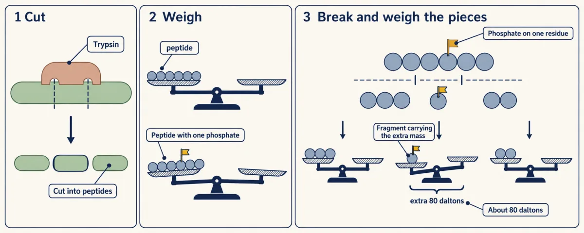
Mass spectrometry finds it by weighing. Cut the protein into peptides with trypsin, an enzyme that cuts after lysine and arginine, usually not when proline follows. The instrument weighs each peptide (strictly, it measures mass divided by charge) closely enough that one phosphate shows.
mass of a phosphopeptide = mass of the peptide + about 80 daltons for each phosphate
In tandem mass spectrometry (MS/MS) the instrument then selects one peptide, breaks it along its backbone and weighs the fragments. The fragments spell out the sequence, and the ones carrying the extra 80 daltons show which residue holds the phosphate, provided enough fragments break between the candidate residues. Sometimes they do not, and the site can only be narrowed to two or three residues. No antibody is needed and no site has to be known in advance. Once the rare phosphopeptides have been enriched, one experiment on a whole extract can report thousands of sites. A site that is not reported may still exist, because some peptides are never detected.
A worked case in which these methods were used to map and test phosphorylation sites on one transcription factor, BATF, is under Going deeper.
Which kinase? Give one kinase an ATP of its own
Read the transcript of “Give One Kinase an ATP of Its Own”
Film transcript: Give One Kinase an ATP of Its Own
What if you have a kinase but do not know the substrates? The approaches each have a weakness. A purified kinase in a test tube shows what it can phosphorylate, which is more than it does in a cell. Prediction from sequence gives candidates. Finding the kinase bound to a protein shows contact, which is not catalysis. Removing the kinase with an inhibitor, RNA interference or a knockout changes many proteins. The direct substrates look the same as those three steps downstream. Kevan Shokat's laboratory found a way through, which Chris introduces as organic chemistry meets protein manipulation. Kinases share an ATP pocket. One bulky residue in it, the gatekeeper, limits what fits.
Replace the gatekeeper with glycine or alanine, and the pocket gains a hole. An ATP with a bulky group added to its adenine fits the enlarged pocket, and fits normal kinases poorly. Such a kinase is called analog-sensitive. The laboratory introduced the approach in 1997 with the tyrosine kinase v-Src.
“What if you have a kinase but don’t know the substrates?” The approaches he lists each have a weakness. A purified kinase in a test tube shows what it can phosphorylate, which is more than it does in a cell. Prediction from sequence gives candidates. Finding the kinase bound to a protein shows contact, which is not catalysis. Removing the kinase with an inhibitor, RNA interference or a knockout changes many proteins, and the direct substrates look the same as those three steps downstream.
Kevan Shokat’s laboratory found a way through, where organic chemistry meets protein manipulation. Kinases share an ATP pocket, and one bulky residue in it, the gatekeeper, limits what fits. Replace the gatekeeper with glycine or alanine and the pocket gains a hole. An ATP with a bulky group added to its adenine fits the enlarged pocket and fits normal kinases poorly. Such a kinase is called analog-sensitive. The laboratory introduced the approach in 1997 with the tyrosine kinase v-Src.
What an inhibitor proves, and what “necessary” and “sufficient” mean
“How do we determine what effect a phosphorylation event is having on a protein?” The answer has two parts: mutate the site to imitate the gain or the loss of the phosphate, and inhibit or activate the kinase. The second part has misled many laboratories.
Most kinase inhibitors work by plugging the ATP pocket, and about five hundred kinases share that pocket. Staurosporine, a natural product, binds most of the kinases it has been tested against. In 2000 Philip Cohen’s group in Dundee tested compounds sold as selective inhibitors of named kinases against panels of purified kinases, and apart from rapamycin and PD 184352 even the most selective inhibited at least one other kinase. Larger surveys followed, and the 2007 paper judged many widely used compounds too non-specific for any conclusion except one: ruling a kinase out.
So an inhibitor result has a direction. If a dose that shuts off kinase X leaves the response intact, the activity of X is not needed. If the response stops, X or any other target of the compound may be the cause. To pin it on X, use two inhibitors of unlike structure, which are unlikely to share their other targets. Better, express a mutant of X that the drug cannot bind: if the response returns while every other target stays blocked, the drug was acting through X. Then add genetics, a knockout or a knockdown, which goes wrong in different ways.
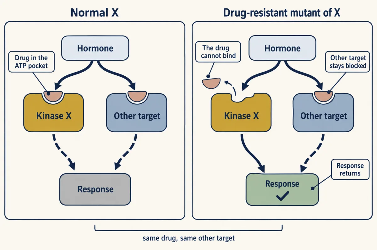
Two words organise all of this. A step is necessary if removing it removes the response: the knockout, the controlled inhibitor, the site changed to alanine. A step is sufficient if turning it on by itself produces the response without the signal: a kinase that is always active, or, more weakly, a site changed to aspartate, which imitates the phosphate only in part. A step can be one without being the other. Every method has its own way of misleading you, so a claim earns belief when two methods that fail differently agree.
Design problem
The human genome encodes about five hundred protein kinases, a cell expresses hundreds of them, and you want the direct substrates of one. Say why adding radioactive ATP to a cell extract cannot give the answer. Then design a version of your kinase that can use a form of ATP that the other kinases cannot, and say what control proves that the labelled proteins are its direct substrates.
Saved in this browser only.
One way to do it
Every kinase in the extract uses ordinary ATP, so radioactive ATP labels the substrates of every active kinase at once. Nothing marks which kinase put each phosphate on.
Find the gatekeeper of your kinase by lining its sequence up with kinases whose pockets are known, and change it to glycine. Check that the mutant still phosphorylates a known substrate. Make an ATP analog with a benzyl group on the N6 nitrogen of adenine and a radioactive gamma phosphate, the one that is transferred. Add the analog to an extract that contains the mutant kinase. Only the mutant uses it well, so a labelled protein received its phosphate from your kinase in one step.
The control is the same extract with the normal kinase in place of the mutant. It should give no labelled bands. A band that appears there comes from a kinase that can use the analog unaided, and is discarded. Each candidate still has to be confirmed in living cells. The enlarged pocket helps there too: a bulky inhibitor built for it shuts off the mutant kinase and spares the normal ones.
Going deeper
How the insulin antibody was found
In the mid-1950s Rosalyn Yalow, a physicist, and Solomon Berson, a physician, worked at the Veterans Administration hospital in the Bronx. They were testing the proposal that people with adult-onset diabetes destroy insulin too fast. They tagged insulin with radioactive iodine, injected it, and followed the radioactivity as it left the blood.
It left more slowly, but only in one group: people who had already been treated with insulin. When their serum was sorted by paper electrophoresis, the radioactivity travelled with a globulin, the class of serum protein that includes antibodies. Something in the blood of treated patients was holding the injected insulin. Immunologists of the day doubted that a protein as small as insulin, 51 amino acids, could raise antibodies. Science rejected the paper. The Journal of Clinical Investigation rejected it at first, then printed it in 1956 with the word “antibody” removed from the title.
A worked case: BATF
A 2003 paper in the Biochemical Journal by Christopher Deppmann, Tina Thornton, Fransiscus Utama and Elizabeth Taparowsky asks where a protein is modified, and what the modification does, of the transcription factor BATF. BATF from cells ran on a gel as shifted bands that calf intestinal alkaline phosphatase removed, which showed phosphorylation in vivo. A gel run with reversed polarity showed that serine 43 and threonine 48, both in the DNA-binding domain, carry phosphate. The paper reports phosphate on several serines and threonines and on at least one tyrosine; phosphoamino acid analysis on a thin-layer plate, which shows what kind of amino acid carries the phosphate, and kinase assays were among its methods. Then serine 43 was changed to aspartate, whose negative charge partly imitates a phosphate, to model what the modification does. Such phosphomimetic mutants are imperfect: aspartate carries less charge than a phosphate and has a different shape, so a mutant that does nothing is weak evidence. Each method is explained in the phosphorylation switch.
The inhibitor surveys
In 2000 Philip Cohen’s group in Dundee took compounds sold as selective inhibitors of named kinases and tested each against a panel of purified kinases. Apart from rapamycin and PD 184352, even the most selective inhibited at least one other kinase. Jenny Bain led further rounds in 2003 and 2007. In 2008 Mazen Karaman, Patrick Zarrinkar and colleagues at the company Ambit Biosciences measured thirty-eight inhibitors against 317 kinases and concluded that “small assay panels do not provide a robust measure of selectivity.”
Your notes
Select any text in the lesson to highlight it or add a note to it. Your highlights and notes are saved in this browser and collected in your notebook.
This browser is not letting the site save anything (a private window or strict privacy settings), so highlights and notes will be lost when you leave the page.
How we measure it
Radioimmunoassay
Mix a fixed, limiting amount of antibody with a fixed amount of radiolabelled hormone and the sample. Unlabelled hormone in the sample displaces label from the antibody, so the bound radioactivity, read against standards of known concentration, gives the amount of hormone.
SDS-PAGE, western blot and immunoprecipitation
Unfold proteins in SDS, separate them by size in a polyacrylamide gel, move them to a membrane and detect one with an antibody. With a phosphospecific antibody the blot reads one phosphorylated site. Immunoprecipitation uses an antibody on beads to pull a protein, and whatever is bound to it, out of an extract.
Tandem mass spectrometry of phosphopeptides
Digest proteins with trypsin, weigh the peptides, then fragment each one and weigh the pieces. A phosphate adds about 80 daltons, and the fragments that carry the extra mass can place it on one residue, with no need to know the site beforehand.
Analog-sensitive kinase
Replace the gatekeeper residue in a kinase's ATP pocket with glycine or alanine so that it accepts a bulky ATP analog that other kinases use poorly. Proteins labelled from that analog are candidates for direct substrates of the engineered kinase.
Method films · the methods library
- Analog-sensitive kinases (chemical genetics) 1:28
- Binding assays: immunoprecipitation, pull-down and gel shift 3:43
- Engineered mutants: site-directed, dominant-negative and constitutively active 4:25
- Gel electrophoresis, SDS-PAGE and western blot 4:01
- Radioactive and stable isotope tracers 4:03
- Radioimmunoassay 2:47
Practice questions on this lesson (3) → MCAT-style, original, each with an explanation
Where this lesson fits
Before thisReview first
- Binding: Affinity, Occupancy and Dose–ResponseThis course · Lesson 3
Competition between labelled and unlabelled hormone follows the binding and occupancy rules taught there.
- Phosphorylation: Kinases and PhosphatasesThis course · Lesson 6
You need to know what kinases and phosphatases do and how phosphosites are tested before judging the evidence here.
- Finding a Signal: NGF, EGF and the BioassayThis course · Lesson 2
Bioassays measure what a hormone does; this lesson contrasts that with counting hormone by antibody.
After thisStudy next
- Hacking the Cell: Reporters, Actuators and Genetic ToolsThis course · Lesson 44
Blots average millions of cells, and reporters and actuators let you watch or start a signal in one living cell.
Suggested by an AI model from the lessons themselves, then checked. How we use AI
Finish line
Test yourself
8 questions
Practice and MCAT-style, with explained answers.
Next · Lesson 44
Hacking the Cell: Reporters, Actuators and Genetic Tools
How do you watch a signal inside a living cell, and how do you start one in a cell of your choosing?
Continue →