Organismal Biology · Unit 3: Supply and exchange
Digestive Systems and Nutrition
Rewritten on : new text, flashcards and film. Your notes and highlights stay, but the text around them may have moved.
Why would a rabbit eat some of its own droppings?
Pause when the reading reaches a film, and offer to play it. Saved on this device.
Watch the film now, or keep listening
Builds on:Animal Body Plans: Similar Bodies, Different BranchesTissues, Organs and HomeostasisNervous Control: A First LookWhy these?
 ▶ Watch Why Order Matters in a Gut · 3:21
Read the transcript of “Why Order Matters in a Gut”
Film transcript: Why Order Matters in a Gut
A rabbit eats some of its own droppings. Why would an animal do that? The answer lies in the shape of the gut itself. Many animals, from earthworms to us, have a complete gut. It is a tube, with a mouth at one end and an anus at the other. Food moves one way along it. So each stretch can do its own job, in order. The mouth and stomach break food up. At the duodenum, the first part of the small intestine, bile and pancreatic juice pour in. Digestion goes on along the small intestine, the main absorbing stretch. Enzymes there do most of the digesting. Transporters in the wall carry the small pieces across. Simple sugars and amino acids cross into blood capillaries.
Most fat is packed into tiny particles that enter the lacteal, a small lymph vessel. That is the first half of a rule. Large food molecules must be broken down before or within the stretch that can absorb them. Here is the second half. Whatever a stretch does not absorb moves on. Watch the water. The small intestine takes up most of it. The colon takes up most of what remains. Only a small leftover reaches the end, and it leaves in the feces. Now feed the tube grass. Its main fiber, cellulose, has bonds no vertebrate is known to break with enzymes of its own. Microbes do it, by fermenting the fiber without oxygen. Fermentation makes short-chain fatty acids.
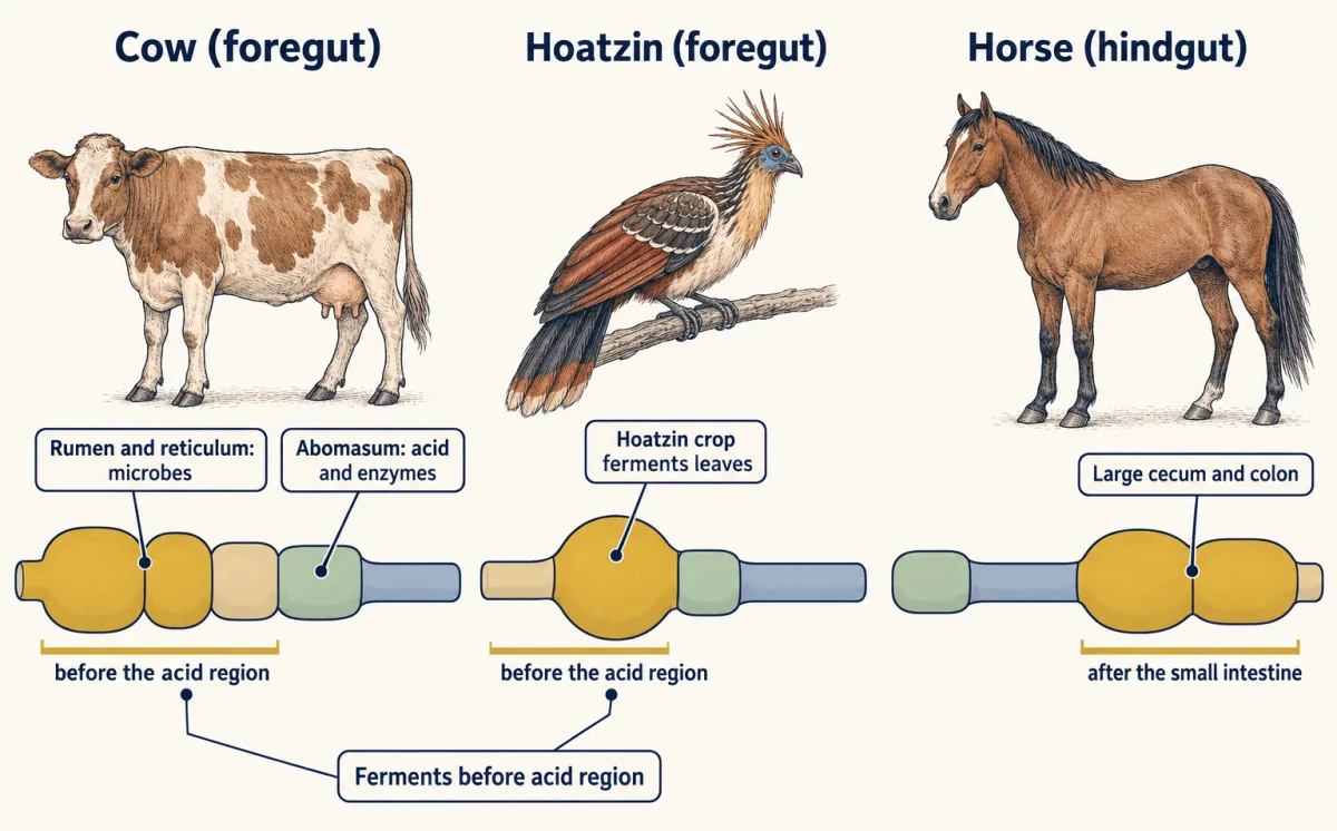
The fermenting chamber's own wall absorbs them on the spot, wherever that chamber sits. But the microbes themselves are food, rich in protein. The acid stomach starts digesting protein. The small intestine finishes the job, and absorbs the amino acids. A cow ferments in front of its acid stomach. Its microbes wash onward, into the acid and then the small intestine. There they are digested. A horse ferments in its cecum and colon, after the small intestine. Its microbes are full of protein, and its small intestine is already behind them. Where does that protein go?
Most of it leaves in the droppings. The stretch that absorbs amino acids is already behind it. A rabbit ferments mainly in its cecum, after the small intestine too. So it makes soft pellets from its cecum, and eats them. The microbes go round again, through the stomach and small intestine. That is why a rabbit eats some of its droppings. Food must be broken down before or within the stretch that can absorb it.
From the film
- A complete gut is a one-way tube, so each stretch can specialize and works on what the stretch before it hands on.
- Large food molecules must be broken down before or within the stretch that can absorb them; what a stretch does not absorb moves on, to be taken up downstream or lost in the feces.
- In humans the body's own digestion starts in the mouth and stomach and is mostly done in the small intestine, where pancreatic enzymes and bile salts act and most nutrients are absorbed; the colon takes back most of the water left.
- Microbes ferment fiber, and their fatty acids are absorbed where they are made. Their protein is recovered as amino acids mainly when it passes through the stomach and small intestine: a cow's microbes do, a rabbit sends them round again, and most of a horse's is lost.
Why would a rabbit eat some of its own droppings? The first film answered with a one-way tube. This text puts numbers on a second puzzle: on round figures, about 9 L of water a day enter an adult human’s small intestine, yet only about a tenth of a litre leaves in the feces. Where does all that water come from, and why does so little leave?
The one idea. A complete gut is a one-way line, so its stretches can specialize. Large food molecules must be broken down before or within the stretch that can absorb them. What a stretch does not absorb moves on, to be taken up or changed further down or lost in the feces, unless the animal sends it round again. For water in any stretch: what leaves = what came in + what the gut adds − what is absorbed.
Scope, stated once for the whole lesson: complete guts at steady state. Water, salts and single sugars (monosaccharides) need no breakdown; sugars of two or more units, starch included, must be split first. Digestion and absorption overlap along the small intestine; microbes change some molecules into others; and other stretches absorb particular things. The water balance counts no exit but absorption and onward flow; a balance for one nutrient must also count what digestion and microbes make or use up. Some newborn mammals absorb whole antibodies from milk, an immune exception, not nutrition.
One tube, four jobs and two helpers
A gut has four jobs.
- Mechanical breakdown tears and grinds food into smaller pieces.
- Enzymatic hydrolysis splits large molecules (starch, proteins, fats) into small subunits by adding water across their bonds. The enzyme chemistry is in the Biochemistry lesson on digestion.
- Absorption moves those subunits across the gut lining, an epithelium (a sheet of tightly joined cells), mostly through the transporters in membrane transport.
- Elimination gets rid of what is left.
Two helpers reach what the host’s enzymes alone cannot:
- Bile salts emulsify fat, breaking large drops into small droplets that lipase, the fat-digesting enzyme, can reach.
- Fermentation is the breakdown of food by microbes living in the gut, without oxygen. With their own enzymes the microbes split bonds the host has no enzyme for, then ferment the products.
In Hydra, a small freshwater cnidarian, and in a planarian flatworm, the gut is incomplete: a sac with one opening, the gastrovascular cavity, where food is digested and the leftovers go out the way they came in. The cavity may branch, but nothing flows through it from mouth to anus. A complete gut, as in earthworms, insects and vertebrates, is the first film’s one-way tube (animal body plans).
Mouth to anus in a human
The first film named the stations. In an adult human, food passes from the mouth to the pharynx, the esophagus, the stomach and the small intestine (duodenum, then jejunum, then ileum). It then enters the large intestine (the cecum, a blind pouch where the small and large intestine meet, then the ascending, transverse, descending and sigmoid colon), the rectum and the anal canal, which opens at the anus.
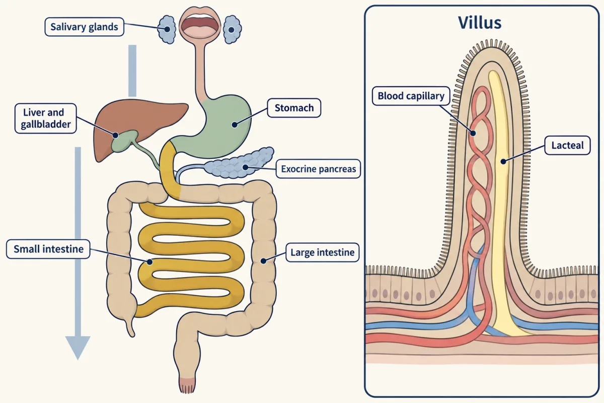
- Mouth and salivary glands. Teeth reduce particle size. Three pairs of major salivary glands (parotid, submandibular, sublingual) add saliva, whose amylase starts digesting starch.
- Esophagus. Waves of muscle contraction, called peristalsis, push the swallowed lump (the bolus) to the stomach.
- Stomach. It mixes food with acid and enzymes into a soup called chyme. Parietal cells secrete hydrochloric acid and chief cells secrete pepsinogen, the inactive form of pepsin, which starts protein digestion. The layers of its wall are in tissues and homeostasis.
- Small intestine. The main site of digestion and of nutrient absorption. Circular folds, finger-like villi and the microvilli on each cell multiply its surface.
- Liver and gallbladder. The liver makes bile; the gallbladder stores and concentrates it. Besides emulsifying fat, bile salts gather the products of fat digestion into tiny clusters, micelles, that carry them to the cell surface; without enough bile salts, fat absorption is greatly reduced. Bile is not an enzyme.
- Exocrine pancreas. It sends digestive enzymes and bicarbonate, which neutralizes the stomach’s acid, into the duodenum.
- Large intestine. It absorbs water and salts, and its microbes ferment what escaped digestion.
Blood from the villus capillaries the first film showed drains first to the liver, through the hepatic portal vein; the fat particles it sent into lacteals are chylomicrons.
The water budget: the one idea in numbers
Take an adult human at steady state (the gut holds the same water from day to day), with round, illustrative figures (textbooks give somewhat different splits). About 9.0 L of water a day enters the small intestine: food and drink, plus saliva, gastric juice, bile, pancreatic juice and intestinal juice. Most of it is the gut’s own secretions, poured in and taken back. About 90% is absorbed in the small intestine.
- Small intestine. Absorbed = 9.0 L per day × 0.90 = 8.1 L per day. Passed on to the colon = 9.0 − 8.1 = 0.9 L per day.
- Colon. Say the colon absorbs 0.8 L per day. Fecal water = 0.9 − 0.8 = 0.1 L per day. Total absorbed = 8.1 + 0.8 = 8.9 L per day, so overall absorption = 8.9 / 9.0 = 98.9%.
Predict. Now halve the colon’s absorption, to 0.4 L per day. Does fecal water double?
Check your prediction
No, it rises fivefold. Fecal water = 0.9 − 0.4 = 0.5 L per day, five times 0.1. Overall absorption = 8.5 / 9.0 = 94.4%, a drop of only 4.4 percentage points. Check: 8.5 + 0.5 = 9.0 L per day, as 8.9 + 0.1 did.
Why so sensitive? Fecal water is a small leftover after two large subtractions, 9.0 − 8.1 − 0.8 = 0.1 L per day, so a small change in either large flow is a large change in the leftover. If the colon still takes exactly 0.8 L per day, a fall in the small intestine’s share from 90% to 89% raises the flow into the colon from 0.90 to 0.99 L per day and fecal water from 0.10 to 0.19 L, 1.9 times as much. These are model results: a real colon has spare capacity and partly compensates. And since most of the water is the gut’s own, a gut that stops taking it back drains the body.
- Small intestine
- Colon
- Overall absorption
- Check: absorbed + fecal water = input
In this sketch: adult human at steady state, no change in the water held in the gut; textbook figures used as illustrative values; bar and arrow widths proportional to flow. Slider ranges are chosen for the sketch; only 9.0 L, 90%, 0.8 and 0.4 L per day come from the lesson.
Rule to keep: absorbed water plus fecal water equals the water that entered. Fecal water is what is left of what reaches the colon after the colon absorbs; in the lesson’s numbers, a small difference between two large flows.
Where the microbes sit: cow, horse, rabbit, hoatzin
The first film’s fiber was cellulose, the main fiber of plant cell walls; humans make no cellulase, the enzyme that breaks it. The microbes ferment cellulose and other plant polysaccharides into short-chain fatty acids (acetate, propionate, butyrate), which the host uses for energy. The partnership is a mutualism, treated in mutualism, parasitism and coevolution.
Foregut fermenters ferment before the acid-secreting region. A cow has one stomach with four compartments: the rumen and reticulum hold the microbes that break down fiber, the omasum follows, and the abomasum is the glandular compartment that secretes acid and enzymes, equivalent to a single stomach. Kangaroos ferment in an enlarged front part of the stomach that evolved separately in marsupials. The hoatzin, Opisthocomus hoazin, a leaf-eating bird of South America, ferments leaves in its crop, the enlarged pouch of the esophagus where many birds store food. A chicken’s crop mainly stores food, and the chicken grinds it in a muscular gizzard.
Hindgut fermenters, the film’s horse and rabbit, ferment after the small intestine; a rabbit takes its soft pellets directly from the anus.
In a cow, as in a human, several stretches work at the same time on different parts of a meal, so they must be coordinated. How does the stomach know a meal is coming? The second film follows the dogs that first answered that.
Read the transcript of “How Does the Stomach Know Food Is Coming?”
Film transcript: How Does the Stomach Know Food Is Coming?
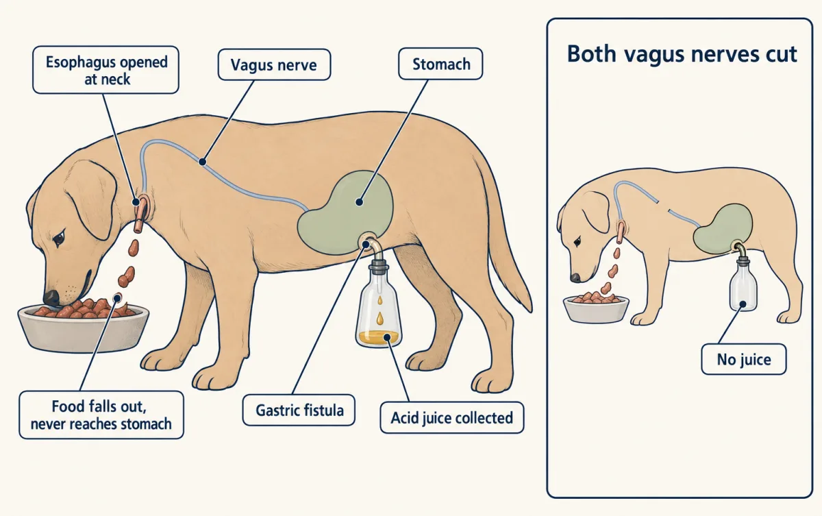
How does a stomach know a meal is coming? Can it start work before any food arrives? The first film showed the gut as a one-way line, with several stretches working at once. Some signals ready a stretch before food arrives. Others answer once it is there. Ivan Pavlov's laboratory, in Saint Petersburg, asked this of dogs. The dogs had recovered from surgery, and were awake and eating. So one gland at a time could be studied in a whole, living animal. In one dog, the esophagus was opened at the neck. Food the dog swallowed fell out into a dish. It never reached the stomach. A second opening, called a fistula, let the workers collect juice straight from the stomach into a flask.
Suppose stomach juice flows only when food touches the stomach. What should the flask collect while this dog eats? After a delay of some minutes, acid juice flowed. It came from the stomach, through the fistula. The dog was eating, but no food had reached its stomach. Then both vagus nerves were cut. These nerves run from the brainstem to the stomach. Now the dog ate, and the flask stayed empty. So in the dog, nerves start the stomach early. This is the cephalic phase, set off by the sight, smell, taste and chewing of food. This rules in a nerve-driven start. It does not rule out later steps, once food is inside the gut. Here are two later steps.
Chyme, the stomach's acid soup of food, moves on into the duodenum, the first part of the small intestine. These steps use hormones. A hormone is a substance released into the blood, which acts on distant target cells. The blood carries the message to the next gland. Acid arriving there makes the duodenal lining release a hormone, secretin, into the blood. The pancreas answers with fluid rich in bicarbonate, which neutralizes the acid. Fat and protein arriving release a second hormone, cholecystokinin, called CCK. The pancreas sends enzymes, and the gallbladder squeezes out bile. So nerves start the stomach before food arrives. And a stretch that food has just reached signals the glands that serve it.
The duodenum reports acid, fat and protein, and the right glands answer.
From the film
- Pavlov's sham-fed dogs poured acid juice from the stomach while they ate, though the food left at the neck and never reached the stomach.
- Cutting both vagus nerves abolished that juice, so in the dog the first, cephalic phase is driven by nerves that ready the stomach before food arrives; this does not rule out later phases.
- Acid reaching the duodenum releases the hormone secretin, which brings bicarbonate-rich fluid from the pancreas to neutralize it.
- Fat and protein reaching the duodenum release CCK, which brings pancreatic enzymes and bile from the gallbladder.
Nerves and hormones run the line
Cutting the vagus nerves did not leave the dog’s gut without nerves. Its wall holds the enteric nervous system, a network of nerve cells that runs local reflexes, such as peristalsis, by itself. The vagus nerves connect the brainstem with the gut and add parasympathetic signals, from the rest-and-digest division of the autonomic nervous system, that increase secretion (the nervous system map, the hypothalamus and autonomic system). The second film’s cephalic phase is that vagus-driven flow, in the dog.
Later phases are partly chemical, carried by hormones, substances released into the blood that act on distant target cells (the endocrine system).
Predict. Take a dog whose vagus nerves have been cut. Meat is placed through the fistula straight into its stomach. Will any juice flow?
Check your prediction
Yes, some, in experiments like the MCAT item below. Food in the stomach can drive secretion without the vagus, for example through gastrin, a hormone from G cells of the lower stomach lining.
Three gut hormones:
| Hormone | Source | Trigger | Main targets and effect |
|---|---|---|---|
| Gastrin | G cells of the stomach lining | Food (especially protein) in the stomach; vagal input | Stomach: more acid, partly by releasing histamine from nearby cells |
| Secretin | Lining of the duodenum | Acid chyme entering the duodenum | Pancreatic duct cells: bicarbonate-rich fluid |
| CCK (cholecystokinin) | Lining of the duodenum | Fats and proteins in the duodenum | Pancreas: enzymes; gallbladder: contracts, releases bile |
Secretin was the first gut hormone found, by Bayliss and Starling, a story told in signals and distance. Gastrin and CCK act on receptors of the type described in GPCRs and G proteins. Nerves and hormones also act on the same glands (Going deeper).
Essential depends on who is eating
An essential nutrient is one an animal must get from food because its body cannot make it, or cannot make enough. For humans, nine amino acids are essential: histidine, isoleucine, leucine, lysine, methionine, phenylalanine, threonine, tryptophan and valine (amino acids). Two fatty acids are essential for humans, linoleic acid and alpha-linolenic acid. Vitamins are organic molecules needed in small amounts; minerals are inorganic elements, such as calcium and iron.
Most mammals make their own vitamin C, but humans, some other primates and guinea pigs cannot and need it in the diet. The domestic cat is an obligate carnivore: it depends on animal tissue and needs taurine, a small sulfur-containing compound related to amino acids, in its diet; adult humans and many other mammals make taurine for themselves. “Essential” is a statement about an animal and its partners, not about a molecule.
Predict. Would a cow need dietary vitamin B12 to the same degree as a human?
Check your prediction
No. Ruminant microbes make B vitamins, including B12 when the diet contains cobalt, and the microbial protein a cow digests supplies much of its amino acids. So a ruminant’s dietary needs differ from a human’s. They do not vanish: a ruminant still needs cobalt, and high-producing cattle benefit from protein that bypasses the rumen.
The one idea again: on a one-way line, a nutrient must be supplied, by food or by microbes, before or within the stretch that can absorb it.
Is the herbivore gut always longer? Not simply. Humans are omnivores, eating plant and animal foods, with a modest cecum and colon where microbes ferment fiber. Larger animals have longer intestines whatever they eat, and related species resemble each other through shared ancestry, their phylogeny. With both accounted for, gut length tracks diet only loosely, and by region.
Design problem
Across many mammal species, herbivores have longer large intestines than carnivores. A classmate concludes that eating plants makes the large intestine grow longer. What would you need to check before accepting a link between diet and gut length, and what kind of evidence would test cause?
Saved in this browser only.
One way to do it
First, scale by body mass: compare length at a given body mass, for example with a regression of log gut length on log body mass, with diet as a second predictor.
Second, account for phylogeny (reading phylogenetic trees): fifty closely related herbivores are not fifty independent tests. A phylogenetic regression asks whether diet explains length beyond shared ancestry. Duque-Correa and colleagues (2021) did this for 519 mammal species, with body mass in the model, and found strong phylogenetic signal.
Third, test each region separately. In that study diet was associated with large-intestine length, but adding diet did not improve the small-intestine model, so “herbivores have longer guts” is too broad.
Fourth, a correlation across species does not show cause: diet might change the gut during a lifetime, or lineages with long large intestines might have taken up plant diets. Feeding experiments within one species would test cause, though a change within one life can mislead: in many frogs, plant-eating tadpoles have a long coiled gut that shortens at metamorphosis when the animal turns to eating animals, but thyroid hormone drives that whole transformation, so diet and gut change are tangled.
Going deeper
Pavlov’s preparation
From about 1889 Pavlov’s St Petersburg laboratory gave dogs an esophagotomy (the esophagus opened at the neck) and a gastric fistula (a stomach opening for draining juice).
Cellulose is not the only fiber
Cellulose is the main fiber of plant cell walls, though pectins and hemicelluloses make up much of the rest.
Gastrin and secretin: discovery dates
Bayliss and Starling reported secretin in 1902. J. S. Edkins proposed gastrin in 1905, but it was purified only in 1964.
How much of CCK’s effect needs nerves?
In healthy human volunteers given CCK-8 at one dose, atropine, a drug that blocks the acetylcholine receptors nerves use on many glands, cut the pancreatic trypsin response by 84%. So at that dose much of the hormone’s effect depended on acetylcholine, and tests on rat pancreas suggested the acetylcholine came from nerves. At a higher, above-normal dose, atropine blocked less of the response.
Hormone cells that talk to nerves
In mice, Kaelberer and colleagues reported in 2018 that some hormone-releasing cells of the intestine form synapses on vagal neurons and signal with glutamate, a fast nerve-like route alongside slower hormones.
Your notes
Select any text in the lesson to highlight it or add a note to it. Your highlights and notes are saved in this browser and collected in your notebook.
This browser is not letting the site save anything (a private window or strict privacy settings), so highlights and notes will be lost when you leave the page.
How we measure it
Chronic fistula and sham feeding
A surgically made opening to the outside lets juice be collected from a gland or organ of an animal that has recovered from surgery, awake and eating. Combined with an opening of the esophagus at the neck, it separates what eating does from what food in the stomach does.
Pharmacological blockade
Give a drug that blocks one signaling route, such as atropine for the acetylcholine receptors that nerves use on many gut glands, and measure how much of a response remains. The fraction that disappears shows how much of the response depends on that route under those conditions; other tests are needed to show where along the route the signal acts.
In vitro fermentation cultures
Gut or fecal microbes from donor animals are grown without oxygen at body temperature with a chosen food added, and pH and organic acids (measured by high-performance liquid chromatography) are followed over hours. It shows what a community can do with a substrate, not what happens in the intact animal.
Comparative method with phylogenetic regression
Measure a trait such as intestine length across many species, scale it by body mass, and fit a model that accounts for how the species are related. It asks whether a pattern holds beyond what shared ancestry and body size already explain.
Practice questions on this lesson (4) → MCAT-style, original, each with an explanation
Where this lesson fits
Before thisReview first
- Animal Body Plans: Similar Bodies, Different BranchesThis course · Lesson 4
It separates complete one-way guts from sac-like incomplete guts, which this lesson uses straight away.
- Tissues, Organs and HomeostasisThis course · Lesson 6
You need the layers of the gut wall and the idea of control loops from there.
- Nervous Control: A First LookThis course · Lesson 7
It introduces autonomic and reflex pathways, which this lesson uses for vagal and enteric control of digestion.
After thisStudy next
- Osmoregulation and Nitrogenous WasteThis course · Lesson 15
It takes the gut's water and salt handling further, showing how animals balance water and nitrogenous waste.
- The KidneyThis course · Lesson 16
It uses the same large-flow, small-leftover water accounting, applied to filtering plasma and taking almost all of it back.
- Soil, Nutrients and PartnersThis course · Lesson 24
It asks what an organism must take in from outside, here minerals from soil with microbial partners.
Elsewhere on the siteFilms, simulations and method films in other courses
- Build a big molecule from small blocksSimulation · Biology
It shows large food molecules being broken into small blocks by adding water, which is the hydrolysis step of digestion.
- Take It Apart, Build It AgainFilm · Biology
It shows digestion breaking starch and protein into small pieces by adding water, one water per broken link.
Suggested by an AI model from the lessons themselves, then checked. How we use AI
Finish line
Test yourself
9 questions
Practice and MCAT-style, with explained answers.
Next · Lesson 15
Osmoregulation and Nitrogenous Waste
A goldfish in a bowl is soaking up water all day, and a cod in the sea is losing it. How does each one stay the same size?
Continue →