Biochemistry · Amino Acids
New to this? Review it at high school level: What Am I Made Of?
Amino Acids: How One Side Chain Decides Everything
Rewritten on : new text, figures and flashcards. Your notes and highlights stay, but the text around them may have moved.
The R group sets what makes each amino acid different — its charge, its pKa, and the Glu→Val swap behind sickle cell.
Pause when the reading reaches a film, and offer to play it. Saved on this device.
Watch the film now, or keep listening
Builds on:Water, Weak Bonds, and the Generation of OrderWhy these?
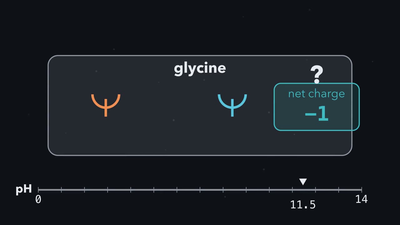 ▶ Watch Add Up the Proton Switches · 3:05
Read the transcript of “Add Up the Proton Switches”
Film transcript: Add Up the Proton Switches
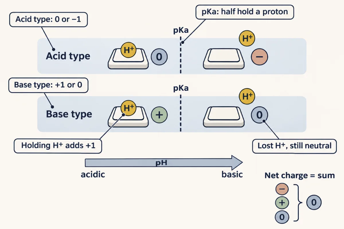
One amino acid, glycine, can carry +1, 0, or −1. What decides which? One comparison, made at every proton switch. A proton switch is a group that holds a hydrogen ion, a proton, or lets it go. Holding it is protonated. Letting go is deprotonated. Each switch has a set point, its pKa. That is the pH where half the groups hold a proton. Below it, most hold on. Above it, most let go. Each pH unit from the pKa changes the ratio of the two forms tenfold. One unit above, about 91% percent have let go. A proton carries +1, so holding it makes a group one charge more positive. An acid-type switch, like a carboxyl, goes from 0 to −1. A base-type switch, like an amino group, goes from +1 to 0.
So letting go is not always negative. Glycine has two backbone switches. The carboxyl's pKa is about 2.3; the amino group's, about 9.6. Here is the whole idea: net charge is the sum of the switches. Your turn. At blood pH, about 7.4, check each switch against its pKa. What is glycine's net charge?
7.4 is above the carboxyl's pKa, so it lets go: minus −1. It is below the amino group's, so it holds on: plus +1. The sum is zero: a zwitterion, with both a plus and a minus. Sweep the pH upward, and the predominant charge steps down: +1, 0, −1. Each drop is centred on a pKa. The exact zero crossing is the isoelectric point, the pI. For glycine, averaging its two pKa values gives the pI, about 5.97. In its simplest form, average the two pKa values around the ladder's net-zero rung. A side chain adds a third switch, or none. Glutamate's acid switch, near 4, has let go at blood pH: negative.
Lysine's base switch, near 10.5, holds on: positive. Histidine's, near 6, is mostly neutral. Inside a protein, neighbours can shift these values. So list the switches, compare each pKa with the pH, and add. That sum is how sickle-cell hemoglobin was first told apart. A valine had replaced a glutamate on each beta chain.
From the film
- Each ionizable group is a proton switch: below its pKa it mostly holds its proton, above it mostly lets go.
- Holding a proton makes a group one charge more positive: acid-type switches go from 0 to −1, base-type from +1 to 0.
- An amino acid's net charge is the sum of its switches; at pH 7.4, glycine is a net-zero zwitterion.
- The pI is the pH of zero net charge; for glycine it is the average of its two pKa values, about 5.97.
In 1949 Linus Pauling, Harvey Itano, S. J. Singer and Ibert Wells put hemoglobin from healthy people and from people with sickle-cell anemia in an electric field, and the two moved differently: at the same pH they carried different net charges.
For two molecules of the same size and shape, only a charge difference explains that. The difference turned out to be one amino acid: at position 6 of each beta chain, a valine where a glutamate belongs. Why does that swap change the charge, and why does it make red cells sickle? The answers differ, and by the end you can work out both.
The film above builds the charge half of the answer: proton switches, pKa and pI. This page adds the numbers behind them and a second property, polarity, which the sickle story also needs.
One backbone, twenty side chains
Every amino acid is a central carbon, the alpha-carbon, carrying four groups: an amino group (written −NH₂), a carboxyl group (written −COOH), a hydrogen, and a side chain, the R group. That is how it is drawn on paper; in water near neutral pH the ends are −NH₃⁺ and −COO⁻ (the next section shows why). Three groups are shared (proline’s side chain loops back onto its backbone nitrogen, making a secondary amine). The R group carries the differences: size, charge, comfort in water, reactivity.
With four different groups, the alpha-carbon is chiral: it has two mirror images, L and D, that cannot be superimposed, like your two hands. Proteins are built almost exclusively from the L form. The exception is glycine, whose R group is just another hydrogen, so it is the only achiral amino acid. That tiny side chain lets it take backbone angles other residues cannot, so it turns up in tight turns.
Proton switches: the one rule for charge
Both ends of the backbone are proton switches, as the film shows: groups that hold a hydrogen ion, H⁺ (protonated), or let it go (deprotonated), depending on how acidic the surroundings are. Each has a set point, its pKa, where half of those groups hold a proton.
The one idea to carry through this lesson (the film’s, in three lines).
- Below a group’s pKa it mostly holds its proton; above it, it mostly lets go. Each pH unit from the pKa changes the ratio of the two forms tenfold.
- Holding a proton always makes a group one charge more positive.
- Net charge is the sum of the switches: two on the backbone, one or none on the side chain. The pI is where they sum to zero.
This holds for free amino acids in water; inside a protein, neighbors can shift a pKa (Going deeper). Point 2 matters most, because switches come in two kinds:
| Kind of switch | Holding H⁺ | Without H⁺ | Examples (pKa) |
|---|---|---|---|
| Acid type | 0 (−COOH) | −1 (−COO⁻) | backbone carboxyl (about 2), Asp and Glu side chains (about 4) |
| Base type | +1 (−NH₃⁺; His and Arg have a protonated ring or guanidinium) | 0 (−NH₂; the neutral ring or guanidine) | backbone amino (about 9.5), His (about 6), Lys (about 10.5), Arg (about 12.5) |
So “deprotonated” does not mean “negative”: a lysine side chain that has lost its proton is neutral.
How much is “mostly”? The Henderson–Hasselbalch equation gives the ratio.
In words How far the pH sits above the pKa is the log of the ratio of the form without the proton to the form with it.
- pH“p H”
- how acidic the solution is; lower means more H⁺
- pKa“p K a”
- the group's set point, where half the groups hold a proton
- [without H⁺]“”
- the form that has let go (written A⁻ in most textbooks, even when it is neutral)M
- [with H⁺]“”
- the form holding the proton (written HA)M
Sanity check At pH = pKa the split is 50:50. One unit above gives 10:1 (about 91% without H⁺), two units 100:1 (99%). Within about half a unit of the pKa the minority form is about a quarter or more, so there the short form of the rule (above the pKa, most let go) is not enough.
Try one number
Glutamate’s side chain at blood pH: 7.4 − 4.25 = 3.15 units, more than two, so over 99% have let go: negative for all practical purposes. (Exactly, 10 to the power 3.15 is about 1400 to one.)
Histidine’s side chain: 7.4 − 6.0 = 1.4, and 10 to the power 1.4 is about 25. About 25 are neutral for every one that is positive, so it is about 4% positive.
Need a refresher? log scales · powers and roots
Textbooks and the MCAT write the same equation as pH = pKa + log([A⁻]/[HA]).
Glycine has just the two backbone switches: carboxyl pKa₁ = 2.34 and amino pKa₂ = 9.60.
Predict. What is glycine’s net charge at pH 1, at pH 6 and at pH 12?
Check your prediction
- pH 1. Below both pKa values, so both hold their protons: −COOH (0) and −NH₃⁺ (+1). About +1.
- pH 6. Above the carboxyl’s pKa, so −COO⁻ (−1); below the amino group’s, so −NH₃⁺ (+1). 0.
- pH 12. Above both, so −COO⁻ (−1) and −NH₂ (0). About −1.
Stack the three forms from most positive to most negative, with a pKa between each pair, and you have glycine’s charge ladder:
| Net charge | Form | Main form when |
|---|---|---|
| +1 | H₃N⁺–CH₂–COOH | pH below 2.34 |
| pKa₁ = 2.34 | ||
| 0 | H₃N⁺–CH₂–COO⁻ | pH between 2.34 and 9.60 |
| pKa₂ = 9.60 | ||
| −1 | H₂N–CH₂–COO⁻ | pH above 9.60 |
A molecule carrying both a positive and a negative charge is a zwitterion (which does not mean net zero: aspartate at 7.4 has both charges and is net −1). Glycine’s net-zero zwitterion is more than 90% of it from about pH 3.3 to 8.6, one unit inside each pKa. That includes blood pH, about 7.4 (the inside of a cell runs a little lower).
The pH of zero net charge is the isoelectric point, pI: the average of the two pKa values around the net-zero rung. For glycine, (2.34 + 9.60) / 2 = 5.97.
Why the average? Near the pI almost all glycine is the zwitterion, with a little +1 and a little −1 form, and zero net charge needs equal amounts of those two. By the rule, the +1 form’s ratio to the zwitterion is 10 to the power (pKa₁ − pH), and the −1 form’s is 10 to the power (pH − pKa₂). Set them equal and pH = (pKa₁ + pKa₂) / 2.
Predict. Aspartate has three switches: backbone carboxyl 1.88, side-chain carboxyl 3.65 and backbone amino 9.60. What is its net charge at pH 7.4, and what is its pI?
Check your prediction
At 7.4 both carboxyls have let go (−1 each) and the amino group holds on (+1): −1.
Walk down the ladder: +1 below pH 1.88, 0 past 1.88, −1 past 3.65, −2 past 9.60. The net-zero rung sits between 1.88 and 3.65, so pI = (1.88 + 3.65) / 2 = 2.77. The common error is to average the two backbone values, giving 5.74, where aspartate carries −1, not 0. Find the net-zero rung first.
Four side-chain families, read from the switches
The two charged families are switch states at pH 7.4; the other two are sorted by polarity, whether a side chain is comfortable in water. Recall: below its pKa a group holds its proton and is one charge more positive.
- Acidic (Asp, Glu). Acid-type switches near pKa 4, far below 7.4, so they have let go: negative.
- Basic (Lys, Arg, His). Lys and Arg switches sit far above 7.4, so they hold on: positive. His is a base whose switch sits near 6, so free histidine is mostly neutral at 7.4; it is grouped with the bases by convention, which is why the classifier files it as “Basic (+)”.
- Polar, uncharged. The hydroxyls of Ser, Thr and Tyr, the amides of Asn and Gln, and the reactive thiol of Cys. Tyr (pKa about 10) and Cys (about 8.2) have switches, but at 7.4 they mostly stay neutral.
- Hydrophobic (Gly, Ala, Val, Leu, Ile, Met, Pro, Phe, Trp). Greasy hydrocarbon and aromatic chains with no switch, which bury themselves in a protein’s core, away from water.
The seven ionizable side chains, Asp, Glu, His, Cys, Tyr, Lys and Arg, include the residues that form salt bridges, ion pairs between opposite charges (Asp and Glu with Lys and Arg), bind metals (His, Cys) and do acid-base catalysis (His above all).
Predict. Which family belongs on the face of a membrane-spanning helix that is surrounded by lipid?
Check your prediction
Hydrophobic: the lipid interior has no water, so charged and polar side chains are out of place and greasy ones are at home.
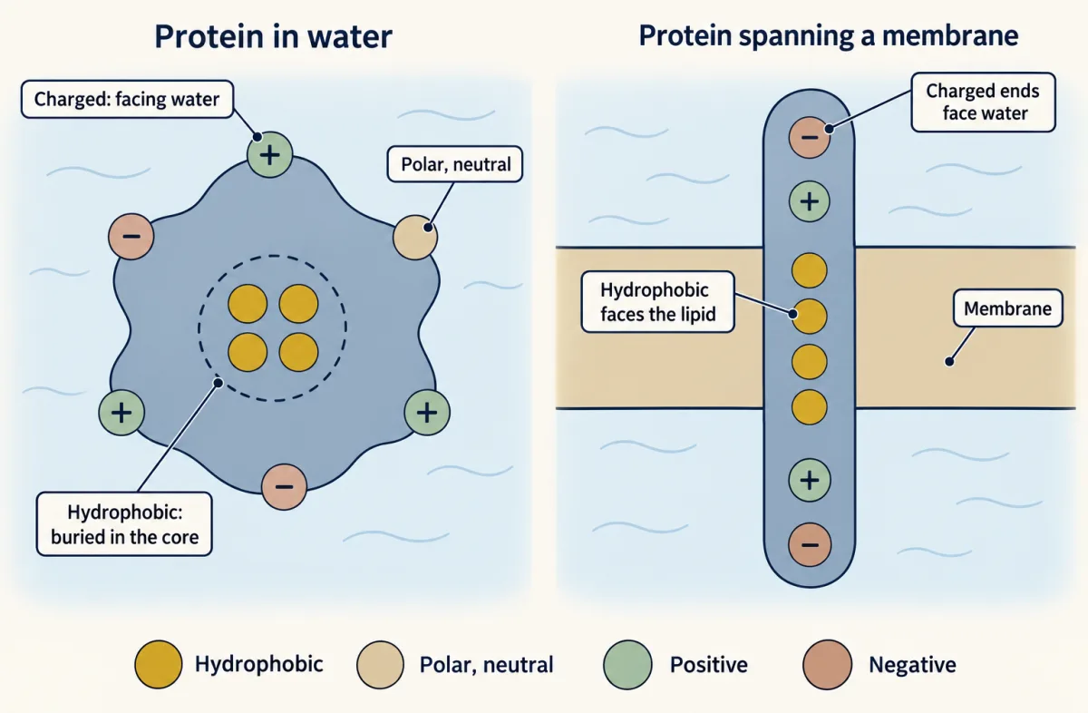
The classifier sorts all twenty, with the aromatic rings (Phe, Tyr, Trp) marked; click any residue to see its job.
Each color is a side-chain family. Click a family above to isolate it, or click any residue for its role.
Side-chain chemistry is destiny: nonpolar residues bury in the water-fearing core, polar and charged ones face the water or line active sites, and the three "specials" (glycine's flexibility, proline's kink, cysteine's disulfide) bend the rules on purpose.
Reading the curve: titration, pKa and pI
A titration curve is the measured record of the charge ladder: add base drop by drop, with equivalents of base (base added per amino-acid molecule) along the bottom and pH up the side. Each flat part is a buffering region, where added base barely moves the pH because one switch is busy letting go of its protons.
For well-separated pKa values, each pKa sits at the middle of a flat part, the half-equivalence point. When the two pKa values on either side of the net-zero rung are far apart, as in glycine (flat parts at 2.34 and 9.60), the pI of 5.97 sits in the steep jump between them: no switch is mid-change there, so almost every molecule is the net-zero form and nothing buffers. When the two are close, as in aspartate (1.88 and 3.65) or lysine (8.95 and 10.53), their flat parts merge and the pI falls inside one broad buffering region, so find it from the ladder, not from the shape of the curve. Histidine, with its extra imidazole switch, has three (pKa₁ = 1.82, pKa₂ = 6.00, pKa₃ = 9.17) and a pI of 7.59.
Drag the pH cursor to see the main form and net charge at each point. Then switch to histidine and move its imidazole slider.
Raise the imidazole pKa from 6.0 to 7.0, as a protein pocket can, and the protonated share at pH 7.4 goes from about 4% to about 28%; raise it to 7.4 and it is half and half, ready to give a proton or take one. Free cysteine’s thiol (about 8.2) actually sits a little closer to body pH, and it does its own catalytic work as a reactive nucleophile. But histidine’s neutral-to-positive switch is the one enzymes use most to pass protons, which is why it turns up in active site after active site: a residue that must give and take protons works best with its pKa near the enzyme’s working pH.
Electrophoresis: which way does it move?
Pauling’s experiment applied the rule. In electrophoresis, molecules in a buffer sit between two electrodes: the negative cathode and the positive anode. Below its pI a molecule is net positive and moves to the cathode; above its pI it moves to the anode; at its pI it stays put.
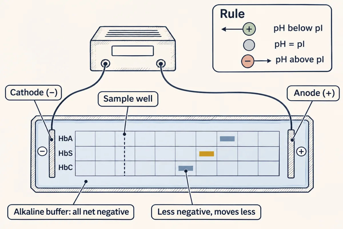
Predict. In a buffer at pH 6.0, which way do glycine (pI 5.97), aspartate (pI 2.77) and histidine (pI 7.59) move?
Check your prediction
Glycine barely moves: the pH is almost exactly its pI. Aspartate, far above its pI, is about −1 and moves toward the anode. Histidine, below its pI, moves toward the cathode; with its imidazole pKa at exactly 6.0, half those side chains are protonated, for a net charge of about +0.5.
The payoff: one swap, two properties
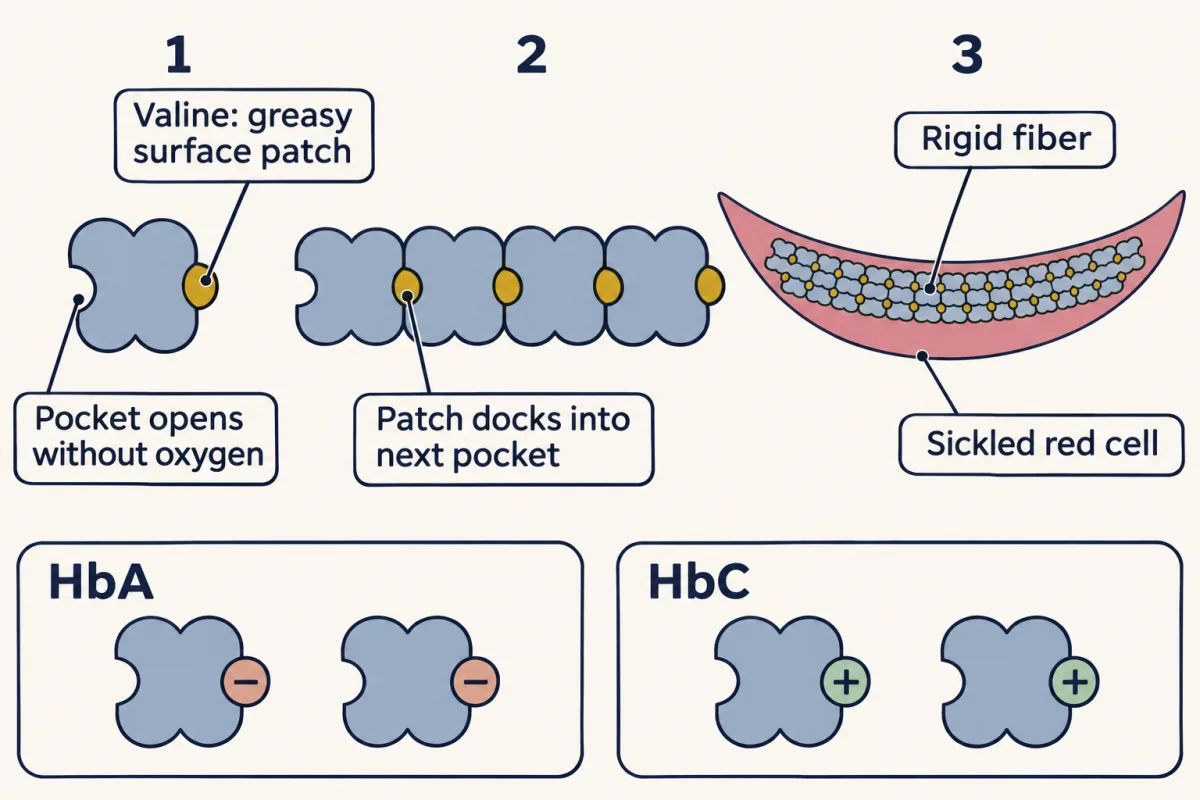
Now the opening question: sickle-cell hemoglobin (HbS), with valine for glutamate at beta position 6.
Charge explains the gel. Glutamate’s side chain is negative at 7.4; valine has no switch. Each of hemoglobin’s two beta chains loses one negative charge, so HbS is +2 per molecule relative to normal hemoglobin (HbA). In an alkaline buffer, where hemoglobin is net negative, HbA runs toward the anode faster than HbS.
Predict. Hemoglobin C (HbC), a real variant, replaces the same glutamate with lysine. How much does that change the charge of each hemoglobin molecule, compared with normal HbA? Would you expect HbC to form fibers?
Check your prediction
Lysine is +1 where glutamate was −1: +2 per chain, +4 per molecule, twice HbS’s change. On the same gel HbC runs slower still, so toward the anode the order is HbA, HbS, HbC.
Yet HbC does not form sickle fibers. It forms crystals and causes a much milder anemia. Lysine, like glutamate, is charged and hydrophilic, so it plants no greasy patch.
So charge explains how Pauling saw the defect; polarity explains why the cells sickle. Hydrophilic glutamate is at home on the protein’s water-facing surface; hydrophobic valine plants a greasy patch there. When hemoglobin gives up its oxygen, its shape shifts and a matching hydrophobic pocket opens on its surface. The valine patch of one molecule sticks into that pocket on the next, chaining hemoglobin into rigid fibers that deform the red cell into a sickle. That is where the disease starts; hemolysis, inflammation and blocked vessels follow from it. Charge and polarity, both set by structure, run from the membrane helix to the active site to the clinic.
Going deeper
The main line above does not depend on these details.
Assigning L and D: the CORN rule
View the alpha-carbon with the H pointing toward you and trace CO → R → N. Clockwise reads L, counterclockwise D; view from the other side and the handedness flips. The D residues in some peptides are made by enzymes after the ribosome. In R/S terms, L is S for every chiral amino acid except cysteine, whose sulfur-bearing side chain makes it R.
Thalidomide
Chirality has a body count. Thalidomide was sold in the 1950s as a sedative; one mirror-image form is thought to carry its sedative effect and the other its power to cause severe birth defects in roughly ten thousand children. Because the body converts each form into the other within hours, even the pure “safe” form would not have been safe.
pKa values inside proteins
A protein’s local environment can shift a pKa substantially: a buried Asp or a Lys in a hydrophobic pocket can move several pH units, and histidine’s is often shifted nearer to 7. Such shifts underlie pH-dependent catalysis and shape changes.
Small print on the numbers
- The uncharged form. The −NH₂/−COOH form is a tiny minority at every pH in water.
- The exact pI. Strictly, the pI is the pH where the net-charge sum is zero; the averaging shortcut matches it to within about 0.02 for the common ionizable amino acids, with tyrosine furthest off.
- Real curves. At very low pH, part of the added base goes to free H⁺ in the water, so on a measured curve the carboxyl’s half-equivalence point reads its pKa only approximately; the plotter draws the idealized curve.
- Tyr and Cys. At pH 7.4 tyrosine’s side chain is about 99.8% neutral and free cysteine’s about 86% (14% thiolate), so neither normally forms salt bridges.
- Numbering. Clinical genetics counts beta-globin from the starting methionine, which the finished chain lacks, so the sickle change Glu6Val is written p.Glu7Val.
Detecting amino acids in the lab
Chromatography sorts a mixture of amino acids by charge and hydrophobicity, the two side-chain properties this lesson is built on, and ninhydrin stains free alpha-amino groups purple (proline turns yellow) so each spot shows. The Techniques panel has more.
Your notes
Select any text in the lesson to highlight it or add a note to it. Your highlights and notes are saved in this browser and collected in your notebook.
This browser is not letting the site save anything (a private window or strict privacy settings), so highlights and notes will be lost when you leave the page.
How we measure it
Titration to find pKa and pI
Add base in measured steps and track pH. Each plateau is a buffering region centered on a pKa; the steep jumps mark the equivalence points. The half-equivalence point reads the pKa directly, and pI sits at the pH where net charge is zero — the two numbers that fingerprint an amino acid's acid-base behavior.
Assigning chirality (the CORN rule)
View the alpha-carbon with the H pointing toward you and trace CO → R → N around the carbon. Clockwise reads L, counterclockwise reads D. Commit to one camera angle: look from the opposite side and the handedness flips. Ribosomes build proteins from L-amino acids (plus achiral glycine); D residues in some peptides are made by later enzymes.
Detecting amino acids (ninhydrin and chromatography)
Ninhydrin reacts with free alpha-amino groups to produce a purple color (yellow for proline), so a separated mixture lights up spot by spot. Paper or ion-exchange chromatography sorts amino acids by charge and hydrophobicity — the same two side-chain properties this lesson is built on.
Practice questions on this lesson (29) → MCAT-style, original, each with an explanation
Where this lesson fits
Before thisReview first
- Water, Weak Bonds, and the Generation of OrderThis course · Lesson 3
You need its account of water and weak bonds to see why polar side chains face water and greasy ones hide.
After thisStudy next
- Protein Three-Dimensional Structure: The Sequence Sets the ShapeThis course · Lesson 5
It uses these side-chain properties to explain how a sequence folds, and why one swap can break the shape.
- Protein Binding and Allostery: How Hemoglobin Loads and Delivers OxygenThis course · Lesson 9
It takes the Glu→Val swap and histidine's proton switching further, using hemoglobin's oxygen binding and release.
- Techniques in Protein Biochemistry: Purify It, Then Prove ItThis course · Lesson 6
It uses each protein's charge at a given pH and its pI to separate proteins on columns and gels.
Elsewhere on the siteFilms, simulations and method films in other courses
- Build a big molecule from small blocksSimulation · Biology
Building a big molecule from small blocks shows how amino acids link into chains before side chains matter.
- Take It Apart, Build It AgainFilm · Biology
It shows amino acids as the building blocks that digestion splits and the body rejoins into protein.
- Speed up and slow down an enzymeSimulation · Biology
Enzyme activity changes with pH because charged side chains in the active site gain or lose protons.
Suggested by an AI model from the lessons themselves, then checked. How we use AI
Finish line
Test yourself
28 questions
Practice and MCAT-style, with explained answers.
Next · Lesson 5
Protein Three-Dimensional Structure: The Sequence Sets the Shape
One sequence, one working shape. Change a single letter and the shape, and the job, can break.
Continue →