Cell Signaling · Unit 1: How a cell knows anything
Signals and How Far They Travel
Rewritten on : new text and flashcards. Your notes and highlights stay, but the text around them may have moved.
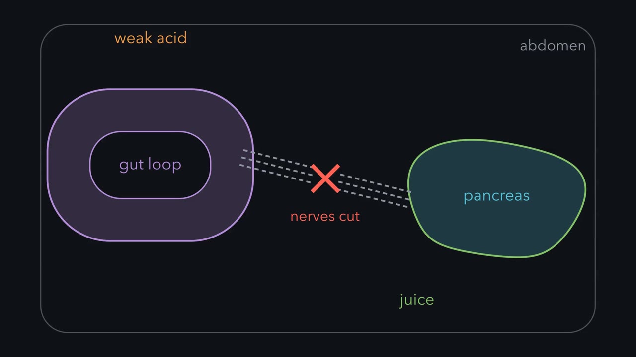
Cut every nerve to a loop of gut, drip acid on it, and the pancreas still answers. Who told it?
Pause when the reading reaches a film, and offer to play it. Saved on this device.
Watch the film now, or keep listening
Builds on:Crossing the MembraneWhy these?
 ▶ Watch Why Far Signals Ride the Blood · 2:38
Read the transcript of “Why Far Signals Ride the Blood”
Film transcript: Why Far Signals Ride the Blood
Cut every nerve to a loop of a dog's gut, then drip weak acid onto its lining. The pancreas still secretes its juice. It sits in a different part of the abdomen, and the leading view said the gut instructs it by nerve. But no wire joins the two organs now. What carried the message? The answer was a chemical, later named secretin. Acid released it from the gut lining, and the blood carried it to the pancreas. An extract of the lining, filtered and injected into a vein, made the pancreas secrete even more. But molecules can also drift on their own. So why ride the blood at all? That drift is called diffusion. A diffusing molecule is not heading anywhere.
It wanders. The typical time to wander a distance grows with the square of that distance. So diffusion punishes distance: a far target waits much longer than a near one. Take a small molecule in water, and change only the distance. It crosses a synaptic gap, 20 nanometers wide, in a fraction of a microsecond. It crosses a millimeter in about 8 minutes. It crosses a centimeter in about 14 hours. The centimeter is only 10 times farther than the millimeter, yet it costs hours instead of minutes. Past a few hundred micrometers, diffusion cannot deliver a message that must act within a minute. So a gland releases its hormone into the blood, which circulates around the body in about a minute.
Fast local signals do the opposite. A nerve cell releases its message into a gap only tens of nanometers wide. The message arrives almost at once, though some spills out to nearby cells. The extract reached every organ in the dog, and the pancreas poured out juice. A signal does not choose its audience. The receiver does. Epinephrine, a hormone, makes liver cells break down glycogen, fat cells break down fat, and the heart beat harder. So keep this rule: far messages ride the blood, and the receptor decides who answers.
In January 1902, two London physiologists cut every nerve running to a loop of a dog’s small intestine and dripped weak acid onto its lining, as stomach acid would reach it after a meal. The pancreas, a separate organ in a different part of the abdomen, began to secrete its digestive juice anyway. With no wire between the two organs, what carried the message?
The answer was a chemical. That answer shapes this whole course, because every cell is sealed inside a membrane and learns about the world only from what reaches its surface. How a message gets there is a transport problem, and this lesson asks what distance costs.
A message with no wire
The leading view in 1902, held by Ivan Pavlov among others, was that the gut instructed the pancreas by nerve. Nerves are fast and measurable, and nerve reflexes were the accepted explanation for how the gut controlled digestion. William Bayliss and Ernest Starling tested it by taking the nerves out, on 16 January 1902 at University College London. After the acid test, the same afternoon, they cut off a further piece of intestine, rubbed its lining with sand in weak acid, filtered the liquid and injected it into a vein.
Predict. For each test, what does a nerve-only theory predict, and what does a theory of a blood-borne chemical predict?
Check your prediction
| Test | Nerve-only theory predicts | Blood-borne-chemical theory predicts | Result |
|---|---|---|---|
| Acid on the gut lining, every nerve to the loop cut | No secretion, unless a local reflex runs through nerves in the gut wall that the cut missed | Secretion | The pancreas secreted |
| Extract of the gut lining injected into a vein | No secretion, because the injection bypasses every gut nerve | Secretion | The pancreas secreted even more |
Others at the time argued for that local reflex, but only the chemical theory fits both results.
Something in the gut lining, later named secretin, was released by acid and carried to the pancreas in the blood.
Two terms name what the dog showed. Signal transduction is the process by which a cell converts a signal from outside into a change inside, in its behavior or in the genes it reads. A first messenger is the signal itself, the molecule or physical stimulus that arrives from outside before the cell has done anything with it. Secretin is a first messenger. (The overview lesson signal transduction follows one signal’s relay inside the cell. This lesson stays outside.)
The experiment leaves a puzzle. The extract went everywhere the blood goes, so why doesn’t every organ respond? The section on receptors answers that.
Otto Loewi showed the same logic across a much shorter gap. In 1921 he stimulated the vagus nerve of a beating frog heart, which slowed it, then took the salt solution that had been in contact with that heart and transferred it to a second heart. The second heart slowed too, though no nerve to it had fired. He called the unknown substance Vagusstoff, vagus substance. A nerve cell had given an order by releasing a chemical that drifted to its target, not by passing a current. The chemical was later identified as acetylcholine.
Three questions and one idea
Three questions run through the lesson. How does a message get from sender to receiver? How long does it last? Who answers it? The first two share one answer, which needs two terms. A released molecule that wanders through fluid diffuses, and how fast it wanders is its diffusion coefficient, D. Sooner or later every messenger is destroyed or captured, and the average time one molecule survives is its lifetime, τ (tau).
The one idea to carry through this lesson. Lifetime buys reach and costs tempo. For a messenger that diffuses through roughly uniform tissue and is removed by first-order clearance, the steady gradient has a decay length of about √(D × τ). After release stops, its concentration falls on the timescale τ; building the gradient out to its decay length also takes on the order of τ. Shortening the lifetime reduces reach and speeds decay. Lengthening it increases reach but slows decay, and the trade is a poor one: the decay time grows in step with lifetime, while reach grows only as its square root. When a job needs more reach than diffusion can give in the time allowed, the body carries the messenger in flowing blood, which is bulk flow.
Which part applies where: the rule can describe autocrine, paracrine and synaptic signals when extracellular transport is mainly diffusion and removal is approximately first-order. Blood flow carries endocrine signals, so √(D × τ) does not set their range, although clearance still helps set how quickly their concentration falls after secretion stops. Contact modes lie outside this extracellular diffusion-clearance rule.
Diffusion punishes distance
Why ride the blood, when molecules drift on their own? Because diffusion is a random walk, and the typical time to wander a distance grows with the square of that distance.
In words The characteristic time is the distance squared, divided by twice the diffusion coefficient.
- t“t”
- the typical time to wander the distances
- x“x”
- the distancecm
- D“D”
- the diffusion coefficient, about 10−5 cm²/s for a small molecule in watercm²/s
Sanity check Double the distance and the time goes up four times, not two. Units: cm² divided by cm²/s leaves seconds.
Try one number
A small molecule, D = 10−5 cm²/s, wandering 1 mm (0.1 cm).
x² = 0.01 cm², and 2D = 2 × 10⁻⁵ cm²/s.
t = 0.01 / (2 × 10⁻⁵) = 500 s, about eight minutes.
Need a refresher? powers and roots · units and prefixes
Predict. A small molecule wanders 1 mm in about eight minutes. How long does it take to wander 1 cm, ten times farther?
Check your prediction
About fourteen hours, not eighty minutes: ten times the distance costs 10² = 100 times the time. At the other end, the same molecule crosses a 20 nm synaptic gap in a fraction of a microsecond.
Now run the law the other way and ask how far a small molecule gets in one minute: x = √(2 × 10−5 × 60) cm, about 0.035 cm, or 350 µm. Past a few hundred micrometers, diffusion cannot deliver a message that has to act within a minute. That is why hormones ride the blood, which circulates around the body in about a minute, and why fast local signals need the gap to be tiny. The film at the top of the page walks through the same three distances.
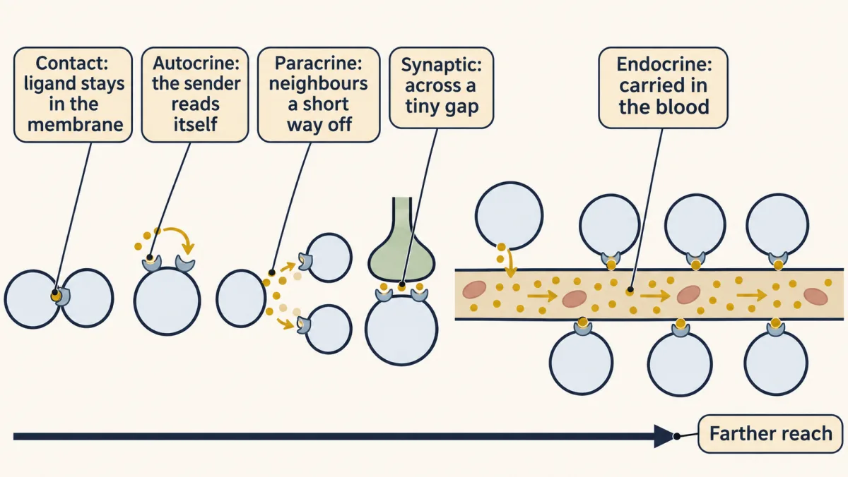
Five modes, five answers to the same physics
Each mode of signaling answers that law differently: how to cross a distance in the time the job allows, and how to keep the message from the wrong cell.
| Mode | Distance | How the message gets there | Typical arrival | What keeps it private |
|---|---|---|---|---|
| Contact | Touching cells | No extracellular trip: an anchored ligand binds across the contact, or ions and small molecules pass through a shared channel | No long transport delay; response kinetics still vary | The contact is the address |
| Autocrine | The sender itself | Released and bound at its own surface | Fast | Nothing inherently; nearby receptor-bearing cells may also respond |
| Paracrine | Neighbors a short way off | Diffusion through the fluid between cells | About a second or less for a small molecule a few cells away; minutes to hours for a protein that sticks or travels farther | A short life and capture along the way |
| Synaptic | A gap tens of nanometers wide | Diffusion across the gap | About a microsecond or less to cross | The tiny gap and fast clearance |
| Endocrine | The whole body | Bulk flow in the blood | Around the body in about a minute | Not the route; mainly which cells carry the receptor |
Contact. Two cells touch, and nothing is released into the space around them. In juxtacrine signaling, a ligand (a molecule that binds a receptor) is anchored in one cell’s membrane and binds a receptor on the cell pressed against it. In a gap junction, protein channels link the interiors of two cells, and ions and small molecules pass straight through. Strictly, what passes through is shared between interiors and never meets a surface receptor, so it is an awkward fit for a first messenger.
Autocrine. The cell releases a signal that binds receptors on its own surface, which lets a cell reinforce its own decision.
Paracrine. The signal is released into the fluid between cells and acts on neighbors a short way off. Histamine released during inflammation is the standard example. The signal is diluted as it spreads and destroyed or captured along the way.
Synaptic. A neuron releases a neurotransmitter into a gap only tens of nanometers wide, and the receiving cell sits right across it. This is a paracrine signal with the geometry built for speed and privacy: the distance is so small that the message arrives almost at once, and most of it acts on the cell across the gap, though some can spill out to receptors nearby. Some transmitters, serotonin among them, are often released away from a tight synapse and drift farther, a pattern called volume transmission.
Endocrine. A cell, often but not always in a gland, releases a hormone, a signal carried in the blood, and every cell the blood reaches is exposed to it. Insulin and estradiol work this way. So does secretin, which comes from cells in the gut lining, not from a gland. (The film’s line that a gland releases its hormone describes the common case, not the rule.)
Lifetime buys reach and costs tempo
Now the one idea, with numbers. A diffusing signal has no sharp edge; it fades. Molecules wander from the sender and are destroyed by enzymes, taken up by cells or stuck to the extracellular matrix. In its lifetime τ, a molecule wanders about √(D × τ), the distance over which the signal fades:
In words The reach is the square root of the diffusion coefficient times the lifetime.
- reach“reach”
- the one-dimensional decay length over which steady concentration falls by a factor of e (about 2.7), to roughly a third of its level at the sourcecm
- D“D”
- the diffusion coefficient: how fast the molecule wanderscm²/s
- τ“tau”
- the average lifetime of one molecule before it is destroyed or captureds
Sanity check This has the same square-root scaling as diffusion, but it comes from balancing diffusion with first-order removal. Units: cm²/s times s is cm², and its square root is cm.
Try one number
A secreted protein in water, D = 10−6 cm²/s, that survives τ = 100 s.
D × τ = 10−4 cm², and √(10−4 cm²) = 10−2 cm = 100 µm, about five to ten cell diameters.
Need a refresher? powers and roots · units and prefixes
Predict. A growth factor reaches about 100 µm from the cell that makes it. A mutation lets each molecule survive nine times longer. How far does it reach now? And how long does the wider gradient take to build?
Check your prediction
Three times farther, about 300 µm, because reach grows with the square root of lifetime: √9 = 3. And about nine times longer to build, because build time scales with the lifetime itself. Near the source the gradient fills in within a fraction of one lifetime, and farther out it takes longer still, so nine times the lifetime means nine times the wait. Three times the reach costs nine times the time.
Tissue is not water. Binding to matrix and to receptors slows a protein’s spread, because a molecule that is stuck part of the time covers less ground. Measured values in tissue are lower, from about 10−9 to 5 × 10⁻⁷ cm²/s, so the water value of 10−6 is an upper bound. Holding lifetime fixed, here is what a thousandfold lower D does:
| Lifetime | Reach with D = 10−6 cm²/s (water) | Reach with D = 10−9 cm²/s (sticky tissue) |
|---|---|---|
| 4 seconds | about 20 µm, one or two cells | under 1 µm |
| 100 seconds | about 100 µm, five to ten cells | about 3 µm |
| 3 hours | about 1 mm | about 30 µm |
At the slow end every reach is about thirty times smaller (√1000 ≈ 32), as much as a thousandfold drop in lifetime would cause. In real tissue the two seldom change alone: binding that slows a protein’s spread often also protects it and lengthens its effective lifetime. Either way, a paracrine signal stays local because its neighborhood destroys it, captures it or slows its spread.
The synapse pushes the same tradeoff to an extreme. Acetylcholinesterase removes acetylcholine within about a millisecond. Substituting a small-molecule D and that effective lifetime gives √(D × τ) = √(10−5 × 10⁻³) cm, about 1 µm. This is only an order-of-magnitude scale, not a literal boundary: the cleft is thin, the enzyme is not distributed uniformly, and its kinetics can saturate. Rapid clearance still helps make the signal brief and limit spillover.
The blood changes the bargain. Circulation, not molecular diffusion, sets an endocrine signal’s range. Clearance helps set the fall after secretion stops, but it does not by itself set how quickly secretion can rise or a target can respond. Epinephrine and insulin each fall to half their level in the blood within minutes, epinephrine in roughly two to three minutes, so ten minutes after the adrenal gland stops releasing it, about a tenth or less remains. The message fades unless the gland keeps sending it, while onset also depends on delivery, secretion and receptor-response kinetics.
Dilution has its own cost. Once mixed through the circulation, a hormone is distributed through about five liters of blood (about three of plasma), although its effective distribution volume depends on the hormone. Many endocrine receptors therefore bind low concentrations, and many surface-receptor pathways amplify what they detect through second messengers, the subject of later lessons.
When a cell answers its own call. Disrupting paracrine control is one route to cancer. A growth signal can be constrained in part by its short reach and short life. A cell that makes a growth factor and carries its receptor has created an autocrine loop, which can reduce its dependence on growth signals from neighboring cells and contribute to inappropriate proliferation. A landmark case came in 1983, when the cancer-causing gene of simian sarcoma virus, v-sis, turned out to encode a form of the B chain of platelet-derived growth factor (PDGF), a factor that blood platelets release at wounds to make nearby cells divide. Cells transformed by the virus make the factor and are driven by it through their own PDGF receptors.
Ending the message: clearance sets τ
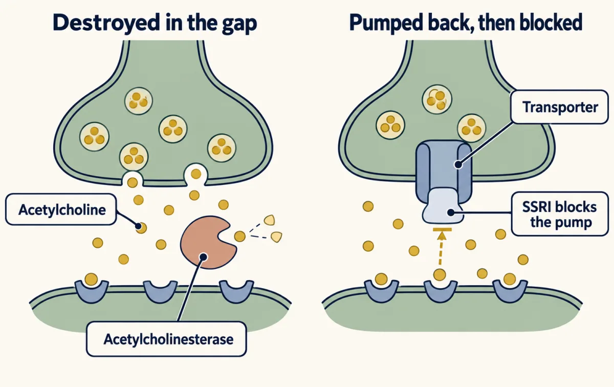
A signal that cannot end is a switch stuck on. One way cells end signals is by clearing the messenger, and each route of clearance is a way of setting τ. Enzymes destroy the molecule. Transporters pump it back into the cell that released it or into neighboring glia. Or blood flow carries it off, and from a small space such as a synaptic gap, diffusion out of the gap does the same job. A cell can also stop listening by turning down its receptors, which later lessons take up.
Acetylcholine shows the fastest case. Acetylcholinesterase sits in the gap between nerve and muscle, and its millisecond speed is the point: if the transmitter lingered, the next message could not be told apart from the last, so the gap has to be wiped clean after every impulse. That same speed made acetylcholine almost impossible to catch, until Dale and Feldberg blocked the enzyme and used a living muscle as the detector.
Predict. Eserine blocks acetylcholinesterase. Using the one idea, what happens to acetylcholine’s lifetime at the synapse, and what does the synapse gain and lose?
Check your prediction
The lifetime goes up, so each release has a longer effect. That is the gain, and Loewi showed it in 1926, when eserine prolonged the vagus substance’s effect. The cost is tempo: the gap is no longer wiped clean after each impulse, so the next message is harder to tell from the last. Reach grows too, but only as the square root of the lifetime.
Many transmitters are cleared another way. Serotonin is pumped back into the sending neuron by a transporter protein. A selective serotonin reuptake inhibitor (SSRI), such as fluoxetine, blocks that transporter, so serotonin lingers longer in the gap and the fluid around it and keeps its receptors occupied longer. That block is where the drug’s action begins, within hours. Its benefit in depression takes weeks to appear and is thought to come from slower changes in the brain that follow, so it is not simply a matter of topping up a missing chemical. Blocking acetylcholinesterase prolongs acetylcholine with the same result, by a different route, which is why some Alzheimer’s drugs and nerve agents both act on this enzyme.
Who answers: the receptor
Back to the dog’s puzzle: why doesn’t every organ the extract reached respond? The 1902 experiment recorded the pancreas and was not designed to survey other organs. The answer comes from tissues watched side by side.
The receptor, not the signal, sets who responds. Epinephrine, a hormone from the adrenal gland, drives glycogen breakdown in liver cells, fat breakdown in fat cells and a stronger beat in the heart, and a cell without the receptor ignores it. Acetylcholine slows the heart and contracts skeletal muscle, because the two cell types carry different receptors for the same molecule.
This answers the lesson’s third question, and it is the film’s closing rule: far messages ride the blood, and the receptor decides who answers.
Can the messenger cross? Where the receptor sits
Whether a messenger can cross predicts where its receptor sits. First messengers range from a push on the membrane to a whole protein, listed here roughly from small to large:
- Physical stimuli: mechanical force, sound, temperature and light.
- Ions and small molecules: Ca2+, Na+, Cl-, O2, CO2, CO and NO.
- Metabolites: glucose, adenosine, lipids and amino acids.
- Hormones: estradiol and ghrelin, which are a steroid and a peptide.
- Secreted proteins: interleukins, growth factors, death factors and chemokines.
- Extracellular matrix: collagen, elastin, fibronectin and laminin.
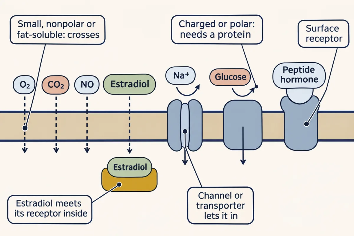
The categories overlap. A hormone is defined by how it travels, in the blood, not by what it is made of, so estradiol, uncharged and fat-soluble, behaves more like O2 and NO than like the peptide ghrelin. That is why the useful sort is by crossing, not by category.
Predict. Glucose is small and uncharged. Estradiol is larger, four fused carbon rings. Which one crosses the lipid core of the membrane on its own?
Check your prediction
Estradiol, which has almost no charged groups. Glucose is covered in polar hydroxyl groups, so it needs a transporter. Charge and polarity decide, not size alone.
The lipid core of the bilayer admits small molecules that are uncharged and not very polar, and fat-soluble ones. O2, CO2 and NO slip through, and so does estradiol. These can be sensed inside the cell, and estradiol meets its main receptor there, though it also acts on some receptors at the surface.
Many other messengers need a protein in the membrane: a surface receptor, or a channel or transporter that lets them in. Ions are charged and cannot pass on their own. Neither can glucose, a peptide, a protein or a matrix fiber. Acetylcholine and epinephrine are small, but the nitrogen of acetylcholine carries a permanent positive charge, and the amine of epinephrine is positively charged at body pH. Carbon monoxide and some other small, uncharged or fat-soluble messengers can cross without such a protein. How surface receptors are built is the subject of lesson 4.
Design problem
You must tell every liver cell in the body to release glucose within a minute, and separately tell only the cell next door to divide. Choose a mode of signaling for each job and defend it.
Saved in this browser only.
One way to do it
The liver job: endocrine. Use a hormone released into the blood. The blood reaches every liver cell within about a minute, while diffusion would need hours to cover even a centimeter. Adrenaline fits. It ends within minutes by dilution and breakdown, and its receptor starts a relay inside each liver cell that amplifies it, so a dilute hormone is enough. The price is that every other cell with that receptor answers too, including fat cells and the heart.
The next-door job: juxtacrine or paracrine. If only a touching cell must divide, use a ligand anchored in the membrane, so the contact is the address. If a small neighborhood may divide, use a paracrine factor that is destroyed quickly, so that √(D × τ) is about a cell diameter or two. An endocrine signal would be wrong here: through the blood, a command to divide reaches every cell that carries the receptor.
Going deeper
Catching acetylcholine with a leech muscle
By the time fluid from around a stimulated nerve reached a test tube, the enzyme had destroyed the acetylcholine, and what was left was far below anything a chemical test of the 1930s could detect. Eserine, which protects the transmitter, also makes a strip of leech muscle respond to far smaller amounts, and in 1934 Henry Dale and Wilhelm Feldberg combined the two. The height of the leech muscle’s slow contraction, read against contractions from known amounts, became the measurement. Fluid collected from a stomach while its vagus nerve was stimulated made the muscle contract. A nerve had been caught releasing the chemical.
Diffusion in three dimensions
The lesson and the film use the one-dimensional estimate, t ≈ x²/2D. In three dimensions a molecule reaches a given distance about three times sooner, t ≈ x²/6D. Around a point source the steady signal falls as e−r/λ/r rather than e−x/λ, but the decay length λ = √(D × τ) is the same. Neither change alters a conclusion: time still grows with the square of distance, and reach with the square root of lifetime.
Where the reach rule bends
The rule assumes roughly first-order removal (a fixed fraction of the molecules removed per unit time) in roughly uniform tissue. Uptake by receptors that saturate, binding to matrix (which lowers the effective D), cytonemes (thin cell projections that carry signals between cells) and transport through cells (transcytosis) all change the numbers.
Your notes
Select any text in the lesson to highlight it or add a note to it. Your highlights and notes are saved in this browser and collected in your notebook.
This browser is not letting the site save anything (a private window or strict privacy settings), so highlights and notes will be lost when you leave the page.
How we measure it
Severing nerves and injecting a tissue extract
To ask whether a nerve carries a message, cut it and see whether the effect survives. Then grind the suspected source tissue in acid and inject the extract into the blood. If the extract reproduces the effect, a circulating substance carries the message.
Fluid transfer between two beating hearts
Stimulate the nerve of one isolated heart, collect the fluid that was in contact with it, and apply the fluid to a second heart. If the second heart changes, the first nerve released a substance into the fluid.
Bioassay with eserine-treated leech muscle
A living tissue that responds to a molecule can serve as a detector. A strip of leech muscle contracts to tiny amounts of acetylcholine, and the size of the contraction is read against contractions from known amounts. Eserine stops the enzyme that would destroy the sample.
Method films · the methods library
Practice questions on this lesson (3) → MCAT-style, original, each with an explanation
Where this lesson fits
Before thisReview first
- Crossing the MembraneBiology · High school
You need to know which molecules cross the lipid bilayer on their own, which decides where a receptor sits.
After thisStudy next
- Binding: Affinity, Occupancy and Dose–ResponseThis course · Lesson 3
It takes the receptor's answer further, with affinity and occupancy deciding how much of a signal a cell detects.
- A Field Guide to ReceptorsThis course · Lesson 4
It builds on who answers by showing how receptor families let one messenger mean different things to different cells.
- Turning the Signal Off: Arrestins, RGS Proteins and PhosphodiesterasesThis course · Lesson 16
It takes clearance and signal lifetime further by showing how cells switch off the receptor and the second messenger.
Elsewhere on the siteFilms, simulations and method films in other courses
- Counting failuresSimulation · Neuroscience
It shows transmitter release and quantal packets at a synapse, the short-range mode of signaling classified here.
- Half-life and plasma levelSimulation · Organismal Biology
It shows hormone level in plasma over time, the long-range endocrine mode with dilution and clearance.
- Tap, Drain and a SensorFilm · Organismal Biology
It shows a hormone released into blood and carried to distant receptor cells, the endocrine mode.
Suggested by an AI model from the lessons themselves, then checked. How we use AI
Finish line
Test yourself
8 questions
Practice and MCAT-style, with explained answers.
Next · Lesson 2
Finding a Signal: NGF, EGF and the Bioassay
A mouse tumor that never touched a chick embryo's nerves made its ganglia swell. What was it releasing, and why do neurons depend on a signal from their targets?
Continue →