Biochemistry · Digestion & Absorption
Digestion and Absorption: Turning a Meal into Molecules Your Cells Can Use
Rewritten on : new text, figures and flashcards. Your notes and highlights stay, but the text around them may have moved.
Digestion has two halves that only work together: enzymes cut a meal into small pieces, and the gut wall carries those pieces into the blood or lymph.
Pause when the reading reaches a film, and offer to play it. Saved on this device.
Watch the film now, or keep listening
Builds on:Basic Concepts of Enzyme ActionCarbohydrates: One Bond Decides Fuel or FiberLipids: The Building Blocks That Refuse to PolymerizeWhy these?
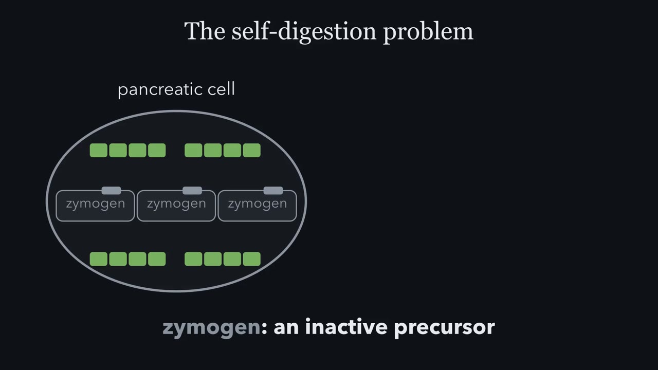 ▶ Watch The master switch of digestion · 2:48
Read the transcript of “The master switch of digestion”
Film transcript: The master switch of digestion
Here is the central puzzle of digestion. Your digestive enzymes are powerful enough to tear proteins apart, yet the cells that make them are themselves built from protein. So how do you manufacture a molecular shredder without shredding the factory? The answer is the zymogen, an inactive precursor. The cell builds the enzyme in a folded-but-dormant form, ships it out, and normally switches it on where digestion should happen. This single design principle, activation only where and when needed, recurs everywhere in physiology from blood clotting to apoptosis. Keep it in mind as our organizing idea. Notice that digestion is really one chemical trick repeated three times.
Hydrolysis means breaking a bond by inserting water. Proteases attack the peptide bond, disaccharidases attack the glycosidic bond, and lipases attack the ester bond. In every case, water donates a hydrogen to one fragment and a hydroxyl to the other, capping both freshly broken ends. The substrates look different, the enzymes are different, but the logic is identical. If you understand one row, you understand all three. That bottom summary is the takeaway worth memorizing. Water provides the H and the OH that finish off each broken bond. This is the activation map for the digestive proteases. In the stomach, low pH converts pepsinogen into active pepsin.
In the small intestine, the pancreas delivers a whole family of zymogens. Watch what trypsin does once it is switched on. It activates chymotrypsinogen, the procarboxypeptidases, and proelastase. Trypsin is the master switch. That single arrow pattern, one activated enzyme turning on all the others, is why the note at the bottom matters. It also previews a clinical point. Knock out the ability to make trypsin and the entire downstream cascade never fires, no matter how much of the other zymogens you have. Put the cascade idea to the test. On the left is the normal activation tree. Enteropeptidase switches on trypsin, and trypsin then activates chymotrypsin, the carboxypeptidases, elastase, and procolipase, a lipase helper that works even uncut.
On the right, remove the ability to make trypsinogen. Now the master switch is gone, and the downstream protease zymogens stay locked, no matter how abundant they are. That is why a trypsinogen deficiency is so much worse than losing any single other zymogen. Lose one branch and you lose one enzyme. Lose trypsin, and pancreatic protein digestion collapses at once, while fat digestion is largely spared.
Every meal you eat is a chemistry problem your body has to solve. How do you turn a pizza into you? The pizza is built from someone else’s molecules: wheat protein, starch, cheese fat, none of it yours and none of it small enough to cross into a cell.
Here is the catch. The cheese protein and your own pancreas are held together by the same bond, the peptide bond, and the pancreas makes enzymes whose whole job is to cut it. So why does the pancreas cut the meal and not itself? The answer explains most of digestion.
Digestion has two halves that only work together. First, enzymes break the meal into small pieces. Second, the lining of the small intestine takes those pieces up and passes them on, to the blood or, for long-chain fats, to the lymph. Break it down, then take it up: almost nothing reaches glycolysis or any later pathway until both are done.
One reaction, downhill but slow
The single chemistry of digestion is hydrolysis: a water molecule splits a covalent bond and caps the two new ends, donating an -H to one side and an -OH to the other. One water molecule is consumed for every bond broken, spent as a reactant rather than idled as solvent. Three bonds hold our food together. Proteases hydrolyze the peptide bonds in protein. Amylases and the disaccharidases of the brush border (the dense microvilli on the gut cells’ surface) hydrolyze the glycosidic bonds in starch and sugars. Lipases hydrolyze the ester bonds in fat. One cut, three cutters: each enzyme family decides which bond it can reach.
If water is the reactant, and the gut is full of water, what are the enzymes for? Hydrolysis is downhill but very slow. Downhill (ΔG is negative): left long enough, almost every peptide bond in a dilute solution would end up cut. Slow: with no enzyme, a peptide bond in neutral water lasts years to centuries, and a glycosidic bond in starch lasts millions of years. In a meal’s six hours or so in the gut, plain water would break fewer than one peptide bond in a thousand. An enzyme makes a cut in milliseconds.
Predict. If hydrolysis is downhill, why doesn’t a steak left in a glass of water overnight fall apart into amino acids?
Check your prediction
Downhill tells you where the reaction ends up, not how fast it gets there. On a bond’s timescale of years to centuries, one night in water does almost nothing. (A real steak does change overnight, through the meat’s own enzymes and bacteria: enzymes again.)
The one idea to carry through this lesson. Breaking a food bond with water is downhill but, on its own, extremely slow. So a bond is cut only where an active enzyme meets an exposed bond, and digestion is the body controlling those two meetings. In the gut it exposes bonds (chewing, acid unfolding protein, bile breaking fat into droplets) and lets each enzyme’s shape pick which bonds it can reach. In the gland it does the opposite: any secreted enzyme whose target sits beside it in the secretory granule (every secreted protease, since the granule is packed with protein, and the phospholipase that can attack the granule’s own membrane) is made switched off and turned on only in the gut. So the pancreas makes trypsin switched off and amylase switched on.
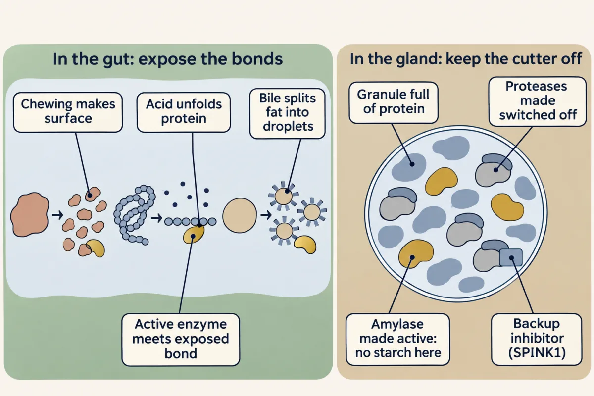
The rule tells you which enzymes must be built switched off; it does not forbid switching others off too. Absorption needs only a hand-off: the small pieces cross the gut wall through doors built for them.
Exposure starts with mechanical work. Teeth and stomach muscles crush; they break nothing covalent, but by shredding food they multiply its surface area so enzymes have more bonds to reach. Crush, then dissolve.
What comes out is exactly what the gut wall can absorb: amino acids plus di- and tripeptides from protein, monosaccharides from starch and sugars, and fatty acids with monoacylglycerols from fat. The lanes below follow each from the mouth to the blood or lymph, and the next three sections take them from easiest to hardest: carbohydrate, protein, then fat. Click any stage to see the enzyme working there and the bond it breaks, and use Show the water to see where each water molecule is spent.
Click any stage above to see the enzyme, the bond it breaks, and where each macronutrient becomes absorbable.
Three classes, three sets of enzymes, one reaction: hydrolysis. Carbohydrate ends as monosaccharides, protein as amino acids, fat as fatty acids and monoglycerides — all small enough to cross the gut wall. Carbs and protein head straight to the blood; fat is too bulky for capillaries, so it takes the scenic route through the lymph. And the protein side balances on a single switch: enteropeptidase flips trypsin on, trypsin flips the other proteases on. Knock that switch out and an infant can starve at a full table.
Carbohydrate: the enzyme’s shape picks the bond
Starch digestion begins in the mouth with salivary alpha-amylase and continues with pancreatic alpha-amylase. Amylase reads two things about a glycosidic bond: which way it points (alpha, not beta) and where it joins (carbon 4, not carbon 6). It cleaves only alpha-1,4 bonds. Fail either test and the bond is invisible to it.
Predict. Amylopectin, the branched form of starch, is chains of alpha-1,4 links joined at alpha-1,6 branch points. Cellulose is glucose joined by beta-1,4 links. What does amylase leave from each?
Check your prediction
From amylopectin, oligosaccharides and stubby limit dextrins, not free glucose: amylase cuts the chains but cannot touch the branch points. From cellulose, nothing: every link is beta. That is why we live on starch but not on cellulose, though both are pure glucose polymers.
Brush-border enzymes finish the job: alpha-glucosidase and alpha-dextrinase, plus sucrase and lactase splitting the disaccharides.
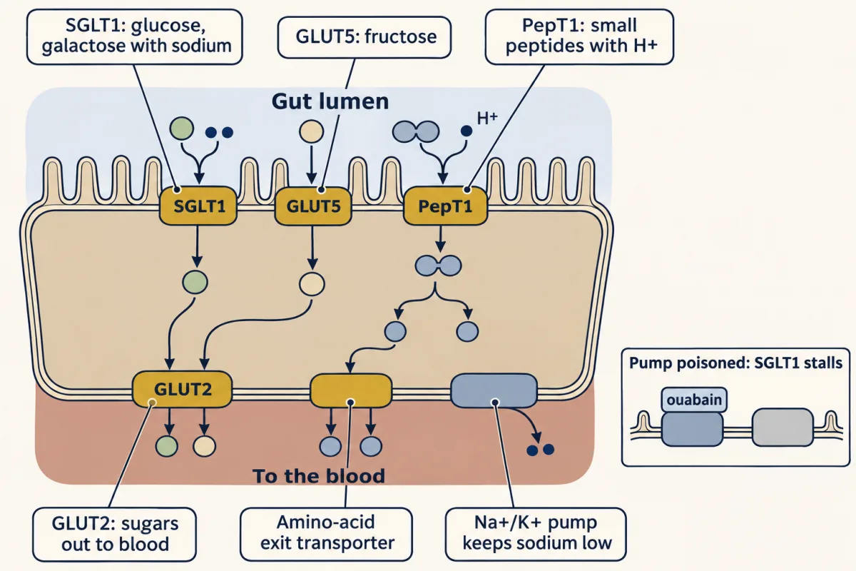
Then the doors into the gut cell, the enterocyte. Sodium-glucose cotransporter 1 (SGLT1) carries glucose and galactose in by secondary active transport: uphill for the sugar, paid for by sodium flowing downhill. The sodium gradient is kept up by the basolateral Na+/K+-ATPase (poison that pump with ouabain and SGLT1 stalls). Each glucose rides in with two sodium ions, and each is pulled in by both the sodium gradient and the cell’s negative inside, so the push is squared. In principle SGLT1 can pile glucose up inside the cell to thousands of times its lumen level, and the gut keeps pulling glucose in even when little is left. GLUT5 carries fructose by facilitated diffusion, so it can move fructose only down its gradient.
Leaving is the easy step. GLUT2, in the basolateral membrane, is the door all three sugars leave through toward the blood. It is passive too, and that works because SGLT1 has already piled glucose up inside the cell: from there, the blood is downhill.
Protein: switch the enzyme on only in the gut
The mouth contributes only mechanical work; there is no significant salivary protease. The chemistry starts in the stomach, where the gastric H+/K+-ATPase, the proton pump, drives the pH down to 1.5 to 2.5.
That acid does one job from each half of the rule. It denatures dietary proteins, unfolding them so their peptide bonds are exposed. And it switches on the stomach’s protease. The chief cells store pepsinogen, an inactive precursor, at neutral pH. As the surrounding pH falls, and fastest below about pH 3, the extra segment that holds it off lets go. The first few pepsin molecules then cut more pepsinogen, so activation is autocatalytic and snowballs. No separate enzyme is needed: acid is the trigger.
When the partly digested protein reaches the small intestine, its lining answers with two key hormones. Secretin triggers a flood of bicarbonate that neutralizes the stomach acid. CCK (cholecystokinin) triggers release of the pancreatic enzymes and squeezes bile from the gallbladder. Into the lumen come the pancreatic proteases, and with them the opening puzzle.
Why the pancreas does not digest itself
The pancreas makes these proteases in bulk and stores them in granules surrounded by protein: the other enzymes in the granule, its membrane proteins, the cell itself. A peptide bond is safe for years in plain water. It is not safe next to an active protease.
Predict. What must be true of a protease the pancreas can safely make and store?
Check your prediction
It must be inactive everywhere inside the pancreas and switch on only after it leaves. The bonds in the gland are exposed; the only thing the gland can control is whether the enzyme works.
The pancreas does this in three steps. The film at the top of the page walks through the same relay.
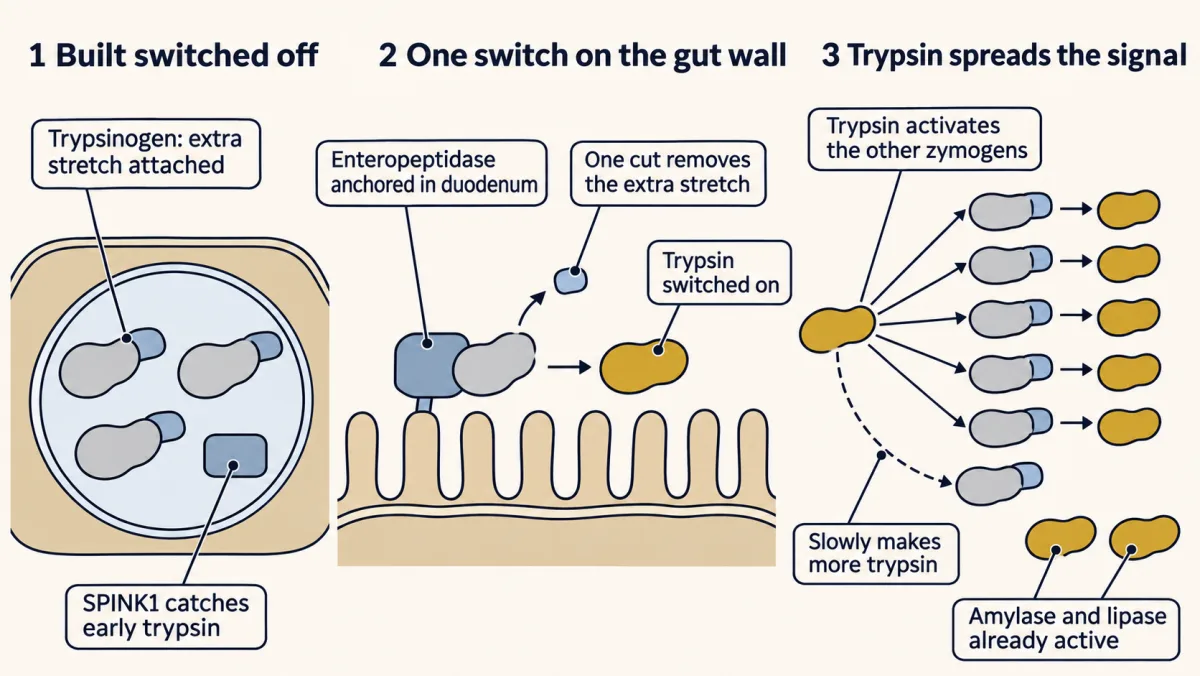
Step 1: build it switched off. Each protease is made as a zymogen (or proenzyme), an inactive precursor with an extra stretch of polypeptide. In trypsinogen, while that extra stretch at the start of the chain is attached, the active site is misshapen: the pocket that holds the substrate and the spot that steadies the reaction’s charged intermediate (the oxyanion hole) are not yet formed. Each zymogen travels through the cell and sits in a secretory granule, still inactive, with a trypsin inhibitor made in the same cells (SPINK1) as backup to catch the odd molecule that switches on early.
Step 2: put the one switch outside the gland. Enteropeptidase is anchored to the lining of the duodenum, the first part of the small intestine. It recognizes a rare sequence at the start of trypsinogen, four aspartates followed by a lysine, and makes a single cut just after the lysine. The new end of the chain tucks into the protein and the active site snaps into shape. Because the switch sits on the gut wall, the first trypsin normally appears only where pancreatic juice meets the intestine.
Step 3: let trypsin spread the signal. Trypsin cuts peptide bonds just after a lysine or an arginine, and the activating bond in every pancreatic zymogen follows one: chymotrypsinogen, proelastase and the procarboxypeptidases (three more protein cutters), prophospholipase A2 (the enzyme that attacks membrane lipids) and procolipase (a lipase helper). Trypsinogen has such a bond too, so trypsin also makes more trypsin, though slowly, because the four aspartates next to that bond repel trypsin. Trypsin is the master switch: one cut by enteropeptidase becomes a flood of active enzymes, all of it in the gut.
Predict. Step 3 is a loop: trypsin makes more trypsin. What danger does that loop carry?
Check your prediction
A loop that amplifies in the gut amplifies anywhere. If a few trypsinogen molecules switch on inside the pancreas faster than the inhibitor can catch them, trypsin activates more trypsin and then the other zymogens, and the gland starts to digest itself. Premature trypsin activation inside the gland is thought to be a key early step in acute pancreatitis, alongside inflammation that does not need trypsin; gallstones and heavy alcohol use are its commonest triggers. In one inherited form, a single change in cationic trypsinogen (PRSS1 R122H) removes the spot where stray trypsin is normally cut and destroyed, so the loop loses its brake.
Now take the switch away. In infants born without enteropeptidase, the duodenal fluid has almost no protease activity, but add enteropeptidase to it in a tube and trypsin activity appears: the zymogens were there all along, and only the switch was missing. Pancreatic protein digestion largely fails. Pepsin and the brush-border peptidases still do some work, but not enough, and the infant fails to thrive and swells from low blood protein despite eating. Triacylglycerol and carbohydrate digestion are largely spared: lipase is secreted active and uncut procolipase already helps it, and the carbohydrate enzymes never needed trypsin. Try it with Knock out enteropeptidase / trypsin in the lanes above and watch carbohydrate carry on while fat only slows. The film removes trypsinogen rather than enteropeptidase; either way no trypsin forms, and the disorder seen in clinics is enteropeptidase deficiency.
Why are amylase and lipase secreted active? Run the rule. The granule holds no starch, and the cell’s own glycogen lies in the cytoplasm, on the other side of the granule membrane. There is no triacylglycerol in the granule either, and pancreatic lipase works only at an oil-water surface, helped by colipase. Their targets are not inside the granule, so they need no off switch. The same logic covers pancreatic ribonuclease and deoxyribonuclease, also secreted active: the granule holds no RNA or DNA. Pepsinogen is the one exception to the enteropeptidase relay: the same design, with acid as the switch.
Three sites of cutting
Once switched on, trypsin, chymotrypsin, elastase, and carboxypeptidase chop proteins in the lumen into short oligopeptides and some free amino acids. The second and third sites are the brush border and the cell itself. Brush-border peptidases trim peptides to amino acids plus di- and tripeptides; the small peptides are absorbed intact through the transporter PepT1 and finished off by peptidases inside the cell, and the freed amino acids exit to the blood through basolateral uniporters and antiporters.
Acid in the stomach, bicarbonate in the duodenum: each enzyme in this chapter works best at the pH of its own compartment. Slide along the gut below and watch which enzymes switch on and which fall silent.
There is no single "best" pH for digestion — each enzyme is tuned to the compartment it works in. Salivary amylase starts on starch at the near-neutral pH of the mouth, then the stomach's acid bath denatures it for good while switching pepsin on. Once the chyme reaches the duodenum, pancreatic bicarbonate neutralizes the acid back to ~8, which is exactly where pancreatic amylase, trypsin, chymotrypsin, and lipase do their best work. Match the enzyme to its pH, and you have matched it to its place. pH sets how well each enzyme works; only pepsin uses pH as its on-switch. The pancreatic proteases are switched on by a cut, and amylase and lipase arrive already on.
Fat: the exposure problem
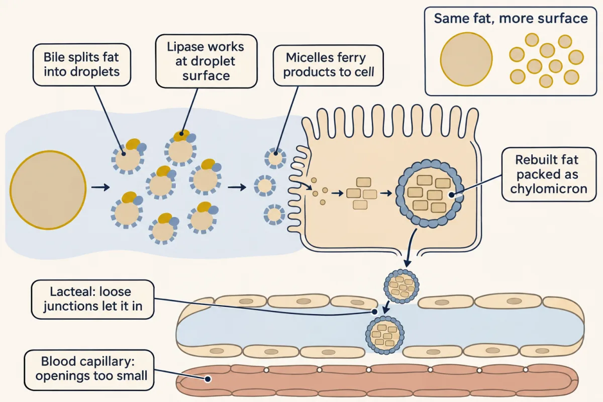
Fat is the hard lane because of exposure: it will not dissolve in the watery gut, so lipase reaches only the bonds at a droplet’s surface. The solution has four steps.
- Bile salts emulsify the fat the way soap cuts grease, breaking it into tiny droplets. This is geometry: splitting one 1 mm droplet into droplets 1 µm across, with the same total volume, multiplies the surface area 1,000-fold, and the surface is where lipase works.
- Pancreatic lipase, helped by colipase and by a gastric lipase that starts the job in the stomach, works at the droplet’s surface. It cleaves the ester bonds at the two outer positions (sn-1 and sn-3) to leave a 2-monoacylglycerol and two free fatty acids.
- Those products are packed into micelles, tiny bile-salt bundles that ferry them through the watery layer to the cell.
- Inside the enterocyte, the cell rebuilds triacylglycerols from the parts, IKEA furniture reassembled after shipping, then exports them as chylomicrons.
Why spend energy undoing digestion? Fatty acids and monoacylglycerol are what can cross the membrane. Rebuilding uses them up as they arrive, so their level inside stays low and more keep flowing in; it stops free fatty acids acting as detergents; and it turns them into cargo that can be wrapped in protein and shipped. The rebuilt molecule need not be the original one.
Chylomicrons are far larger than the openings in the wall of a blood capillary, but they can slip through the loose junctions of the lymph vessel at the center of each villus, the lacteal. So they take the scenic route through the lymph. Long-chain fats, along with cholesterol and the fat-soluble vitamins, travel this way.
Predict. In cystic fibrosis, thick secretions can block the pancreas, so little pancreatic enzyme reaches the duodenum, while stomach acid and bile are normal. Which lane fails worst, and why?
Check your prediction
Fat. Carbohydrate has salivary amylase to start and brush-border disaccharidases to finish. Protein has pepsin and the brush-border peptidases. Fat has no brush-border backup: bile still emulsifies the droplets, and gastric lipase starts the job, but pancreatic lipase is the principal triacylglycerol-digesting enzyme. Undigested fat passes into the stool (steatorrhea).
Taking it up: where each piece goes
The hand-off sorts the products. Monosaccharides, amino acids, and short- and medium-chain fatty acids are water-soluble enough (medium-chain fatty acids ride on albumin) to leave for the portal blood and the liver; long-chain fat leaves as chylomicrons through the lymph.
Clinical connections
The same routes predict what goes wrong, and where drugs act.
- Lactase deficiency (carbohydrate lane). Undigested lactose stays in the gut and draws water in after it, so the diarrhea is osmotic, with no immune attack involved. Gut bacteria then ferment the lactose, producing gas and bloating.
- Pancreatitis and pancreatic insufficiency (protein and fat lanes): trypsin switched on in the wrong place, or too little enzyme reaching the right one (the predictions above).
- Acid reflux (GERD) (protein lane). The stomach’s proton pump is the target of omeprazole, which is activated by acid and forms a covalent bond to the pump. The bond outlasts the drug in the blood, so acid stays suppressed until the cell makes new pumps.
- Celiac disease is an autoimmune reaction, not an allergy, to proline- and glutamine-rich gluten peptides that resist complete digestion (proline kinks the backbone), and it develops only in genetically susceptible people.
That is the full machine: foreign polymers in, absorbable pieces out. The monosaccharides are where the next chapter begins, as glucose steps onto the conveyor belt of glycolysis.
Going deeper
How slow is slow?
The half-lives above are literature values for bonds in neutral water at 25 °C with no enzyme. Published estimates for the peptide bond run from about 7 to about 450 years; a starch glycosidic bond is about 4.7 million years, and a simple carboxylic ester (ethyl acetate at pH 7) about 2 years. Body temperature and stomach acid speed these up only modestly; lab hydrolysis of protein takes 6 M HCl at about 110 °C for a day. The downhill side: ΔG°′ for peptide hydrolysis is between about −8 and −17 kJ/mol, so a 1 mM peptide at equilibrium is more than 99.99% cut. An enzyme with a turnover of about 50 per second makes a cut every 20 ms.
Why the gut wall’s own peptidases do not digest it
Here the rule is only a partial fit: the cell’s own surface proteins face active pancreatic proteases and brush-border peptidases. Heavy glycosylation of the surface proteins shields their peptide bonds, the brush-border peptidases work on small unfolded peptides (the exposure half of the rule), enteropeptidase cuts one rare sequence, and mucus and the lining’s turnover every 3 to 5 days add protection.
How a zymogen is held off
The extra stretch works differently in different zymogens. In trypsinogen it holds the active site misshapen (Step 1); in pepsinogen it physically plugs the active site until acid releases it. The stomach’s pepsinogen also comes in two groups, PGI and PGII, with the same acid switch.
Colipase and why fat digestion survives without trypsin
Trypsin converts procolipase to colipase, which helps pancreatic lipase grip the fat droplet. Lipase itself is secreted already active, and uncut procolipase already does much of colipase’s job, so trypsin’s cut tunes fat digestion rather than enabling it. That is why losing enteropeptidase or trypsin cripples protein digestion while triacylglycerol digestion is largely spared.
Why omeprazole outlasts itself
Omeprazole is a prodrug: it stays inert until it reaches the acid-filled channels of the parietal cells, right where the pumps sit. It lasts only about an hour in the blood and can catch only pumps that are actively secreting, so each daily dose blocks part of the pump population, and full effect builds over about three to five days. After a dose, acid secretion recovers only as the cell slowly breaks the covalent bond and makes new pumps.
SGLT2 and the gliflozins
SGLT1’s kidney cousin, sodium-glucose cotransporter 2 (SGLT2), is another sodium-coupled glucose door, run by the same Na+/K+-ATPase gradient. It is the target of the gliflozin diabetes drugs: block it and glucose the kidney would have taken back is dumped into the urine.
GLP-1 drugs: cure and side effects from one physiology
Glucagon-like peptide 1 (GLP-1), an incretin hormone from intestinal L-cells, slows gastric emptying and signals fullness. The blockbuster drug semaglutide (Ozempic, Wegovy) amplifies the GLP-1 signal. Its famous nausea comes partly from slowed gastric emptying and partly from direct action on the brainstem. Most of the weight loss comes from the drug’s action on appetite circuits in the brain, and it blunts the glucose spike by boosting insulin release from the pancreas.
What turns gluten resistance into celiac disease
Proline’s kinks let gluten fragments survive intact, but resistance alone does not cause the disease. In people who carry the HLA-DQ2 or DQ8 genes, the enzyme tissue transglutaminase modifies those fragments so they bind tightly to HLA-DQ2 or DQ8 on antigen-presenting cells, which activate T cells, and the resulting immune attack damages the gut lining. The damage flattens the villi, so absorption of many nutrients suffers, and celiac damage to the villi can cause a secondary lactase deficiency.
Your notes
Select any text in the lesson to highlight it or add a note to it. Your highlights and notes are saved in this browser and collected in your notebook.
This browser is not letting the site save anything (a private window or strict privacy settings), so highlights and notes will be lost when you leave the page.
How we measure it
Cell-free enzyme isolation (the Kühne move)
Take a digestive organ, grind it, and recover the catalytic activity in a tube where you can control pH, substrate, and time. Kühne studied trypsin in pancreatic extracts this way and coined the word enzyme for the worker itself. The whole chapter is this trick made routine: proteases, amylases, and lipases studied as molecules, not as an appetite of the living gut.
Protein crystallization and solubility-curve purity (Northrop)
Salt a protein out of solution with ammonium sulfate, collect crystals, and test purity not with a melting point (proteins have none) but with solubility curves grounded in the Gibbs phase rule. Northrop crystallized pepsin and trypsin this way and showed the catalytic activity tracked the crystal exactly, helping end the colloidal theory of enzymes.
Zymogen activation assays
To prove an enzyme is born inactive, you measure activity before and after a specific cleavage. Add enteropeptidase to trypsinogen and watch protease activity switch on; show pepsinogen turns itself on below pH 3. The assay helped order the activation cascade and is how enteropeptidase deficiency was first diagnosed; today genetic testing can confirm it.
Membrane-transport pharmacology
Block one transporter and read the consequence. Ouabain poisons the Na+/K+-ATPase and SGLT1 glucose uptake collapses, proving the cotransporter runs on the sodium gradient. SGLT2 inhibitors spill glucose into urine; this is how transporter identity is mapped and turned into drugs.
Practice questions on this lesson (5) → MCAT-style, original, each with an explanation
Where this lesson fits
Before thisReview first
- Basic Concepts of Enzyme ActionThis course · Lesson 7
You need to know that enzymes speed up possible reactions without changing their direction to follow why hydrolysis is downhill but slow.
- Carbohydrates: One Bond Decides Fuel or FiberThis course · Lesson 10
The alpha versus beta glycosidic bonds from there explain why amylase cuts starch but not cellulose.
- Lipids: The Building Blocks That Refuse to PolymerizeThis course · Lesson 11
Triacylglycerol structure and the ester bonds from there are what lipase cuts and the enterocyte rebuilds.
After thisStudy next
- Glycolysis: The Investment-and-Payoff PathwayThis course · Lesson 16
The monosaccharides absorbed here are what enter glycolysis as the first step of fuel use.
- Integration of Metabolism: How the Body Switches FuelsThis course · Lesson 30
Absorbed sugars, amino acids and chylomicron fats feed the fed-state fuel switching described there.
- Fatty Acid Degradation: The β-Oxidation SpiralThis course · Lesson 25
Fats delivered by chylomicrons and fatty acids from digestion are the fuel burned by this pathway.
Elsewhere on the siteFilms, simulations and method films in other courses
- The gut's daily water budgetSimulation · Organismal Biology
This lesson's own gut-tube ideas come from the animal-level view of how a one-way gut specializes.
- Why Order Matters in a GutFilm · Organismal Biology
It shows why a one-way gut lets each stretch work on what the previous one hands on.
- Take It Apart, Build It AgainFilm · Biology
It shows digestion as hydrolysis splitting starch and protein into small pieces by adding water.
- The gut keeps its own timeFilm · Physiology
It shows how the gut times its secretions and emptying, which controls when digestive enzymes act.
Suggested by an AI model from the lessons themselves, then checked. How we use AI
Finish line
Test yourself
10 questions
Practice and MCAT-style, with explained answers.
Next · Lesson 15
Metabolism: Basic Concepts and Design
Metabolism is an energy economy; learn its grammar before any pathway teaches its vocabulary.
Continue →← Lesson 13 · Signal-Transduction Pathways: How Cells Compute With Molecular Switches