Neuroscience · Unit 1: Neurons and their signals
The Map: How the Nervous System Is Organized
Rewritten on : new text, figures, flashcards and film. Your notes and highlights stay, but the text around them may have moved.
Inject a protein tracer into a mouse's blood and, in the cerebral cortex, it goes no farther than the cells lining the capillaries. Why there?
Pause when the reading reaches a film, and offer to play it. Saved on this device.
Watch the film now, or keep listening
Builds on:Tissues, Organs and HomeostasisNervous Control: A First LookWhy these?
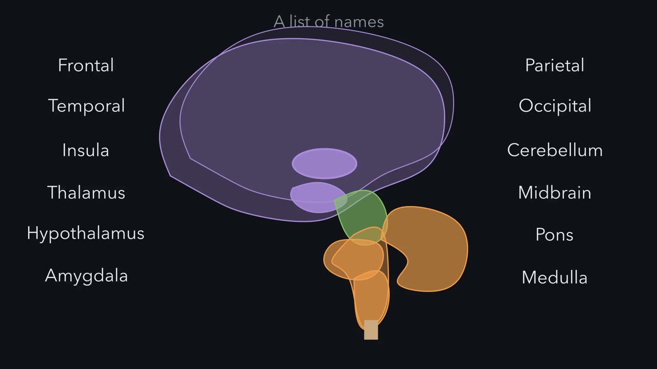 ▶ Watch One Tube, Bent Once · 3:05
Read the transcript of “One Tube, Bent Once”
Film transcript: One Tube, Bent Once
Your brain has a great many named parts. Is one shape behind them all? Most neurons and most glia of your brain and spinal cord come from the wall of one tube. It forms along the embryo's back. The one idea: picture the central nervous system as one hollow tube, bent once. Its walls became the brain's parts and the spinal cord. Its canal became the ventricles. And the tube's own long axis, not the body's, sets what the direction words mean. As it grows, the front end swells into three bulges: forebrain, midbrain and hindbrain. The forebrain and the hindbrain each divide in two. The rest becomes the spinal cord. The front of the forebrain buds out on each side, as the two cerebral hemispheres.
Their canals become the two lateral ventricles. The back of the forebrain becomes the thalamus and hypothalamus, around the third ventricle. The midbrain keeps a narrow canal, the cerebral aqueduct. The hindbrain becomes the pons and medulla, around the fourth ventricle. The cerebellum grows from the hindbrain's dorsal side, so the fourth ventricle lies beneath it. In the spinal cord, many neurons that receive sensory signals develop in the tube's dorsal half, and motor neurons in its ventral half. It is a plan, not a complete split: sensory fibres also reach ventral neurons. So sensory fibres enter through dorsal roots, and motor axons leave through ventral roots.
The brainstem keeps this plan, with sensory nuclei moved to the side. The forebrain does not follow it. In a rat, the tube runs nearly straight from nose to tail. Dorsal is toward its back. Stand the rat up, face pointing forward. The body, cord and brainstem turn. Only the forebrain keeps its frame. In a human, the tube bends by roughly a right angle where the midbrain meets the diencephalon. The embryo bends in three places. This one matters for naming. You are standing. In your spinal cord, which way does dorsal point?
Toward your back. The brainstem uses the same frame. In your forebrain, dorsal points to the top of your head. Three planes name the sections: sagittal, coronal and horizontal. A horizontal cut runs along the forebrain's axis. How does it pass through the brainstem?
Roughly across it, because the brainstem's axis runs up and down. So the tube tells you where things are. What each place does is another question.
From the film
- Picture the central nervous system as one hollow tube, bent once.
- The tube's walls became the brain regions and spinal cord, and its canal became the ventricles.
- Dorsal and ventral are set by the tube's own axis, so the words change direction where the human tube bends.
Most of the neurons and glia in your brain and spinal cord descend from the wall of one tube, a few millimetres long, that formed along the back of the embryo. Its open ends had closed by about four weeks after fertilization, about six weeks into pregnancy as it is usually counted.
Learn that tube, and the map of the nervous system stops being a list of names. The map comes first because every recording you meet later in this course comes from a place on it. How the tube forms and closes is a developmental story of its own.
A map and its puzzles
Start with what has to be explained. The central nervous system (CNS) is the brain and spinal cord. Its parts carry a great many names, and some facts about them look like things you would simply have to memorize:
- Four cavities. The brain holds four fluid-filled spaces, the ventricles, joined in a fixed order. Why four, and why that order?
- A blocked channel. Block one narrow channel between them, and some ventricles swell while others do not. Which ones?
- A word that points two ways. In your spinal cord, “dorsal” points toward your back. In your forebrain, it points toward the top of your head. Why?
- A seal. Inject a protein tracer into a mouse’s blood, and in many tissues, muscle for example, it leaks out of the capillaries. In the cerebral cortex it goes no farther than the single layer of cells lining them. Why there?
- An address for a function. What kind of experiment entitles you to say that a function lives in a particular place?
The sections below take them in order: one shape answers the first three, an experiment answered the fourth, and the fifth is about which experiments entitle you to which claims.
The one idea to carry through this lesson. Picture the central nervous system as one hollow tube, bent once. That is a working picture of how it develops, not a literal adult anatomy. The tube’s walls became the brain’s regions and the spinal cord; blood vessels grew in from outside, and the immune cells called microglia moved in. In the brain its canal became the ventricles, through which cerebrospinal fluid takes a one-way net route to an exit in the hindbrain. And the tube’s own long axis, not the body’s, sets what dorsal, ventral, rostral and caudal mean.
The picture does not tell you what a region does, or where the blood-brain barrier sits on the vessels that grew in; experiments had to find those. And the embryo’s tube in fact bends in three places, but for naming places in the adult brain only one matters: the bend near the midbrain, which this lesson calls the bend in the neuraxis.
One tube, bent once
Picture the tube lying along the back of the embryo, its front end toward the head. As it grows, the front end swells into three bulges: the forebrain, the midbrain and the hindbrain. The forebrain then divides in two, and so does the hindbrain, giving five bulges in a row. The rest of the tube becomes the spinal cord. The front of the forebrain also buds out on each side as the two cerebral hemispheres, so its canal becomes one lateral ventricle in each, and each stays joined to the third ventricle by an opening, the interventricular foramen.
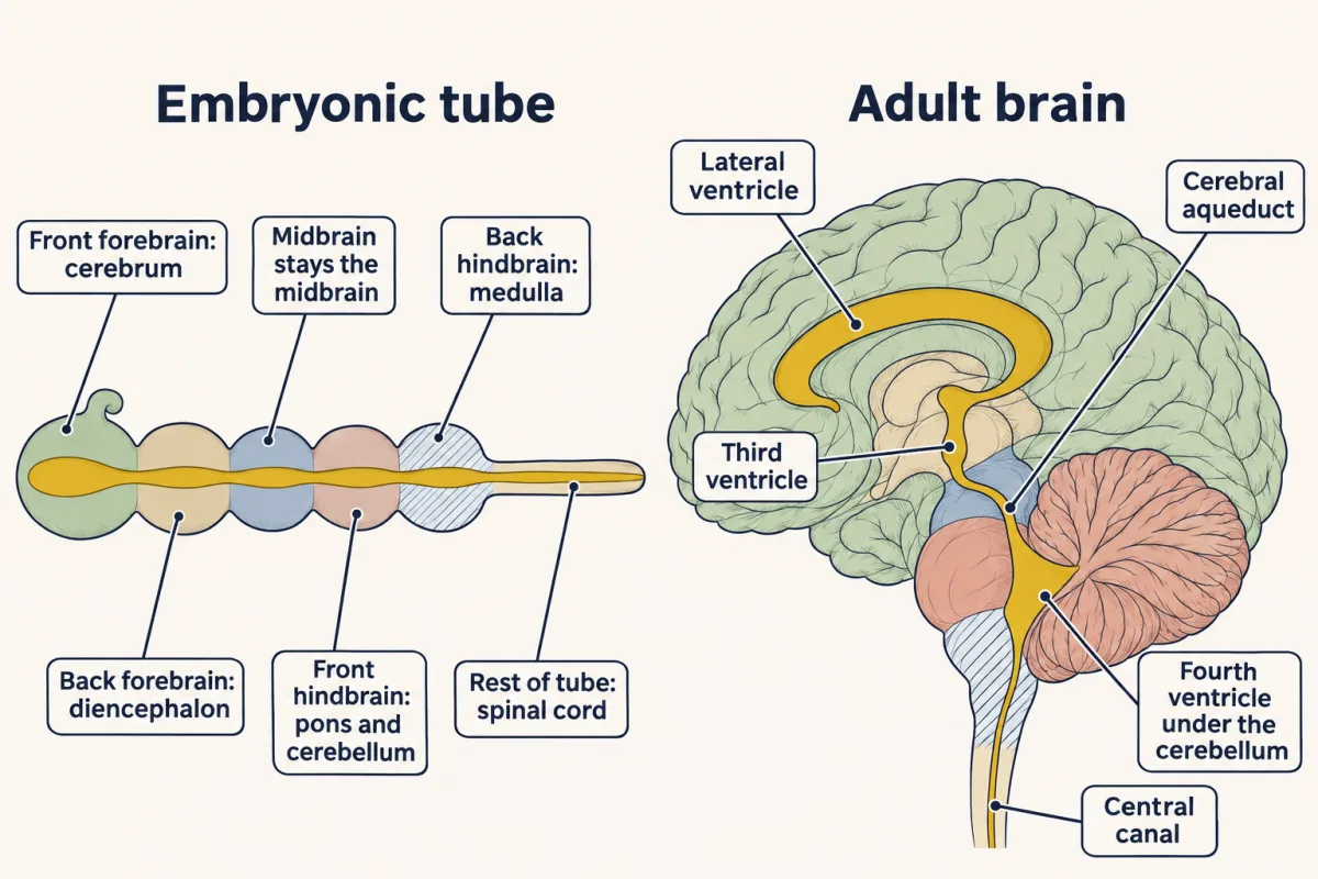
| Part of the embryonic tube | What its walls become | What its canal becomes |
|---|---|---|
| Front of the forebrain (telencephalon) | Cerebrum: cerebral cortex and the basal nuclei (often called basal ganglia) beneath it | the two lateral ventricles, one in each hemisphere |
| Back of the forebrain (diencephalon) | Diencephalon: thalamus and hypothalamus | the third ventricle |
| Midbrain | Midbrain | the narrow cerebral aqueduct |
| Front of the hindbrain | Pons, and the cerebellum growing from its dorsal side | the fourth ventricle |
| Back of the hindbrain | Medulla | the fourth ventricle, narrowing into the central canal |
| The rest of the tube | Spinal cord | the central canal, largely closed in most adult humans |
So the adult brain has four parts: the cerebrum, the diencephalon, the brainstem (midbrain, pons and medulla together) and the cerebellum.
The cerebrum grew most. Its cortex folded into ridges (gyri) and grooves (sulci) and is divided into frontal, parietal, temporal and occipital lobes. A few landmarks you will use all term:
- primary motor cortex is in the precentral gyrus;
- primary somatosensory cortex is in the postcentral gyrus;
- primary visual cortex is in the occipital lobe (Brodmann’s area 17);
- primary auditory cortex is in the temporal lobe.
Predict. The cerebellum grows from the dorsal side of the front of the hindbrain. Which ventricle should lie directly beneath it?
Check your prediction
The fourth ventricle. It is the canal of the hindbrain, so the cerebellum lies over it, and the pons and medulla form its floor. In the adult, the fourth ventricle lies between the pons and medulla in front and the cerebellum behind. You did not need to memorize that; the tube gave it to you.
A map names places, not jobs
Each of these regions takes part in many functions, and most functions involve several regions. The thalamus relays and gates information passing to and from the cortex. The hypothalamus takes part in homeostatic regulation of temperature, feeding, fluid balance and hormone release. The brainstem carries the long tracts between brain and cord, holds most cranial nerve nuclei, and takes part in breathing and cardiovascular control. The basal nuclei take part in selecting and controlling movement, among other roles. The cerebellum takes part in comparing intended movements with sensory feedback and correcting them, among other roles; the cerebellum lesson shows how.
The cortex itself is a mosaic of many smaller areas, and their borders vary between people. How cortex is divided into areas is another lesson.
Many pathways cross the midline, so for movement and touch each cerebral hemisphere mostly serves the opposite side of the body. Not every system works this way: each side of the cerebellum mostly serves the same side of the body, and many systems are bilateral. You will trace where each one crosses, and work out lesions from it, in touch and pain and visual cortex.
Outside the tube: the peripheral nervous system
Everything nervous outside the brain and cord is the peripheral nervous system (PNS): nerves, and ganglia, which are clusters of neuron cell bodies outside the CNS. Inside the CNS, a cluster of cell bodies is called a nucleus (not to be confused with the cell’s nucleus), and a bundle of axons running together is a tract.
Signals travel in two directions. Afferent fibres carry signals toward the CNS, as from a touch receptor in the skin. Efferent fibres carry signals away from it, to a muscle or a gland.
The tube explains where each kind connects. In the spinal cord, the neurons that receive incoming sensory signals develop mainly in the dorsal half of the tube’s wall, which becomes the dorsal horn, and motor neurons develop in its ventral half. So sensory fibres enter the cord through dorsal roots, and motor axons leave through ventral roots.
The first-order sensory neurons themselves sit just outside the cord, in the dorsal root ganglia. These formed from neural crest cells, a band of cells at the edges of the folding sheet that left as the tube closed. The brainstem keeps the same plan for its cranial nerve nuclei, though in the hindbrain the tube splays open to form the floor of the fourth ventricle, so the sensory nuclei lie lateral and the motor nuclei medial.
This is a plan for where groups of neurons develop, not for every tract. In the classical picture it stops at the brainstem. The forebrain does not follow it: primary motor cortex, for example, lies just in front of somatosensory cortex, not ventral to it.
This lesson divides the PNS into three parts:
- the somatic division serves the skin, joints and skeletal muscle;
- the autonomic division (sympathetic and parasympathetic) serves smooth muscle, heart muscle and glands, largely without conscious control;
- the enteric division lies in the wall of the gut and has its own sensory neurons, interneurons and motor neurons, so it can run local reflexes. Many textbooks instead count it as part of the autonomic division; the facts are the same, only the filing differs.
Somatic does not mean voluntary. The knee jerk is a somatic motor response that happens before you decide anything: the command travels out through a ventral root to skeletal muscle. The stretch reflex gets its own lesson, and the cells that make up all this tissue come in the next lesson.
Naming places on a bent tube
The neuraxis is the long axis of the CNS: the tube’s own axis. In a rat it runs nearly straight from nose to tail. In a human it bends by roughly a right angle where the midbrain meets the diencephalon.
Picture a rat on all fours. Its tube runs straight, so the words are easy. Dorsal is toward its back, which is up. Ventral is toward its belly. Rostral is toward its nose, and caudal toward its tail. Now stand the rat up on its hind legs, but keep its face pointing forward, as yours does. The body, spinal cord and brainstem turn, and their direction arrows turn with them. Only the forebrain keeps its old frame. In you, that turn falls inside the skull, not at the neck. That is the bend.
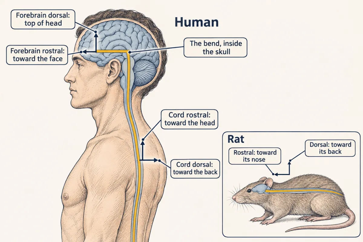
So the direction words change meaning across the bend:
| Part of the CNS | Dorsal points toward | Ventral points toward | Rostral points toward | Caudal points toward |
|---|---|---|---|---|
| Forebrain | the top of the head | the base of the skull | the face | the back of the head |
| Spinal cord | the back of the body | the belly | the head | the feet |
Always say which frame you mean.
Two more pairs of words do not change across the bend. Medial means toward the midline and lateral away from it. Ipsilateral means on the same side of the body, and contralateral means on the opposite side.
Predict. You are standing up. Which way does dorsal point in your spinal cord? In your brainstem? In your forebrain?
Check your prediction
In the spinal cord, toward your back: the side the dorsal roots enter. The brainstem lies caudal to the bend, so it follows the cord’s frame: dorsal there also points toward your back. In the forebrain, dorsal points toward the top of your head. That is why the cerebellum, grown from the hindbrain’s dorsal side, lies toward your back.
Three section planes cut the brain:
- a sagittal plane separates left from right (the midsagittal plane runs down the midline);
- a coronal plane separates front from back;
- a horizontal plane separates top from bottom.
A section of the cord cut across its long axis is a transverse section.
Predict. A horizontal section through the head runs along the forebrain’s long axis. How does the same cut pass through the brainstem?
Check your prediction
Roughly across it, close to a transverse section, because the brainstem’s axis runs up and down, like the cord’s. Roughly, not exactly: the brainstem is not perfectly vertical, so the cut is somewhat oblique. The same logic says that, because of the bend, a coronal section of the forebrain and a transverse section of the cord are both roughly perpendicular to the neuraxis at their level.
The fluid in the tube
Read the transcript of “Follow the Fluid, Find the Seal”
Film transcript: Follow the Fluid, Find the Seal
The tube's canal is full of fluid. Where is it made, and where does it go? In each ventricle, the choroid plexus secretes cerebrospinal fluid. It is the main source, not the only one. The net flow runs one way: from the lateral ventricles to the third, then through the aqueduct into the fourth. The exit is in the hindbrain. Openings in the fourth ventricle let fluid out around the brain and cord. In most adult humans, the cord's own central canal is largely closed and is not a major route. The fluid circulates around the cord instead. On the usual teaching numbers, the fluid is replaced about three times a day. Is it ever completely fresh? Picture a glass of dyed water.
Pour in clean water slowly, stirring as it overflows. After one full glassful, the dye has faded but not gone. About 37% % of the dye is still there. Replacing a volume does not empty it. Now plug the cerebral aqueduct while secretion continues. Which ventricles swell?
Everything upstream: both lateral ventricles and the third. Not the fourth, downstream of the plug. Now the seal. In muscle, for example, a protein tracer in the blood leaks out of the capillaries. In the brain's cortex, it goes no farther than the cells lining them. Three layers could be the seal: the endothelial cells, the basement membrane around them, and astrocyte endfeet outside. In 1967, Thomas Reese and Morris Karnovsky injected horseradish peroxidase into mice. Wherever this enzyme lies, a chemical reaction leaves a dense deposit an electron microscope can see. Which layer stopped the tracer?
The endothelial cells. The deposit filled the vessel and went no farther. That holds for this tracer. Tight junctions between those cells were the probable block. Then Milton Brightman and Reese put the tracer into the ventricles instead. It spread between brain cells, reached the capillaries from outside, and stopped at the same junctions. Two experiments, run in opposite directions, pointed to the same layer. A second seal sits on a specialized part of the tube's lining. In the choroid plexus, leaky capillaries lie under a sealed epithelium. So the net flow runs one way through the tube. Two main seals, both selective, decide what gets in from the blood.
From the film
- Cerebrospinal fluid is made mainly by the choroid plexus and flows from the lateral ventricles through the third and fourth ventricles to the space around the brain and cord.
- Replacing a fluid volume does not empty it, and a lasting block of the aqueduct tends to enlarge the ventricles upstream of it.
- The blood-brain barrier is at the capillary endothelial cells and the blood-CSF barrier is at the choroid plexus epithelium.
Cerebrospinal fluid (CSF) fills the four ventricles. In the adult mammalian brain most of it, though not all, is secreted by the choroid plexus, a frond of capillaries covered by a secretory epithelium that projects into each ventricle. Secretion is an active process, driven by transporters in that epithelium (the evidence, and a debate about the mechanism, are in Going deeper).
The net route runs one way, though the fluid also moves back and forth with each heartbeat and breath:
- from each lateral ventricle through an interventricular foramen into the third ventricle;
- through the cerebral aqueduct into the fourth ventricle;
- out of the hindbrain through the fourth ventricle’s median and lateral apertures, into the subarachnoid space around the brain and cord, where it cushions them;
- out through arachnoid granulations into the venous sinuses, and by other routes, including lymphatic vessels (see Going deeper).
In most adult humans the central canal is largely closed and is not a major route for CSF; the fluid reaches the cord through the subarachnoid space around it.
How quickly is the fluid replaced?
Start with a picture. Take a full glass of dyed water. Pour clean water in slowly, stirring all the time, so the glass overflows as you pour. When you have poured in one full glassful, is the water clear? No. Each spill carries away a mix of old and new water, so the dye fades but does not vanish. If the new water pushed the old out like a piston, one glassful would clear it. Because it mixes, about 37% of the dye is still there.
Now the numbers. Take conventional adult human teaching values, not measurements from one person: a total CSF volume V of about 150 mL and a secretion rate Q of about 500 mL/day, with outflow equal to secretion so the volume is steady. Treat the CSF as one well-mixed compartment, like the stirred glass: the new fluid carries no marker, and the marker leaves only with the outflow.
turnover rate = Q / V; volume-to-flow time τ = V / Q; fraction of an initial marker left = exp(−t / τ)
- Turnover rate: 500 mL/day ÷ 150 mL = 3.33 per day.
- Volume-to-flow time: τ = 150 ÷ 500 = 0.3 day, which is 0.3 × 24 = 7.2 h.
- The same in minutes: 500 mL/day ÷ 1,440 min/day = 0.347 mL/min, and 150 ÷ 0.347 = 432 min = 7.2 h.
- Why an exponential? At every moment the outflow removes the same fraction, 1/τ per unit time, of whatever marker is left, so the marker M falls at a rate proportional to itself: dM/dt = −M/τ. A marker present at time zero therefore falls as exp(−t/τ). At t = 7.2 h that is exp(−1) = 0.368: about 37% of the marker is still there after one full volume of fluid has been made. Replacing a volume does not empty it.
- Do not confuse τ with the half-life. Half the marker is gone at τ × ln 2 = 7.2 h × 0.693, about 5.0 h.
- Turnover rate Q / V
- —
- Volume-to-flow time τ = V / Q
- —
- Check in mL/min
- —
- Fraction of the time-zero marker left
- —
—
In this sketch: the lesson’s conventional adult teaching values (500 mL/day, 150 mL) are the starting point; the sliders vary them and are not measurements from one person; one well-mixed compartment; outflow equals secretion, so the volume is steady; the marker is present at time zero and none is added afterwards. The ventricles are drawn as a schematic, not to scale.
Turnover = Q / V; τ = V / Q; fraction of an initial marker left = exp(−t / τ): after one full volume of fluid has been made, 36.8 percent of the marker is still there. Block the aqueduct and fluid made upstream has no exit, so this one-compartment rule stops applying.
Block the tube
The washout rule describes how a marker fades. A blockage asks a different question: where does the fluid go when its exit is closed?
Predict. Block the cerebral aqueduct while secretion continues. Which ventricles enlarge?
Check your prediction
The lateral and third ventricles. Fluid made in them loses its normal exit, so pressure rises and they enlarge. The fourth ventricle, downstream of the block, does not.
The general rule, in this simplified obstructive model: a lasting, complete block, with secretion continuing and no adequate alternative drainage, tends to enlarge the ventricles upstream of it. Block the fourth ventricle’s apertures, and all four ventricles enlarge, because all four lie upstream of the exit. This rule predicts obstructive hydrocephalus, with a block inside the tube. If absorption fails instead while the pathways stay open, often all four ventricles enlarge together. That is communicating hydrocephalus, and the upstream rule does not predict it.
Where is the seal?
Back to the fourth puzzle. By the 1960s it was known that many dyes injected into the blood stain most tissues but leave the brain pale, so a barrier existed. Where it sat was the open question.
Picture a brain capillary in cross-section. Three layers could be the seal:
- the endothelial cells that line the capillary;
- the basement membrane around them;
- the astrocyte endfeet pressed against the outside of the vessel.
A light microscope could not tell them apart. Thomas Reese and Morris Karnovsky chose a tracer that an electron microscope can see. Horseradish peroxidase is an enzyme, and wherever it lies it drives a histochemical reaction that leaves a dense deposit. They injected it into the veins of mice, fixed the cortex at times from 10 to 60 minutes later, and looked at the capillaries.
Predict. Where did the deposit stop?
Check your prediction
At the endothelial cells. The deposit filled the vessel lumen and went no farther. None was found in the basement membrane or between the astrocyte endfeet. Reese and Karnovsky proposed that tight junctions between endothelial cells were probably what blocked the path between cells. They published in 1967. The result holds for that tracer and that time window.
Then the experiment was run from the other side. Milton Brightman and Reese put the same tracer into the ventricles of mice. It spread through the spaces between neurons and glia, reached the outer surface of the capillaries, and stopped at the junctions between endothelial cells. Two experiments run in opposite directions pointed to the same layer.
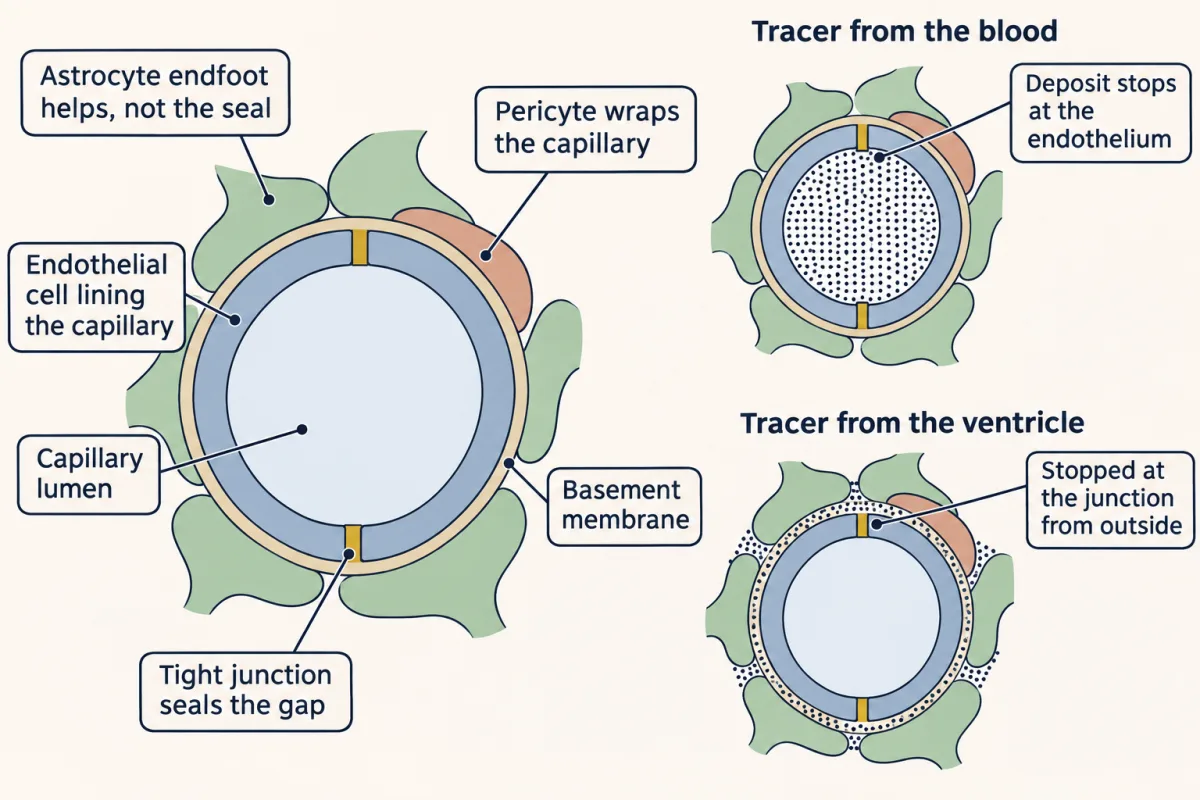
So two main barriers separate the brain from the blood. The blood-brain barrier lies at the endothelial cells of brain capillaries, joined by tight junctions, protein seals between neighbouring cells (the junction itself is covered in the epithelia lesson). These cells also move very little material across in vesicles and pump many drugs back out. Pericytes, cells wrapped around the capillaries, and astrocytes help them keep these properties: astrocytes are not the seal, but they help maintain it. The barrier is selective, not a wall: transporters carry glucose and amino acids across, and small lipid-soluble molecules diffuse through. A few small regions around the ventricles, the circumventricular organs, have leaky capillaries.
The blood-CSF barrier sits on a specialized part of the tube’s own lining. Choroid plexus capillaries are leaky, and the seal is formed by the tight junctions of the epithelium over them. A third barrier, a sealed cell layer in the arachnoid membrane, separates the cerebrospinal fluid (CSF) from blood vessels in the dura.
From where to what: four kinds of evidence
Read the transcript of “From Where to What”
Film transcript: From Where to What
The map tells you where a region is. How do you find out what it does? You need an experiment. Each kind licenses a different claim. First, a lesion. A deficit after damage, compared with controls, shows the tissue contributes to the task. But damage rarely respects borders, so which part mattered can be unclear. Second, a recording. Firing that goes with a choice shows covariation, not that the brain uses it. Third, imaging. Functional MRI measures blood oxygenation, not spikes. In anaesthetized macaque visual cortex, it followed local field potentials better than spiking. Field potentials mostly reflect synaptic and dendritic currents near the electrode.
So an activation does not mark a region as the seat of anything. Fourth, stimulation adds activity. Monkeys judged which way noisy dots moved. An electrode sat among area MT neurons that all fire most for one direction of motion, their preferred direction. On random trials, weak current flowed while the dots were shown. If the monkey uses these neurons, which way should stimulation push its choices?
Toward the neurons' preferred direction. In 86 of the 89 experiments with a significant effect, that is what happened. That shows a causal contribution in this task. It does not make area MT the seat of motion, or show it is required. So a lesion shows contribution, not which part mattered. A recording shows covariation, not use. Imaging shows where a blood signal goes, not which neural signal. Stimulation shows contribution, not that the region is required. Reese and Karnovsky also added something and watched. But their tracer showed only where it stopped, a different kind of claim. Claim only what the evidence licenses.
From the film
- Each kind of evidence licenses a different claim about where a function lives.
- A lesion shows contribution, a recording shows covariation, and imaging shows a blood signal rather than which neural signal.
- Stimulation in area MT shifted choices toward the neurons' preferred direction, which shows a causal contribution in that task, not that MT is the seat of motion.
The tube has told you where everything is. It cannot tell you what a region does. That takes an experiment, and each kind licenses, that is, entitles you to, a different claim.
| Evidence | What it can show | What it cannot show alone |
|---|---|---|
| Lesion | The damaged tissue contributes to the task, as the rest of the brain performs it | Which part of the damage mattered |
| Recording | Activity covaries with a task or stimulus | That the activity is used |
| Imaging (fMRI) | Where a haemodynamic signal goes with a task | Which neural signal, or necessity |
| Stimulation | Adding activity changes behaviour | That the region is required |
A lesion tests necessity, but damage spreads. Scoville and Milner’s 1957 report on bilateral medial temporal resections in human patients is the classic case: the recent-memory loss was striking, and comparing patients with smaller and larger removals pointed to the hippocampal region, but each resection removed several medial temporal structures at once, so the deficit could not be pinned to one structure. Memory systems tells that story.
A recording shows that firing goes with a stimulus or a choice, not that the brain uses it. Imaging has the same limit plus one more: Blood-oxygen-level-dependent (BOLD) fMRI measures blood oxygenation, and in anaesthetized macaque visual cortex it followed local field potentials, which mostly reflect summed synaptic and dendritic currents near an electrode, better than spiking (Logothetis and colleagues, 2001). An fMRI activation does not mark a region as the seat of anything.
Stimulation adds activity and asks whether behaviour follows. That shows a contribution, in that task, from the stimulated population, not from one identified cell type: imaging in anaesthetized animals found that the current sparsely activates a scattered population around the tip, some neurons as far as millimetres away. Tools for recording many neurons and for silencing or driving defined cells come in large-scale neuroscience and circuit experiment design.
Design problem
Rhesus monkeys judge the direction of noisy moving dots, and you suspect that area MT, where neurons are tuned to the direction of motion, contributes to the judgement. Your recordings already show that MT firing covaries with the monkeys’ choices. Design an experiment that tests whether MT activity contributes to the choice, say what result you predict, and say what your result would and would not license.
Saved in this browser only.
One way to do it
Place a microelectrode among MT neurons that share a preferred direction, mapped first. On randomly interleaved trials, deliver weak microstimulation while the dots are on, and compare choices on stimulated and unstimulated trials at the same motion strengths. Interleaving balances slow drifts in attention or motivation across the two kinds of trial.
Predict the sign first: if those signals are used, stimulation should push choices toward the preferred direction, not merely make the monkey worse. Salzman, Murasugi, Britten and Newsome (1992) did this in 139 experiments: stimulation had a significant effect in 89, and in 86 of those the bias was toward the preferred direction.
What it licenses: MT activity contributes causally to this choice in this task. Not that MT is the only place motion is processed, or that it is required. A lesion or reversible silencing would test necessity, though depending on the method a lesion can also damage neighbours and passing fibres. fMRI would show neither, because it observes activity without changing it, and it measures blood oxygenation, not spikes.
Reese and Karnovsky also added something and watched what happened, but the two experiments license different claims: the tracer showed where one tracer stopped, and the stimulation showed a causal contribution to a choice. Claim only what the evidence licenses.
Going deeper
How many neurons and glia?
Counts need their source. One study using the isotropic fractionator on the brains of adult men estimated 86.1 ± 8.1 billion neurons and 84.6 ± 9.8 billion non-neuronal cells (Azevedo and colleagues, 2009), so the claim that glia outnumber neurons ten to one is not supported.
Where pathways cross the midline
The corticospinal tract crosses mainly in the caudal medulla, the dorsal column pathway in the medulla, and the spinothalamic pathway in the spinal cord. At the optic chiasm only fibres from the nasal half of each retina cross.
Brodmann’s areas and a modern atlas
Area 18 lies next to area 17 and is not primary. Maps get revised: one multimodal MRI atlas of healthy young adults divides each hemisphere into 180 areas (Glasser and colleagues, 2016).
The evidence that secretion is active
In isolated mouse choroid plexus, water could be moved against an imposed osmotic gradient, so secretion is an active process (Steffensen and colleagues, 2018). That was one study in isolated mouse tissue, and the mechanism it proposed is debated; the conclusion that secretion is active is the safe part.
Lymphatic exits for CSF
In mice, lymphatic vessels at the skull base clear cerebrospinal fluid (CSF) macromolecules (Ahn and colleagues, 2019). Dural lymphatic vessels have been imaged in five healthy people and three marmosets (Absinta and colleagues, 2017), though that study could not show that they drain CSF.
Does sleep speed clearance?
In 13 sleeping people, slow EEG rhythms were followed by pulses of CSF flow (Fultz and colleagues, 2019). That is a correlation, and mouse tracer studies disagree about whether clearance rises or falls during sleep. Keep the question open until sleep and rhythms.
Your notes
Select any text in the lesson to highlight it or add a note to it. Your highlights and notes are saved in this browser and collected in your notebook.
This browser is not letting the site save anything (a private window or strict privacy settings), so highlights and notes will be lost when you leave the page.
How we measure it
Tracer cytochemistry with electron microscopy
A tracer that can be made visible, such as horseradish peroxidase, is injected into the blood or a ventricle, the tissue is fixed, and a reaction deposits a dense product wherever the tracer lies. Electron micrographs then show which cell layer it crossed and where it stopped, for that tracer and that time after injection.
Lesion studies
You compare behaviour after damage to a region with earlier performance or with matched controls, whether the damage was made on purpose in an animal or caused by surgery, stroke or injury in a person. A deficit shows the damaged tissue contributes to the task, as the rest of the brain performs it, but damage rarely respects boundaries and the rest of the brain may compensate.
Functional MRI (BOLD)
MRI detects a change in blood oxygenation that follows local neural activity by a few seconds, and maps it across the whole brain without opening the skull. It shows where activity goes with a task, not which spikes occurred or whether the region is needed.
Electrical microstimulation
Small current pulses through a microelectrode activate neurons near its tip during a task. If the behaviour shifts in the direction those neurons' tuning predicts, their activity contributes to that behaviour, in that task.
Practice questions on this lesson (3) → MCAT-style, original, each with an explanation
Where this lesson fits
Before thisReview first
- Tissues, Organs and HomeostasisOrganismal Biology · Starting college
It introduces tissues, organs and the control systems that this lesson's map of nervous system divisions assumes.
- Nervous Control: A First LookOrganismal Biology · Starting college
It first traces a reflex route through dorsal and ventral roots, which this lesson explains by the tube's layout.
After thisStudy next
- Neurons and Glia at WorkThis course · Lesson 2
It takes the cells that fill the tube's walls, including the astrocytes that help maintain the barrier, further.
- Touch and PainThis course · Lesson 13
It traces where touch and pain pathways cross the midline, using the sensory dorsal root plan from this lesson.
- Memory SystemsThis course · Lesson 22
It tells the Scoville and Milner lesion story, using the limits of lesion evidence set out here.
Elsewhere on the siteFilms, simulations and method films in other courses
- Narrow First, Then FoldFilm · Developmental Neuroscience
It shows how the embryonic neural tube forms and what happens when part of it stays open.
- Two Segments, Two JobsFilm · Developmental Neuroscience
It shows how the neural tube is divided head to tail into the hindbrain segments that this map names.
Suggested by an AI model from the lessons themselves, then checked. How we use AI
Finish line
Test yourself
8 questions
Practice and MCAT-style, with explained answers.
Next · Lesson 2
Neurons and Glia at Work
Let potassium pile up outside neurons during seizure-like firing and E_K can move by more than 30 mV. Who cleans it up?
Continue →