Organismal Biology · Unit 2: Control and constancy
Nervous Control: A First Look
Rewritten on : new text, flashcards and film. Your notes and highlights stay, but the text around them may have moved.
A doctor taps the tendon below your kneecap and your leg kicks before you decide anything. What route does the signal take, and how fast does it run?
Pause when the reading reaches a film, and offer to play it. Saved on this device.
Watch the film now, or keep listening
Builds on:Tissues, Organs and HomeostasisHomeostasis in ActionWhy these?
 ▶ Watch Every Response Has a Running Time · 3:06
Read the transcript of “Every Response Has a Running Time”
Film transcript: Every Response Has a Running Time
Tap the tendon below your kneecap, and your leg kicks before you decide anything. How long does that take, and where does the time go? The signal runs a route with five stations. First, a receptor in the thigh muscle detects the stretch. Then a sensory neuron carries the signal up to the spinal cord. In the spinal cord, the integrating centre, it crosses one synapse. A motor neuron carries it back down the leg. Then the effector, the thigh muscle, contracts. Zoom in on the motor neuron. Its dendrites receive signals, and the cell body gathers them. One long axon carries the signal away, in a large mammal sometimes a metre or more. Its terminals pass it on.
Along the axon the signal is an impulse. It is all or none: full size, or nothing at all. On many axons it hops between short bare gaps in a wrapping called myelin. At most synapses, the terminal releases a chemical that crosses a narrow gap. But how fast does the impulse run? In 1850, Hermann von Helmholtz asked that of a frog. He stimulated its nerve near the calf muscle and timed the twitch. Then he moved the stimulus farther up the same nerve, and timed it again. Every time, the twitch came a little later than before. Each delay includes time at the junction and in the muscle. One part of these two delays belongs to the nerve alone. Which part?
The difference. The junction and the muscle take the same time in both trials, so they cancel. The extra delay belongs to the extra length of nerve. Divide that extra length by that extra time, and you get the nerve's own speed. Helmholtz found a few tens of metres per second, about a car on a motorway. Now back to the knee. Each axon's time is its length divided by its speed. In this model, each synapse in the cord adds about a millisecond. The junction and the muscle add a little more. The bar's legs add up to just under the measured time, when the thigh muscle's electrical signal starts. The bar leaves out the brief tap and stretch before the spindle fires.
The kick itself comes later. Nearly all of the bar is time on the two axons. So a response is a route, and its time is the sum of its legs. Change a length, a speed, or the number of synapses, and the time changes.
From the film
- A nervous response runs a route: receptor, sensory neuron, integrating centre, motor neuron, effector.
- Along an axon the signal is an all-or-none impulse; at most synapses it crosses as a released chemical.
- Helmholtz timed a frog nerve from two points; subtracting the two delays removed the junction and muscle, leaving the nerve's own speed.
- In a simple model, a response's time is the sum of its legs: each axon's length over its speed, about a millisecond per synapse in the cord, and short delays at the ends.
- In the knee-jerk model, nearly all of the bar is time on the two axons, and its legs add up to just under the measured time at which the thigh muscle's electrical signal starts.
Tap the tendon below your kneecap and your thigh muscle fires about 20 milliseconds (ms, thousandths of a second) later; the leg swings soon after, before you decide anything. Step on a tack and your foot lifts too, but later. Where does the time go?
The one idea. A nervous response runs a route: receptor (which detects the change), sensory neuron, integrating centre (where the spinal cord or brain passes the signal on), motor neuron, effector (the muscle or gland that responds). Its time is the sum of its legs: each axon’s length over its speed, about a millisecond per synapse in the cord, and short delays at the ends.
response time ≈ (L/v for each axon, added up) + (synapses in the cord × about 1 ms) + end delays, where L is an axon’s length, v its conduction velocity (the impulse’s speed along it), and the end delays are at the receptor (from stimulus to firing), the nerve-muscle junction and the muscle.
Scope: a simple model of a chain of neurons; numbers below are model numbers unless marked measured. The 1 ms per synapse is a teaching assumption: real synapses and cord processing vary and often take longer, so a delay suggests a synapse count but cannot prove it. Nor does the sum say what the effector does: whether the heart speeds up or slows down depends on which division reaches it, its transmitter and the receptor that transmitter acts on.
One neuron: what the first film left out
The first film named a neuron’s (nerve cell’s) parts, from dendrites to axon terminals (the cell body, or soma, also holds the nucleus), and its all-or-none impulse, the action potential, a brief, self-renewing electrical change across the membrane. So a stronger stimulus makes the neuron fire more often, and can bring more neurons into action, but cannot make an impulse taller. How ions moving through membrane channels make the impulse is in the action potential and the resting membrane; the resting voltage of -70 mV in many figures is an illustration, and real values vary between cells.
Where the film’s terminal released its chemical is a synapse, a junction between cells; the chemical is a neurotransmitter, and the gap it crosses is the synaptic cleft. Some synapses are electrical, with ions passing straight between cells (synaptic transmission). The film’s wrapping is myelin, layers of membrane from glial cells, the nervous system’s support cells (Schwann cells outside the brain and spinal cord, oligodendrocytes inside); its bare gaps are the nodes of Ranvier (myelination; for neurons as separate cells, the neuron doctrine).
How fast? What Helmholtz’s subtraction can tell you
The first film showed Helmholtz’s 1850 two-point timing of a frog nerve and its answer, a few tens of metres per second.
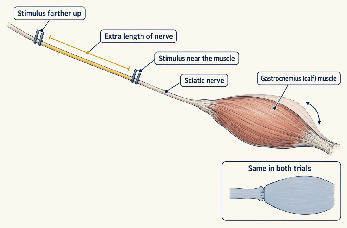
Predict. Had Helmholtz skipped the subtraction and divided whole length by whole delay, what speed would he report for an illustrative nerve with a true speed of 25 m per s, stimulated 40 mm from the muscle, with 2 ms of junction and muscle delay?
Check your prediction
About 11 m per s, less than half the true speed. The nerve takes 0.040 m / 25 m per s = 1.6 ms, so the whole delay is 3.6 ms, and 0.040 m / 0.0036 s is about 11 m per s. The difference between two trials gives 25 m per s whatever the shared delay.
The subtraction rules in a physical process of finite speed in this preparation, not what the signal is. And the number is the frog’s: a frog is an ectotherm, whose body temperature follows its surroundings, and cooling slows conduction in a given fibre, so a room-temperature frog value is not a human value at 37 °C.
conduction velocity v = difference in distance / difference in time
Worked problem. These numbers are illustrative, not Helmholtz’s.
- Velocity from two points. Two stimulus points on a frog nerve are 30 mm apart. Contraction starts 1.2 ms later from the far point than from the near one. v = 0.030 m / 0.0012 s = 25 m per s.
- The knee jerk, in this model. The film’s sensory and motor axons are each about 0.5 m at 60 m per s: 0.5 / 60 = 0.0083 s each, so with 1.0 ms for the synapse, 8.3 + 8.3 + 1.0 = 17.7 ms, and an assumed 1.5 ms at the junction and muscle makes about 19 ms. The measured adult latency, about 20 ms, runs from the tap to the start of the thigh muscle’s electrical response; the sum leaves out the short time from tap to spindle firing, so the near match is not a full accounting. The visible kick begins tens of milliseconds later, while the muscle builds force.
- The same path without speed. At 1 m per s each axon takes 0.5 s, so axons and synapse take 500 + 500 + 1 = 1,001 ms instead of 17.7 ms, about 57 times longer.
- Each axon: time = length / velocity
- Total latency = sensory + synapse + motor
- Compared with the same path at 60 m per s
- Share of the total taken by the synapse
Inset: a two-point velocity measurement (illustrative nerve at the velocity setting above, not Helmholtz’s value)
- Extra delay = distance / velocity
- Velocity from the subtraction: difference in distance / difference in time
In this sketch: the lesson’s 0.5 m adult path, the same velocity in both axons, one synapse of 1.0 ms, illustrative numbers. This is a model sum, not a measured knee-jerk latency; it ignores the time the sense organ, the nerve-muscle junction and the muscle take. Only the direct excitatory limb of the knee jerk is monosynaptic. The inset’s numbers are illustrative, not Helmholtz’s: his frog nerve at room temperature is not a human nerve at 37 degrees C.
Rule to keep: time = distance / velocity for each axon, and the total is the sum of the axon times and the synaptic delay; a two-point measurement removes the delays common to both trials by subtraction.
In this model axon time is the large term, and two main features of the axon set its speed (temperature and other membrane properties matter too). Width: a wider axon offers less resistance to the current spreading ahead of the impulse, so the impulse travels faster. Myelin: the impulse is renewed only at the nodes, not along every bit of membrane, so it skips ahead. The squid Loligo’s unmyelinated giant axon, about half a millimetre across, conducts at roughly 20 m per s near sea temperature; the largest myelinated vertebrate motor axons, about 20 micrometres across, conduct faster still. Myelin buys speed without width, so a vertebrate fits many fast axons into one nerve.
The sum has three kinds of term: axons, synapses and end delays. The second film gives two reflexes the same end delays and races them: does the slower one lose its time on axons or at synapses?
Read the transcript of “One Synapse or Several”
Film transcript: One Synapse or Several
The first film timed the knee jerk, leg by leg. Now you step on a tack: is lifting your foot faster or slower, and why? Put the two reflexes side by side. Both use the same five stations: receptor, sensory neuron, integrating centre, motor neuron, effector. Their circuits differ in the middle. Their fibres and path lengths differ too. In the knee jerk, the spindle's sensory fibre synapses straight onto the motor neuron. That is one synapse in the spinal cord. This direct path is called monosynaptic. But a side branch excites an interneuron, a neuron that stays inside the cord. It quiets the hamstring, the muscle that would oppose the kick. So even the knee jerk is not one wire.
Now the tack. Pain-sensing endings in the skin fire. Their thin sensory fibres excite a chain of interneurons in the cord, right from the start. Some interneurons drive the flexors, which lift the foot. Others quiet the opposing extensors. Charles Sherrington showed this pairing in mammals, and called it reciprocal innervation. Now run both timers together, on the same scale. The withdrawal bar keeps growing after the knee jerk's has stopped. It has more synapses, so it shows more ticks. In this example the withdrawal has three synapses, and the knee jerk has only one. So are the extra synapses most of the extra time?
No. In this model, each synapse adds only about a millisecond. The big difference is the sensory leg. Thin pain fibres conduct slowly, so that leg takes tens of milliseconds. Why is a thin fibre slow? A thin axon offers more resistance to the current spreading ahead of the impulse. So the impulse travels more slowly, and the sensory leg stretches out.
Part of the gap is also the longer motor path. In this example, slow fibres are most of it, not all. In real people, fibre speed and processing in the cord both vary. Both reflexes end on skeletal muscle, and that makes both of them somatic. Somatic does not mean voluntary.
Neither reflex waits for a decision. So in these two reflexes, the synapses set the circuit. The slow fibres set most of the extra time. The sum from the first film still holds.
From the film
- The knee jerk's direct path has one synapse in the spinal cord; it is monosynaptic.
- Interneurons in the cord also quiet the opposing muscle, so even the knee jerk is not a single wire.
- The withdrawal reflex runs through interneurons from the start, exciting flexors and inhibiting extensors.
- In this example, the withdrawal is slower mostly because its pain fibres conduct slowly; each extra synapse adds only about a millisecond.
- Both reflexes are somatic, because their effector is skeletal muscle, not because they are voluntary.
The reflex arc: what the second film left out
Both reflexes run through the spinal cord, which with the brain makes up the central nervous system (CNS). The peripheral nervous system (PNS) is everything outside: nerves and ganglia, clusters of nerve cell bodies. Its somatic division drives skeletal muscle; its autonomic division drives cardiac muscle, smooth muscle and glands (nervous system map).
A reflex is a rapid, automatic response to a stimulus; its route is a reflex arc. The film skipped how the knee jerk starts: the tap stretches the quadriceps through its tendon, so its muscle spindles, stretch receptors, fire through large, fast sensory fibres called Ia fibres. Of the film’s monosynaptic path and interneurons (neurons wholly within the CNS), one detail: in the knee jerk only the direct excitatory path lacks an interneuron (motor units and reflexes).
Counting synapses with a clock
The second film’s model numbers, for reference (illustrative round values; the withdrawal’s thin pain fibre runs from the sole, its motor axon to a flexor lower in the leg):
| Leg of the route | Knee jerk | Withdrawal |
|---|---|---|
| Sensory axon | 0.5 m at 60 m per s: 8.3 ms | 0.9 m at 20 m per s: 45 ms |
| Synapses in the cord | 1, about 1 ms | 3, about 3 ms |
| Motor axon | 0.5 m at 60 m per s: 8.3 ms | 0.9 m at 60 m per s: 15 ms |
| Junction and muscle | about 1.5 ms | about 1.5 ms |
| Total | about 19 ms | about 65 ms |
Measured human withdrawals are usually slower. After electrical stimulation of the skin or a sensory nerve of the foot, leg-muscle responses commonly begin near a tenth of a second (latency varies with stimulus, muscle and task), and the foot lifts later still: pain fibres can be slower than 20 m per s, and cord processing adds time, in shares that vary.
Predict. You can time a reflex and measure its axons, but you cannot see its synapses. How could a clock estimate how many there are?
Check your prediction
Subtract. The central delay, the latency minus axon conduction time and the delays at receptor, junction and muscle, is the time left in the cord. If each synapse takes about 1 ms and little else adds time, near 1 ms fits one synapse and near 3 ms about three in series: an inference, not a count, which anatomy or other recordings must confirm. Measure the conduction speeds too: set a slow fibre’s speed too high and its extra time looks like extra synapses.
Two autonomic divisions and their exceptions
Adding up the legs also works for routes to the heart, gut and glands, but the knee jerk’s numbers do not carry over. Their motor path usually has two neurons in series, a preganglionic neuron in the CNS and a postganglionic neuron whose cell body sits in a peripheral ganglion and whose axon reaches the organ. That adds a synapse in the ganglion and an axon, and cardiac muscle, smooth muscle and glands have end delays unlike skeletal muscle’s. Whether the organ speeds up or slows down depends on the division, not the sum. John Newport Langley, who named the autonomic nervous system, divided its outflow into two and set apart the gut’s own enteric network, the neurons in its wall. In humans, the sympathetic division leaves from the thoracic and upper lumbar spinal cord (thoracolumbar). The parasympathetic division leaves from the brainstem, in cranial nerves such as the vagus, and from the sacral cord (craniosacral). Where the sacral outflow belongs is debated: a 2016 mouse study proposed calling it sympathetic, which is disputed; this course keeps the standard grouping.
| Human target | Sympathetic | Parasympathetic |
|---|---|---|
| Heart pacemaker | Raises heart rate (norepinephrine) | Lowers heart rate (acetylcholine, vagus) |
| Pupil | Dilates (radial iris muscle) | Constricts (circular iris muscle) |
| Gut | Inhibits peristalsis (the waves that push food along) | Promotes peristalsis and digestion |
| Most blood vessels | Main neural control of tone | Little or no direct supply |
| Sweat glands (eccrine) | Supplied, and the fibres release acetylcholine | No supply |
The divisions oppose each other at the heart, the iris and the gut, but not everywhere. Most vessels and the sweat glands have only a sympathetic supply, turned up or down. In the rabbit parotid salivary gland, sympathetic stimulation increased flow on top of a parasympathetic background secretion, so there the two worked in the same direction. For hypothalamic control see autonomic control and the hypothalamus; for transmitters and drugs, transmitter systems. Hormones, the second kind of control, are in the endocrine system, and the pacemaker in circulation and the heart.
Design problem
The numbers below are illustrative, in the pattern of human drug-blockade studies such as Jose and Collison’s (1970). Healthy young adults at rest have a heart rate of 70 beats per minute. After atropine, which blocks the parasympathetic effect on the heart, it is 110. After propranolol, which blocks the sympathetic effect through beta receptors (the pacemaker’s receptors for norepinephrine), it is 62. After both drugs, it is 100. Which division dominates the resting heart rate, and how strong is each?
Saved in this browser only.
One way to do it
With both divisions blocked, the pacemaker runs at its intrinsic rate, the rate with no autonomic input: about 100 beats per minute here. Measure every effect from it. The resting rate, 70, is 30 below, so the net autonomic effect at rest is braking.
With only parasympathetic input left (propranolol), the rate is 62: vagal braking takes it 38 below intrinsic. With only sympathetic input left (atropine), it is 110: sympathetic drive adds 10. So at rest the parasympathetic division dominates, and sympathetic drive adds a little. The two effects, -38 and +10, sum to -28, close to the observed -30 but not exactly, so they do not combine as simple addition.
One check: the conclusion holds only if each drug fully blocks its own division, and nothing else, at the dose used.
Four layouts of neurons
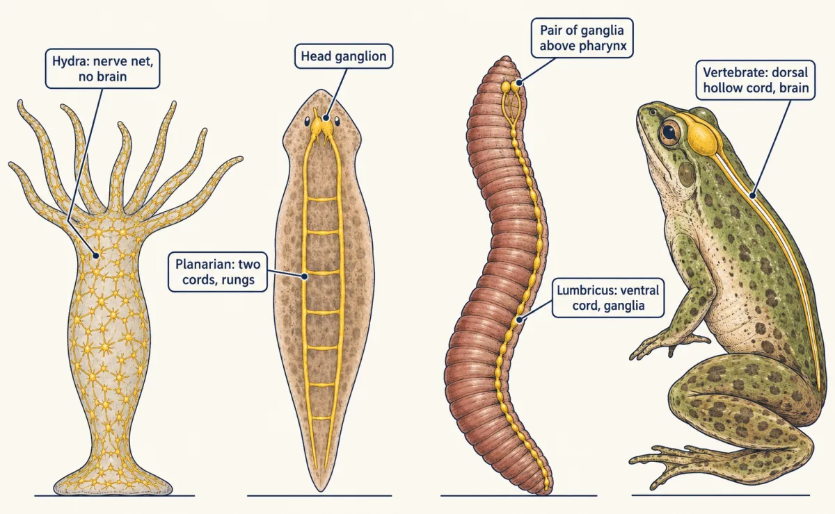
Every animal with neurons links sensing to acting, but the layouts differ, and a nerve net has no separate integrating centre (the groups are in animal body plans).
- Hydra, a freshwater cnidarian, has a nerve net: neurons spread through the body wall and connected in a mesh, with no brain.
- A planarian, a free-living flatworm, has two nerve cords along its underside, joined by cross-connections like the rungs of a ladder, and a ganglion in the head.
- The earthworm Lumbricus, an annelid, has a single ventral nerve cord with a ganglion in each body segment, linked to a pair of ganglia above the pharynx near the front.
- Vertebrates, such as a frog or a human, have a dorsal hollow nerve cord, the spinal cord, running along the back, with a brain at its front end.
Flatworm and earthworm cords are solid. Hydra’s net is a working nervous system in a living lineage, not an unfinished brain.
Going deeper
Helmholtz’s apparatus and the speed numbers
Helmholtz timed these tiny intervals with a galvanometer method and with a recording myograph that wrote the twitch on a moving surface. His teacher Johannes Müller had doubted that the speed could be measured at all. The squid giant axon is sometimes wider than half a millimetre; Hodgkin and Huxley’s 1952 analysis of the impulse used it (action potential). Vertebrate motor axons range from a few to about 20 micrometres.
Your notes
Select any text in the lesson to highlight it or add a note to it. Your highlights and notes are saved in this browser and collected in your notebook.
This browser is not letting the site save anything (a private window or strict privacy settings), so highlights and notes will be lost when you leave the page.
How we measure it
Two-point timing of nerve conduction
Stimulate a nerve at two known distances from the muscle or recording site and measure the delay from each. The difference in distance divided by the difference in time gives the conduction velocity, because delays common to both trials, such as the junction and the muscle's own response, cancel.
Pharmacological blockade of autonomic input
Give a drug that blocks one division's effect on an organ, then the other, then both, and measure the organ's output each time. The change after each block shows how much that division was contributing; with both blocked, the organ shows its intrinsic behaviour.
Microneurography
Insert a fine needle electrode through the skin into a peripheral nerve of a conscious human and record the impulses of single identified nerve fibres, such as the sensory fibres from muscle spindles, while the person moves or the limb is stretched.
Comparative anatomy of nervous systems
Dissect, section and stain the nervous systems of animals from different groups and map where the cell bodies, cords and ganglia lie. Comparing the maps shows which arrangements recur and which are confined to one lineage.
Practice questions on this lesson (3) → MCAT-style, original, each with an explanation
Where this lesson fits
Before thisReview first
- Tissues, Organs and HomeostasisThis course · Lesson 6
It introduces receptor, control centre and effector, the same route this lesson times leg by leg.
- Homeostasis in ActionBiology · High school
It shows a feedback loop with sensor, control centre and effector, which this lesson treats as a timed route.
After thisStudy next
- Hormones and the Endocrine SystemThis course · Lesson 8
Hormones are the second kind of control, so you can compare their slow delivery with the nerve route timed here.
- Muscle, Bone and MovementThis course · Lesson 19
The effector end of the reflex arc is a muscle, and this lesson shows how its fibres shorten.
- Heat Balance and Body TemperatureThis course · Lesson 9
Heat balance uses sensors, a control centre and autonomic effectors such as sweat glands and blood vessels.
Suggested by an AI model from the lessons themselves, then checked. How we use AI
Finish line
Test yourself
8 questions
Practice and MCAT-style, with explained answers.
Next · Lesson 8
Hormones and the Endocrine System
A young cockerel's testis, moved to his belly and cut off from its original nerves, still made him grow into a crowing rooster. What was it sending?
Continue →