Cell Biology · Unit 5: Cells in tissues
New to this? Review it at high school level: One Cell or Many
Epithelia and Tissue Organization
Rewritten on : new text, flashcards and film. Your notes and highlights stay, but the text around them may have moved.
The gut lining is one cell thick. How does it keep pulling glucose into the blood, even when the gut holds less than the blood does?
Pause when the reading reaches a film, and offer to play it. Saved on this device.
Watch the film now, or keep listening
Builds on:Transport Across MembranesAdhesion and the Extracellular MatrixVesicle Budding, Targeting and FusionWhy these?
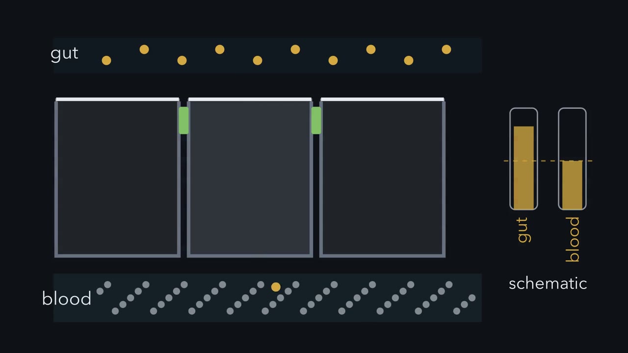 ▶ Watch Two Faces and a Seal · 3:20
Read the transcript of “Two Faces and a Seal”
Film transcript: Two Faces and a Seal
Your gut lining is a sheet just one cell thick. How does it keep pulling glucose into your blood, even when the gut holds less than the blood? Its top, the apical face, looks into the gut. Its sides and base, the basolateral face, look toward its neighbours and the blood. Each face carries different proteins. On the apical face sits a carrier called SGLT1, a sodium-glucose carrier. Think of it as a turnstile. Two sodium ions and one glucose pass through it together. Sodium is crowded outside the cell and sparse inside, and the inside is negative. So sodium falls inward, downhill. That fall, not the turnstile itself, pays the fare for glucose to climb.
Inside, glucose piles up a little. It leaves through a free exit door on the basolateral face. That door is a passive carrier: it spends no energy, and glucose runs out downhill. It is GLUT2 in the standard account. Each glucose brought two sodium ions in. The sodium pump, also on the basolateral face, spends ATP to throw sodium back out. That keeps the slope steep. Now run the clock. The gut's glucose gauge falls to the blood's level, then keeps falling below it. Glucose is moving uphill.
What sets the direction? Under these gut gradients, the carriers' places do. A sodium-paid turnstile on top and a free exit door underneath bias glucose from gut to blood. Now look between the cells. Neighbours are joined near the top by a seal. Try a thought experiment: loosen that seal enough to let glucose slip through. Would the gut still take up glucose?
It depends on the gut. Between the cells, glucose just follows its own gradient. Early in a meal, the gut holds more than the blood, so the open seal adds to absorption. Late in the meal, the gut holds less than the blood. Now the leak runs backwards.
Glucose slides back out between the cells, and the gut level stalls at a floor it cannot sink below. Tighten the seal, and the floor drops. In this thought experiment, what the sheet absorbs is what it carries through the cells, minus what leaks back between them. Here is the one idea. Under the usual gut gradients, where the carriers sit biases glucose flow from gut to blood. The seal does not orient that pathway. It decides what can slip between the cells, and so what can leak back. Our glucose leak was a thought experiment. What the real seal lets through, and how it is measured, is in the text below.
From the film
- In the gut lining, each cell has an apical face toward the gut and a basolateral face toward its neighbours and the blood.
- In these cells, sodium flowing in through SGLT1, the apical turnstile, pays for glucose to come in, even uphill.
- Glucose leaves by a passive exit door on the basolateral face, where the sodium pump throws sodium back out.
- Under the usual gut gradients, apical SGLT1 and basolateral GLUT2 bias glucose flow toward the blood.
- The seal between the cells does not choose the direction; it decides what can slip between them, and so what can leak back.
Film 1 followed glucose through one gut cell; what it left out is the sheet itself and the real seal. The lining is an epithelium, a sheet of cells that covers a surface or lines a tube. In a simple epithelium, one cell thick like this, each cell’s apical face looks toward a lumen (the space inside a tube) or the outside world, and its basolateral face toward its neighbours and the basement membrane, a mat of extracellular matrix (adhesion and the extracellular matrix). The faces’ different proteins and lipids are its apicobasal polarity.
The one idea. For glucose in the gut under the usual gradients, apical sodium-glucose cotransporter 1 (SGLT1) and basolateral GLUT2 bias transport toward the blood. The seal does not orient that pathway; it decides what can slip between the cells, and so what can leak back.
Scope: glucose across a simple vertebrate epithelium. Other epithelia also secrete and place their carriers differently, and a carrier’s direction also depends on the gradients it meets. Film 1 used the simplest case of the seal, the tight junction; the real seal is below.
How far uphill, and at what cost
The film showed sodium paying for glucose; here are the limit and the price. The turnstile is sodium-glucose cotransporter 1 (SGLT1), a sodium-glucose symporter (one carrier moving two solutes the same way); for human SGLT1 expressed in frog oocytes, coupled transport moves two sodium ions per sugar. The exit door, GLUT2 in the standard account, is sodium-independent and works by facilitated diffusion, letting glucose run down its gradient. The sodium-potassium pump never touches glucose. Glucose takes the transcellular route, through the cells; the paracellular route runs between them (membrane transport works out the forces; digestion covers nutrition).
How far uphill?
For an ideally coupled carrier, with the sodium levels and voltage held fixed, net transport stops when the energy sodium releases coming in just matches the energy needed to push glucose up its gradient. Sodium pays twice: it is far more concentrated outside the cell than inside, and as a positive ion it is pulled in by the cell’s negative inside. If the formula is new, start with the worked numbers.
In words The glucose ratio at balance equals the sodium ratio, times a voltage term, both raised to the number of sodium ions per glucose.
- n“n”
- sodium ions carried with each glucose: 2 for human SGLT1
- [Na]_out, [Na]_in“sodium outside and inside”
- about 140 mM and 15 mM in a typical cellmM
- Vm“V m”
- membrane potential, inside relative to outside; about −50 mV here, so the exponent is positivemV
- F/RT“F over R T”
- turns voltage into energy per charge; RT/F is about 26.7 mV at 37 °C1/mV
Sanity check If n = 0 the ratio is 1: an uncoupled carrier can only even glucose out. Each extra sodium multiplies the ceiling by the same factor again.
Try one number
Sodium ratio: 140 / 15 = 9.3. Voltage term for one positive charge at −50 mV, where the two minus signs cancel: exp(50 / 26.7) = 6.5.
One sodium per glucose: 9.3 × 6.5 ≈ 61, about 60-fold.
Two sodium per glucose: the same factor squared, 61 × 61 ≈ 3,700-fold.
Need a refresher? powers and roots · e and ln
A second sodium ion squares the ceiling. An absorbing gut cell is unlikely to approach it: the exit door keeps draining glucose, and real gradients are not fixed. But it means SGLT1 can keep taking glucose long after the gut holds less than the cell. The 2:1 ratio is SGLT1’s, not the family’s: its kidney relative sodium-glucose cotransporter 2 (SGLT2) coupled one sodium per glucose in a study in HEK293T cells, the 60-fold row.
What does it cost?
Each glucose brings two sodium ions in, and the pump throws out three sodium ions for each ATP it spends. In this simple accounting, absorbing one glucose costs about two-thirds of an ATP, against the roughly 30 ATP the body later makes by oxidizing it.
Which face, and why it matters
The film’s leak ran between the cells; here is one through them.
Predict. A gut cell keeps SGLT1 on its apical face but puts its passive glucose carrier, GLUT2, on both faces. Does it absorb more glucose, less, or the same? Does your answer depend on how much glucose is left in the gut?
Check your prediction
It depends on the gut. Early in a meal, when the gut holds more glucose than the cell, apical GLUT2 is a second way in and can raise absorption if the exit on the basolateral face is not limiting. Later, when the gut holds less than the cell, the same carrier lets glucose run back out to the gut and undoes part of SGLT1’s work; how low the gut level can then fall depends on how active each carrier is. A passive carrier moves glucose down whatever gradient it meets; on the basolateral face GLUT2 is an exit under the usual gradient, but it can reverse if the gradient does.
Predict. Cholera toxin makes the gut lining secrete salt and water, and the diarrhoea can kill by dehydration. Oral rehydration solution, used to treat it, is not just salt water: it contains glucose. Why add the sugar?
Check your prediction
The toxin leaves SGLT1 working. Each glucose taken up brings two sodium ions in with it; the sodium gradient, kept by the pump, supplies the energy, and chloride and water follow the absorbed salt. So the glucose lets the gut absorb salt and water even while the toxin drives secretion.
Salt and water follow glucose through the cells, but some also cross between them; the second film turns to the seal that governs that route and the sides that keep the faces apart.
Read the transcript of “The Seal and the Sides”
Film transcript: The Seal and the Sides
What makes the seal between cells selective, and what keeps each cell's two faces distinct? In chapter one, under gut gradients, carrier placement biased glucose toward blood. The seal decided what crossed between cells.
The seal is the tight junction, a belt where neighbouring cells meet near their apical edges. It is not solid caulk. It is a selective gate between cells. Claudin proteins assemble into branching strands around each cell. Narrow routes through those strands can pass small ions.
Meanwhile, the junction strongly restricts bacteria and larger molecules. The small intestine needs some sodium and water to cross between cells, so its junctions are relatively leaky. The bladder needs a tighter barrier. Different epithelia tune this gate for different jobs. How can we measure this gate? Grow cells as a sheet on a porous filter. Put one electrode above and another below, then pass current. If resistance falls, must glucose be leaking through?
No. This reading is transepithelial electrical resistance, or TEER. It reports how easily ions cross the whole sheet. It cannot name the ion or route, or say whether larger molecules leak. Current may cross between cells or pass through them. A low reading can reflect selective ion pores, broader leaks, or breaks. Telling them apart takes other experiments, set as a design problem below. The seal is often called a fence, said to stop membrane proteins drifting between the two faces. How well it fences is tested in the text below.
Cells also mark territories with proteins, and traffic delivers cargo to each face. In the standard scheme, Crumbs marks the apical side, while Scribble marks the basolateral side. The Par complex sits near their boundary. These groups are thought to restrain one another, while polarized traffic places carriers. Below the seal, an adherens junction couples neighbouring actin belts. Desmosomes link keratin networks against stretching.
Gap junctions pass ions and small molecules between cytosols. At the base, hemidesmosomes connect keratin to the basement membrane. Each junction has a different job. Together they let a one-cell-thick sheet stay selective, strong, connected, and anchored. Back in the gut, SGLT1 stays apical while the pump stays basolateral. The selective seal controls the path between cells, while deeper junctions keep the sheet intact. That is the rule: epithelial barriers are built by division of labour. A gate, distinct faces, grips, anchors, and channels work together, but they are not interchangeable.
From the film
- The tight junction is a selective gate near the apical edge, not a watertight plug.
- Claudin proteins build the branching strands of the tight junction.
- TEER measures how freely ions cross the whole sheet, not which ion or route, or whether larger molecules leak.
- Polarity proteins and targeted traffic help keep apical and basolateral faces distinct.
- Other junctions grip neighbours, connect their cytosols or anchor cells to the basement membrane.
The seal: what passes between the cells
Film 2 showed the seal as a selective gate; here is what the experiments behind it show. Much of the water and some of the salt that follow glucose do cross between the cells.
The seal is the tight junction, the most apical junction between neighbours. In 1963 Marilyn Farquhar and George Palade surveyed epithelia from rat and guinea-pig organs by electron microscopy (microscopy and images). In rat kidney tubules filled with hemoglobin, the tracer stopped at the zonula occludens, the junction now called the tight junction. That placed a barrier there for a protein-sized tracer. It did not show that the junction is closed to ions.
Freeze-fracture, which splits membranes down the middle and replicates the inside face in metal, shows tight junctions as branching strands. In 1998 Mikio Furuse, Shoichiro Tsukita and colleagues showed that claudin-1 or claudin-2 alone built such strands in mouse L fibroblasts, cells that normally have none.
You might expect more strands to make a tighter seal. Two strains of a dog kidney cell line, MDCK, test that. They have similar numbers of strands, yet MDCK I resists electric current well and MDCK II is leaky.
Predict. What could make one sheet far leakier to ions than another with the same number of strands?
Check your prediction
What the strands are made of. In 2001 Furuse’s group found that MDCK II cells make claudin-2 and MDCK I cells do not. When they put dog claudin-2 into MDCK I cells, resistance fell more than twentyfold, and strand number did not change significantly.
So for small ions, which claudins a tissue makes largely decides what passes: the gate function. Claudin-2 is thought to form a paracellular pore, a channel through the strands that favours cations; the 2001 experiment fits that idea but does not test it (the design problem below shows how).
Like the small intestine, the kidney’s proximal tubule is a leaky epithelium; the distal colon, urinary bladder and collecting duct are tight. A leaky epithelium uses both routes at once. Claudin pores are narrow, though: claudin-2 pores do not pass even mannitol, an uncharged molecule about glucose’s size. So for glucose the seal mainly governs the ions and water that travel with it, not the direction; only a larger, glucose-permeable leak would let much glucose slip back, as in the film.
Measuring the seal: TEER
The claudin results above rest on film 2’s measurement, TEER (transepithelial electrical resistance), taken on cells grown as a sheet on a porous filter insert. Here are made-up numbers. MDCK monolayers grow on inserts of area 1.12 cm². A blank insert with no cells reads 120 ohm. A control MDCK I monolayer reads 3,000 ohm, and one expressing claudin-2 reads 250 ohm.
- Subtract the blank, because the filter and fluid add resistance of their own. Control: 3,000 − 120 = 2,880 ohm. Claudin-2: 250 − 120 = 130 ohm.
Predict. The next step corrects for the size of the insert. Picture each patch of sheet as a resistor, so a larger sheet has more of them side by side. Does it read a higher or a lower raw resistance? So do you multiply by area, or divide?
Check your prediction
Lower: resistors in parallel pass more current together. So multiply by area, never divide.
- Multiply by area. Control: 2,880 × 1.12 = 3,225.6 ohm·cm². Claudin-2: 130 × 1.12 = 145.6 ohm·cm².
- Compare: 3,225.6 / 145.6 = 22.2-fold drop, the size of effect Furuse and colleagues reported.
- Know the limit film 2 gave: TEER measures how easily ions carry current through and between the cells (often mostly between, in leaky sheets).
TEER (ohm·cm²) = (R monolayer − R blank) × area of insert
Never quote one TEER for “MDCK cells”: MDCK I and MDCK II differ greatly.
- Subtract the blank.
- Multiply by area.
- Compare.
TEER (ohm·cm²) = (R monolayer − R blank) × area of insert. Never divide by area. The numbers are made up, and TEER says how easily ions carry current, not which ion, or whether sugars or proteins leak too. The resistor picture is a sketch of the logic, not to scale.
Design problem: pore or leak?
Claudin-2 lowered MDCK I resistance more than twentyfold without changing strand number. TEER alone cannot say why. Design experiments to decide whether it makes a selective cation pore or a general leak.
One way to do it
Start with selectivity. In parental and claudin-2 MDCK I cells on filters, measure a dilution potential: halve the salt on one side, read the voltage across the sheet, and calculate the sodium-to-chloride permeability ratio. A cation pore predicts a high ratio in claudin-2 cells; a nonselective leak, little change. Bi-ionic potentials, replacing sodium with other cations, add a size and charge profile.
Then test size with uncharged tracers: mannitol, then 4 kDa and larger dextrans. A narrow pore predicts little change in any of them, since even mannitol is too big for claudin-2 pores. A rise points to a larger route: the tight junction’s leak pathway, which is size-selective, or a break in the sheet, which has no sharp size cutoff. Checking for dying or missing cells helps tell those two apart.
Then test the protein. Mutate charged residues in claudin-2’s extracellular loops; if neutralizing a negative charge lowers cation preference, that supports the idea that the loops line the pore.
Controls: parental cells and an empty-vector clone, matched expression, cell density and days in culture, and more than one clone. Limit: this is one dog kidney cell line, so a result about pore behaviour in MDCK I is not yet a result about any intact tissue.
Is the seal also a fence?
The film’s idea needs each carrier to stay on its own face. The tight junction’s other textbook job, as a fence, is said to do that: to stop membrane proteins and lipids drifting between the two faces.
Predict. In 2019 Tetsuhisa Otani, Furuse and colleagues disrupted five major claudins in MDCK II cells. The strands vanished and resistance fell. Did the cells lose their polarity?
Check your prediction
No. Polarity was not disorganized. They did not rule out other claudins, and membrane contacts containing JAM-A remained, so a fence made by other junction proteins was not excluded. Still, in these cells polarity did not need claudin strands. The same study found that it did depend on other junction parts: cells lacking the scaffold proteins ZO-1 and ZO-2 had no tight-junction structure, and their polarity was disorganized.
So the case for the fence is weaker than the slogan, and something else keeps the faces apart.
How each cell knows its top from its bottom
What keeps sodium-glucose cotransporter 1 (SGLT1) on the apical face and the pump on the basolateral one? In the standard scheme, which comes largely from fruit-fly genetics and mammalian cell lines, three groups of proteins mark the sides. The Crumbs complex sits apically. The Par complex, which includes the kinase aPKC, sits at the boundary where apical meets lateral. The Scribble complex sits basolaterally. The groups are thought to restrain one another, so each holds its own territory; aPKC, for example, phosphorylates basolateral proteins and pushes them off the apical membrane. Polarized membrane traffic then delivers the right proteins to each face, as described in the lesson on vesicle traffic.
Holding the sheet together: the rest of the stack
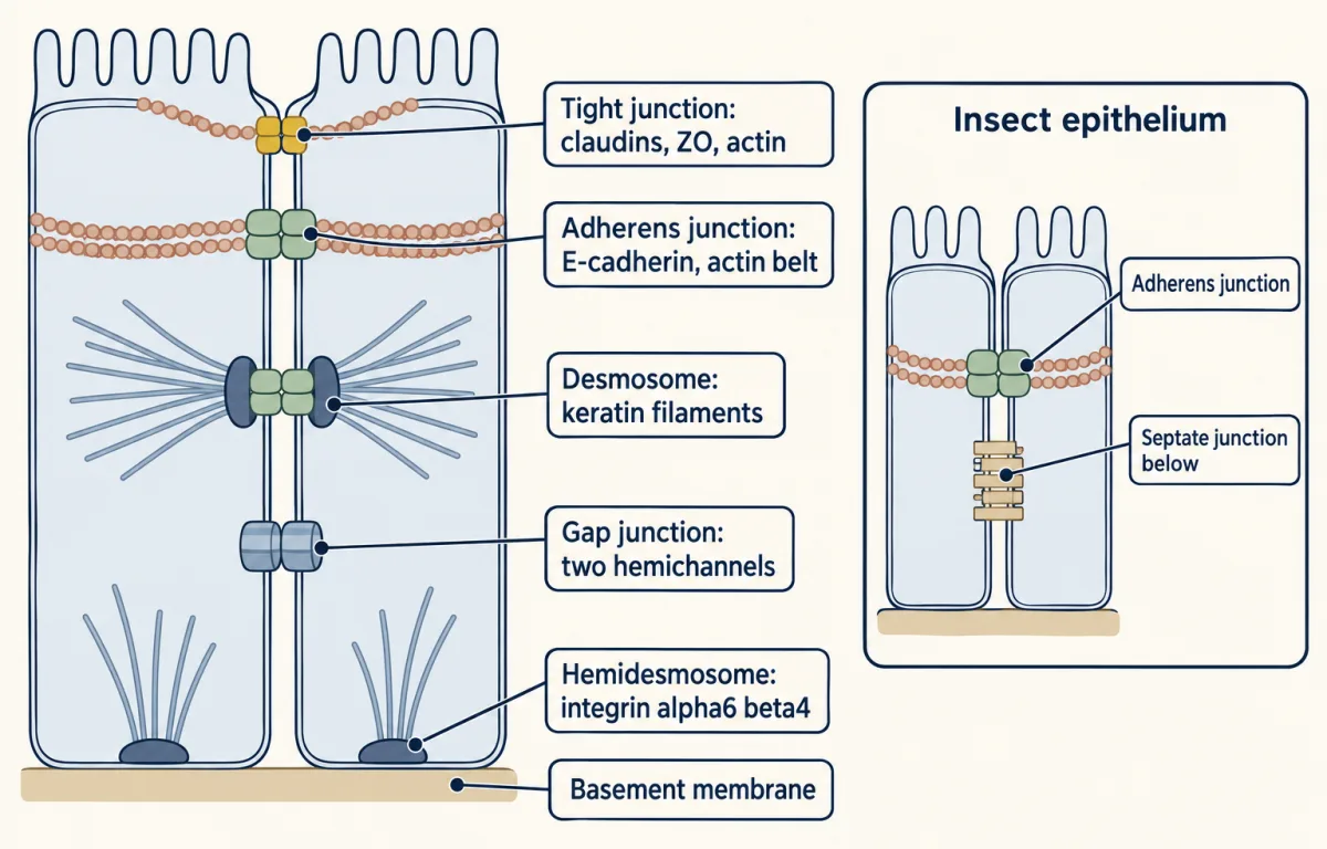
The faces and the seal matter only while the sheet stays in one piece. Below the seal, more junctions are stacked down the side of the cell in a characteristic order: the adherens junction (a belt coupling neighbours’ actin), desmosomes (spot welds), gap junctions (channels between cytosols) and, at the base, hemidesmosomes (anchors to the basement membrane). All but the gap junction link inside to the cytoskeleton.
| Junction, top to bottom | Transmembrane protein | Inside partner | Job |
|---|---|---|---|
| Tight junction (the seal) | Claudins, with occludin among others | ZO proteins, to actin | Seal the gaps; decide which ions pass |
| Adherens junction | E-cadherin, a calcium-dependent adhesion protein | Catenins, to a belt of actin | Couple neighbours’ actin mechanically |
| Desmosomes (spot welds) | Desmogleins and desmocollins, cadherins of their own family | Desmoplakin, to keratin intermediate filaments | Resist stretching and tearing |
| Gap junctions, scattered | Connexins: two hemichannels per channel, one from each cell, each of six connexins in the human connexin 26 crystal structure | No major cytoskeletal anchor in the standard scheme | Pass ions and small molecules from cytosol to cytosol |
| Hemidesmosome, at the base | Integrin alpha6 beta4 | Plectin, to keratin | Anchor the base to the basement membrane |
The order is not universal: many insect epithelia seal with septate junctions, which lie below the adherens junction (the figure’s inset).
Plants: walls and plasmodesmata
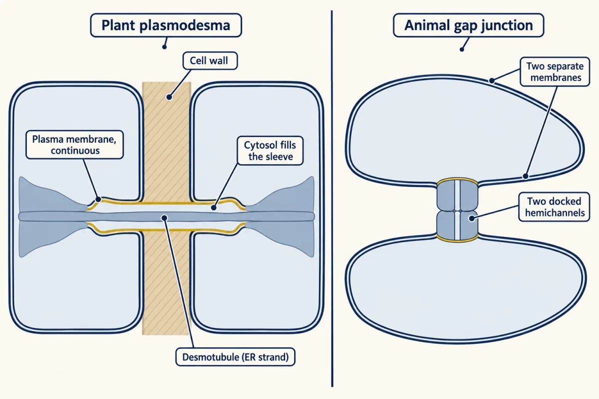
Plant cells have no tight or adherens junctions, so this section is a contrast, not part of the gut story: a plasmodesma differs in kind from a gap junction (figure). Each plant cell is boxed in a cell wall, an extracellular matrix rich in cellulose, and neighbouring walls are glued together. The route between plant cells is the plasmodesma: a channel through the wall lined by plasma membrane that is continuous from one cell to the next. Down its middle runs the desmotubule, a narrow strand of endoplasmic reticulum continuous with the ER of both cells. Cytosol fills the sleeve between the two.
Going deeper
Junction details
Gap junctions are not alike. Channels made of different connexins pass the messenger cAMP very differently relative to potassium: in pairs of cultured cells made to express one connexin, the permeability ratio was 0.180 for connexin 43, 0.027 for connexin 26 and 0.018 for connexin 40. Two cells can be equally coupled electrically and still share a messenger very differently. Even within the fly the order of junctions can flip: in the midgut the adherens junctions lie below the septate junctions.
The claudin knockout numbers
In the five-claudin knockout in MDCK II cells, resistance fell from 52.5 to 14.5 ohm·cm² in one clone. Removing JAM-A along with the claudins caused sporadic polarity defects.
A pore with no visible sleeve
Appearance can mislead at plasmodesmata. In 2019 Dawei Yan and colleagues studied an Arabidopsis root mutant, plm-2. At one interface, between the phloem-pole pericycle and the endodermis, they saw no plasmodesmata of the type with a visible cytoplasmic sleeve. Yet GFP moved across that interface faster than in wild-type roots, by fluorescence recovery after photobleaching. A pore with no visible sleeve still passed protein, at least at this interface.
Your notes
Select any text in the lesson to highlight it or add a note to it. Your highlights and notes are saved in this browser and collected in your notebook.
This browser is not letting the site save anything (a private window or strict privacy settings), so highlights and notes will be lost when you leave the page.
How we measure it
Thin-section electron microscopy with an electron-dense tracer
A tracer that scatters electrons, such as hemoglobin or a heavy-metal salt, is placed on one side of an epithelium, the tissue is fixed and sectioned, and the electron microscope shows how far the tracer got between cells. It localizes the barrier to that tracer, not to ions.
Freeze-fracture electron microscopy
Frozen tissue is cracked so the fracture runs through the middle of membranes, and a platinum-carbon replica of the exposed face is imaged. Tight junctions appear as a network of branching strands, so you can count strands and see whether they exist at all.
Transepithelial electrical resistance (TEER)
Cells are grown as a sheet on a porous filter, electrodes on each side pass a small current, and the resistance of the sheet is read after subtracting a blank filter and multiplying by area. It reports how freely ions cross the whole sheet, through and between the cells (in leaky sheets the route between often dominates), but not which route or which ions, or whether larger molecules leak.
Uncharged tracer flux
A labelled molecule that carries no charge, such as mannitol or a fluorescent dextran of known size, is added to one side and sampled from the other over time. Comparing sizes tells a narrow claudin pore from larger routes: the tight junction's leak pathway, which passes molecules as large as 4 kDa dextran, or a break in the sheet, which has no sharp size cutoff.
Practice questions on this lesson (3) → MCAT-style, original, each with an explanation
Where this lesson fits
Before thisReview first
- Transport Across MembranesThis course · Lesson 5
You need electrochemical gradients and coupled transport from there to work out how far sodium can push glucose uphill.
- Adhesion and the Extracellular MatrixThis course · Lesson 22
It introduces cadherins, desmosomes and the basement membrane that this lesson stacks into the sheet.
- Vesicle Budding, Targeting and FusionThis course · Lesson 11
Polarized delivery of carriers to the apical or basolateral face here builds on targeted vesicle traffic.
After thisStudy next
- Stem Cells and Tissue RenewalThis course · Lesson 24
It uses the gut lining as its example of a sheet that renews while keeping its faces and seals.
- Cell Identity, Cell State and DiseaseThis course · Lesson 25
It builds on the idea of tissue organization, asking what goes wrong in sheets of cells with altered state.
Elsewhere on the siteFilms, simulations and method films in other courses
- Why Order Matters in a GutFilm · Organismal Biology
It shows the gut as a one-way tube where absorption happens across the lining.
- Downhill for FreeFilm · Biology
It shows how molecules cross a membrane downhill for free, as glucose does through the passive exit.
- Which Way Does the Water Go?Film · Organismal Biology
It shows water crossing epithelia toward higher osmolality, the same water movement the seal governs.
- The bile acid budgetSimulation · Physiology
It follows what the gut lining absorbs and how that is budgeted.
Suggested by an AI model from the lessons themselves, then checked. How we use AI
Finish line
Test yourself
8 questions
Practice and MCAT-style, with explained answers.
Next · Lesson 24
Stem Cells and Tissue Renewal
The lining of a mouse's small intestine is replaced every few days, yet it lasts for life. Stem cells do it by making new specialized cells while keeping up their own numbers. How would you prove which cells are the stem cells, and what keeps them that way?
Continue →