Organismal Biology · Unit 5: Defense, movement and reproduction
Animal Development
Rewritten on : new text, flashcards and film. Your notes and highlights stay, but the text around them may have moved.
A frog egg divides into thousands of cells without growing at all. How does each cell come to its fate?
Pause when the reading reaches a film, and offer to play it. Saved on this device.
Watch the film now, or keep listening
Builds on:How Cells DivideThe Cell Cycle and MitosisAnimal ReproductionWhy these?
 ▶ Watch Divided, Not Grown · 3:20
Read the transcript of “Divided, Not Grown”
Film transcript: Divided, Not Grown
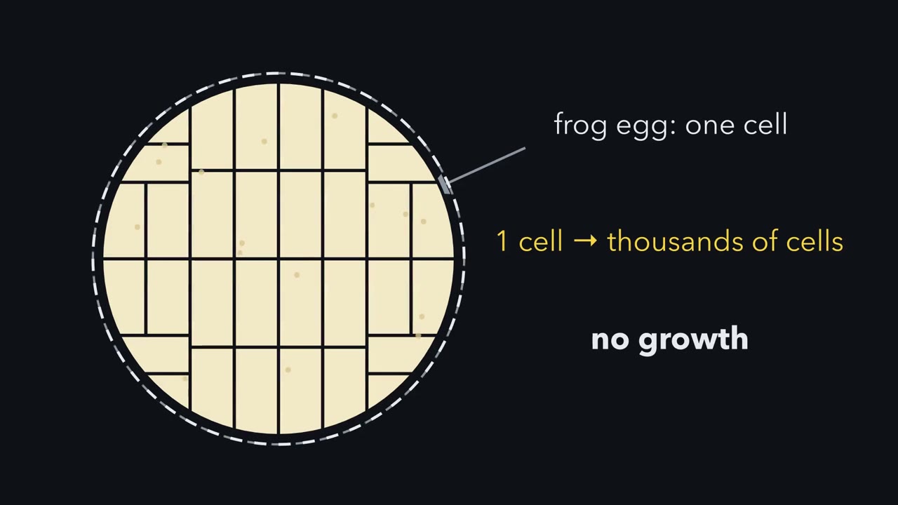
A frog egg divides into thousands of cells without growing at all. So where do the cells come from, and what does each one get? Development starts at fertilization. A sperm fuses with the egg, and a wave of calcium crosses it. A frog egg was paused partway through meiosis, and the calcium wakes it. Only one sperm should get in, and the egg keeps the rest out. Next comes cleavage, a run of divisions with no growth between them. Each new cell is called a blastomere. One cell becomes two, then four, then eight.
Watch the outline. It never gets bigger. Each division cuts the same egg into more, smaller pieces. The egg is divided, not grown. Suppose every division is equal. After five divisions there are thirty-two cells. Is each cell one thirty-second as wide as the egg?
No. In this equal model, each cell holds one thirty-second of the volume. But volume is shared out in three dimensions: length, width and depth. So each cell is about a third as wide as the egg. Something else has changed. There are now thirty-two nuclei, but no new cytoplasm. Each nucleus has far less cytoplasm around it.
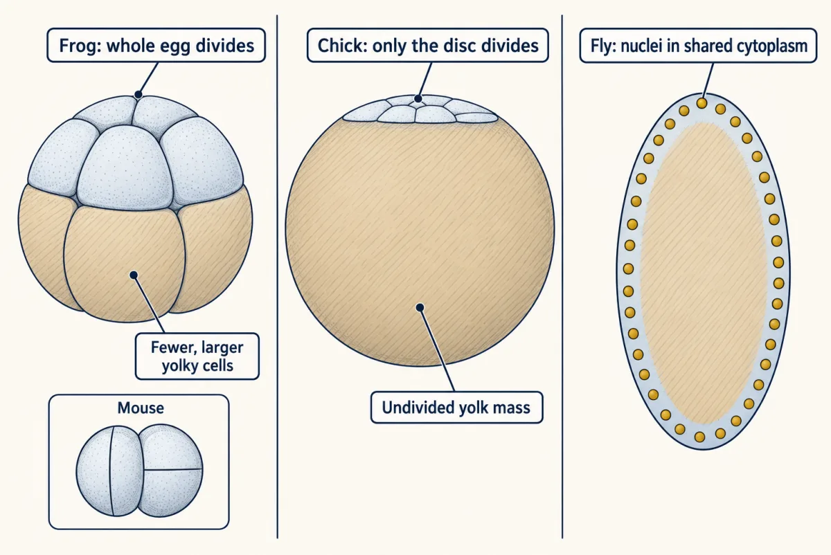
Now suppose the egg is not the same all through. Mark one region in purple. This stands for inherited material, placed in the egg before it divides. Suppose the purple is anchored, so it stays put. The furrows cut through the egg around it. After a few divisions, only some cells hold it. Their neighbours have none. That is what a cell inherits: the slice of egg it was cut from. If an anchored molecule helps set fate, it is a cytoplasmic determinant. Yolk changes the cutting. Yolk is stored food, and it slows the furrows. A frog egg divides completely, with bigger cells in its yolky half. A chick divides only a disc on top of its yolk. A fly first divides its nuclei in shared cytoplasm, with no membranes between them.
Edwin Conklin watched the egg of a sea squirt. Part of its cytoplasm is yellow, and it gathers into a crescent. He followed it cell by cell. It ended up mainly in the cells that build the larva's tail muscle. But following a colour shows where it goes, not what it does. Does the crescent make muscle, or just ride along? That takes a different test. So cleavage shares out one fixed egg. Each cell gets its own slice, and anchored material comes with it.
From the film
- After fertilization, cleavage divides the egg into many smaller cells, called blastomeres, without any growth.
- In the ideal case of equal divisions with no growth, five divisions make 32 cells, each about a third as wide as the egg.
- Material that stays put in one region of the egg goes only to the cells cut from that region: that is what a cell inherits.
- Yolk changes how the egg divides: a frog egg divides completely, a chick egg only in a disc on top of the yolk.
- Following a coloured region of an egg shows which cells inherit it, not whether it causes their fate.
A frog egg divides into thousands of cells without growing at all. How does each cell come to its fate? The first film shows the cutting; this text adds what it leaves out. The gametes of animal reproduction meet here.
The one idea. The egg is divided, not grown. Cleavage shares one fixed egg among smaller cells, and material that stays put in one region goes only to the cells cut from that region: that is what a cell inherits. Gastrulation then moves cells next to new neighbours whose signals can change their fate: that is what a cell is told. A cell’s fate follows from both.
Scope: this picture assumes equal, synchronous divisions and material that stays where it is. Real eggs often cleave unequally or out of step, and can move material (the ascidian’s yellow crescent gathers after fertilization). A localized molecule is a determinant only if it helps set fate.
One sperm, one egg: blocks to polyspermy
Fertilization joins a sperm and an egg into a zygote, the single cell that begins the embryo. The sperm is activated, and its acrosome, a cap of enzymes over the head, opens in the acrosome reaction (in sea urchins, on contact with the egg’s jelly coat). It then binds the egg’s coat and fuses with the egg membrane. Fusion activates the egg: a calcium wave (in mammals, repeated waves) restarts the cell cycle (calcium signaling). Frog and mouse eggs wait partway through meiosis; sea urchin eggs have already finished it.
Polyspermy is fertilization of an egg by more than one sperm. In sea urchins, frogs and mammals it is usually fatal to the embryo: the egg gets an extra set of chromosomes and, in many species, an extra sperm centrosome that organizes an extra spindle pole, so the first divisions go wrong. Sea urchin and frog eggs commonly shut the gate in two stages: a fast block within seconds, then a slow block at the egg’s coat.
A cell’s membrane potential is the voltage across its membrane; an unfertilized egg is negative inside. In a frog or several sea urchin species, sperm fusion swings it positive (depolarizes it), and a second sperm arriving then rarely enters. Timing alone cannot show that the voltage is the barrier.
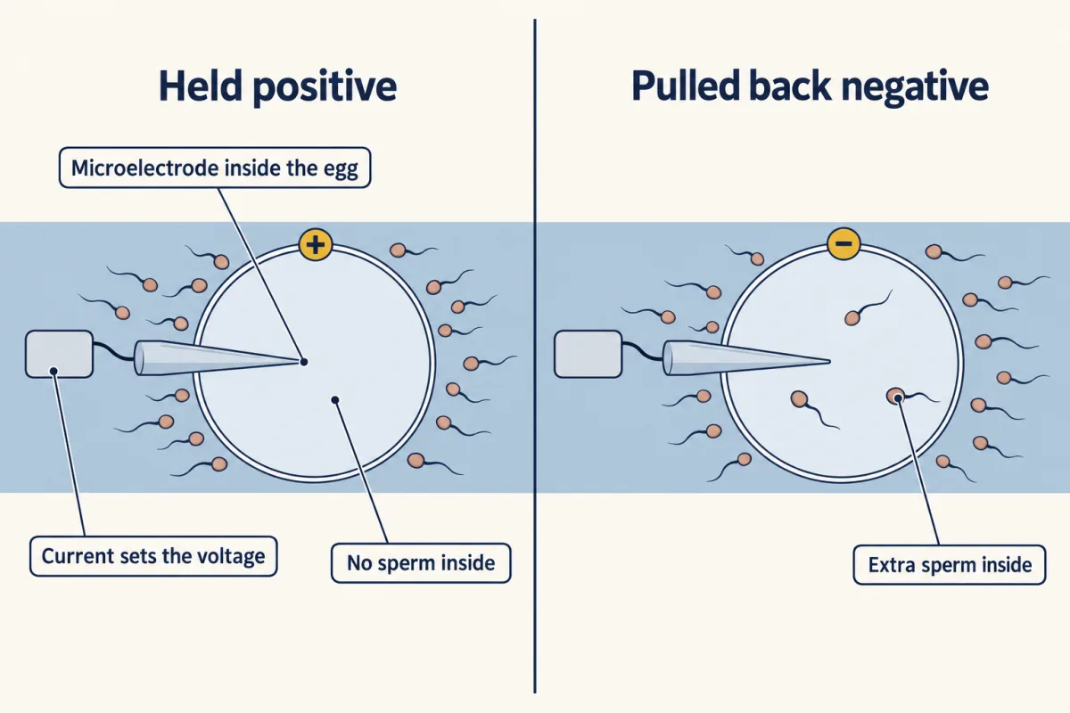
Predict. Suppose you could clamp an unfertilized sea urchin egg, holding it with an electrode at any voltage you chose, then add sperm. What two results would show that the positive voltage itself keeps sperm out?
Check your prediction
Eggs held positive should resist sperm, and eggs pulled back negative just after the first sperm triggers depolarization should let extras in. In 1976 Laurinda Jaffe reported both. Sperm still stuck to eggs held positive but did not fertilize them.
A study of one other sea urchin found no electrical block.
The mouse differs. Its barriers sit at the egg membrane and at the egg’s coat, and both take hold more slowly. Reviews of mammalian eggs conclude that its membrane block is not electrical; one suggested mechanism is that the egg loses Juno, its receptor for a sperm protein, from its surface. All three build the coat block with cortical granules, small vesicles under the egg membrane that fuse with it after activation and release their contents. In sea urchins those contents lift the egg’s coat into a hard fertilization envelope. The mouse egg’s coat is the zona pellucida; Jurrien Dean’s laboratory found that the released protease ovastacin cuts ZP2, one of its proteins, and that sperm kept binding the coat of embryos lacking ovastacin.
Blocks are common, not universal: some animals with very yolky eggs, such as birds and salamanders, normally let several sperm in. Only one sperm nucleus joins the egg nucleus, and the extra sperm nuclei degenerate.
Cleavage: dividing without growing
The first film’s cleavage is rapid in many animals but slow in mammals, and each division is a mitosis. The names, with the animals the first film skipped:
- Holoblastic cleavage divides the whole egg. In the sea urchin it is radial: equal at first, then from the fourth division small micromeres form at one end of the egg, the vegetal pole. In the frog it is radial but unequal, as the first film showed. In snails it is spiral, with division planes at oblique angles to the egg’s axis. In the mouse it is rotational and slow: at the second division one cell divides along the egg’s axis and its partner across it, and divisions fall out of step, giving counts such as three or five.
- Meroblastic cleavage divides only part of the egg. In birds, reptiles and teleost fish (most bony fish) such as the zebrafish, it is discoidal: only a disc of cytoplasm on top of the yolk divides.
- Superficial cleavage, in insects such as Drosophila, starts with nuclei dividing without membranes between them, a syncytium, before membranes form around them at the surface.
Cleavage ends in a blastula, a ball or disc of cells, often around a fluid cavity; the mammalian version, the blastocyst, has an inner cell mass inside an outer layer.
Worked problem: cleavage without growth. The first film showed each cell ending up about a third as wide as the egg. What does that do to membrane? Invented round values: a spherical zygote of 1.000 nL (1.000 x 10⁶ cubic micrometres), five synchronous, complete, equal divisions, no growth, no cell loss, spherical cells.
- Number and volume. Each division doubles the count: 25 = 32 blastomeres, each 1.000 nL / 32 = 31.25 pL (31,250 cubic micrometres). Check: 32 x 31.25 pL = 1.000 nL.
- Width. Width falls as the cube root of volume: 321/3 = 3.17-fold. For a sphere, d = 2 x (3V / 4 pi)1/3: about 124 micrometres for the zygote, 39 for each blastomere.
Predict. No cytoplasm has been added. After five divisions, is the total membrane of all 32 cells less than, equal to, or more than the zygote’s?
Check your prediction
More: 3.17 times as much. Each cell’s area falls about 10-fold (3.17 squared), but there are 32 cells. The zygote’s area, pi x 1242, is about 48,400 square micrometres; 32 blastomeres of about 4,800 each come to about 153,500. This treats each cell as a separate sphere; packed cells share faces. It is the surface-to-volume logic of size and scaling.
Asynchronous mouse divisions break step 1: counts need not be powers of two.
cells = 2n · d = 2 × (3V / 4π)1/3 · area = π × d²
- Blastomeres, 2ⁿ
- —
- Volume each (nL)
- —
- Volume each (pL)
- —
- Volume each (µm³)
- —
- Zygote diameter
- —
- Blastomere diameter
- —
- Diameter ratio, zygote / blastomere
- —
- Zygote membrane area
- —
- Total blastomere membrane area
- —
- Area ratio, blastomeres / zygote
- —
- Nuclei in the same total cytoplasm
- —
- Check: cells × volume each
- —
Rule to keep: cleavage divides the same cytoplasm without growth, so cells × volume each stays equal to the zygote volume while total membrane area rises 2n/3-fold. Circles are drawn to scale with each other, not to a real embryo.
Where a localized molecule goes
Predict. A patch of material fills one eighth of an egg that cleaves equally, and it stays put. After six divisions there are 64 cells. How many carry the patch?
Check your prediction
Eight of the 64 if the division planes follow the patch’s edges; if they cut across it, more cells carry some. In unequal (frog) or rotational (mouse) cleavage, the share depends on where the furrows fall.
Conklin’s yellow crescent in the sea squirt Styela gives a fate map: what cells normally become, not proof that the yellow material causes muscle. The design problem below tests cause.
Cleavage shares out the egg, making new contacts as cells divide. The second film shows how gastrulation moves cells inside on a large scale, among new neighbours.
Read the transcript of “New Neighbours”
Film transcript: New Neighbours
A ball of cells has to become a body with an outside, a middle and a gut. How do surface cells get inside, and does it matter whom they meet? The first film showed cleavage cutting the egg into cells, each with its own slice.
Purple marked what a cell inherits. Cleavage makes new contacts as cells divide, but it does not rearrange the whole embryo. Gastrulation is the stage that does. Cells travel from the surface to the inside, and sort into three germ layers. Ectoderm, in blue, stays outside.
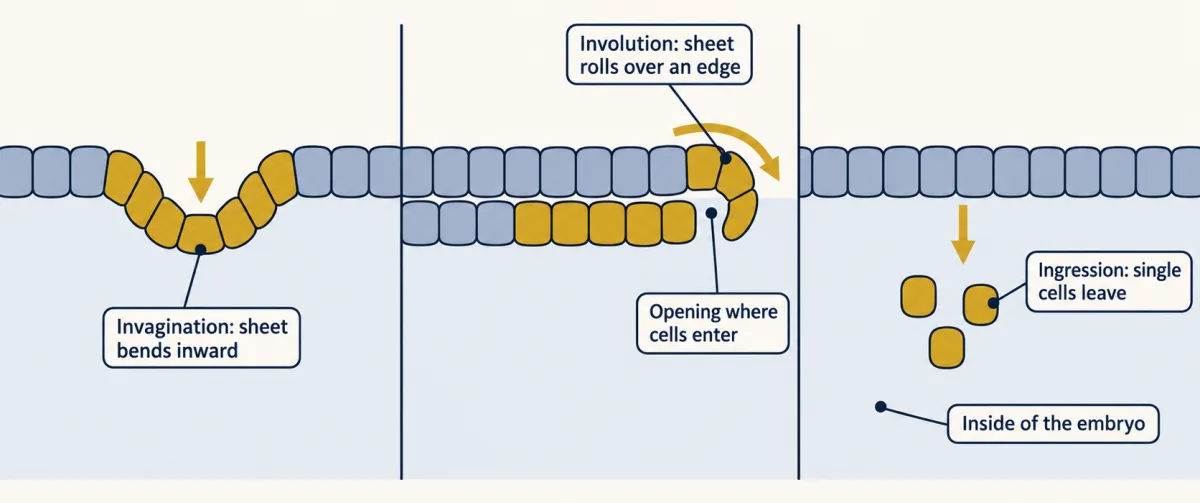
Mesoderm, in red, forms the middle. Endoderm, in yellow, lines the inside. Embryos get cells inside by several movements. In invagination, a sheet bends inward, like a finger pushed into a soft ball. In involution, a sheet rolls over an edge and spreads along the inside. In a frog, cells roll in over the lip of the blastopore, the opening where they enter. In ingression, single cells leave the sheet and move inward, one at a time. Embryos combine these movements to bring cells inside.
Moving inside does something else. Cells now sit next to cells they never touched before. Can a neighbour change what a cell becomes? Hilde Mangold tested this with newt embryos. She cut out a piece of the dorsal lip of the blastopore, the edge on the future back side. The donor came from a pale newt species. She grafted the piece onto the belly side of a darker embryo, from a related species. The host grew a second body axis.
If the graft built that second axis only from its own inherited material, what colour would the new axis be? It was not all pale. Dark host cells were part of it. The graft had recruited them. It told host cells what to become. Remember the crescent: colour showed where it went, not what it did. Here colour still only traced cells. The graft was the test, and colour showed host cells joined in. A signal from one group of cells that changes the fate of its neighbours is called induction. The grafted tissue is called the organizer. So a cell's fate has two sources. Purple: what it inherits from its slice of egg. Green: what its new neighbours tell it.
Most embryos studied closely use both.
From the film
- At gastrulation, cells move from the surface to the inside and sort into three germ layers: ectoderm, mesoderm and endoderm.
- Embryos combine movements to bring cells inside: a sheet bending in, a sheet rolling over an edge, and single cells moving inward.
- Moving inside puts cells next to new neighbours, whose signals can change what they become; this is induction.
- A graft of the newt's dorsal blastopore lip, the organizer, recruited host cells into a second body axis; pigment contrast between the pale donor and dark host showed which cells were the host's.
- Most embryos studied closely use both inherited factors and signals between cells.
Gastrulation and the germ layers
The second film’s three germ layers are two in cnidarians such as sea anemones. Where each of its movements is seen:
- Invagination, a sheet bending in: the sea urchin’s vegetal plate starts the gut this way.
- Involution: the frog’s blastopore lip, as in the film; whether the blastopore becomes mouth or anus is in animal body plans.
- Ingression, single cells moving in: in chick and mammal embryos, cells of the upper layer (the epiblast) ingress through the primitive streak, a thickened midline strip of epiblast, to form mesoderm and endoderm.
Germ layers make tissues, not whole organs. In vertebrates, ectoderm forms the epidermis and nervous tissue. Mesoderm forms muscle, blood, heart, kidneys, the gonads’ supporting tissue (the germ cells arise separately), and most bone and connective tissue (neural crest makes much of the face and skull’s). Endoderm forms the epithelial lining of the digestive and respiratory tracts. A lung or an intestine is built mainly from two layers: endoderm lining, mesoderm muscle and connective tissue, plus neural crest nerve cells.
The nervous system is specified during gastrulation, in some species earlier. In primary neurulation, signals from neighbours, including the dorsal mesoderm, help induce ectoderm to become a neural plate. In frogs, birds and mammals the plate folds and pinches off as a hollow neural tube, the future brain and spinal cord; in zebrafish it forms a solid keel that later hollows out. In secondary neurulation, at the tail end of many vertebrates, a solid cord of cells hollows out instead; how much of the tube forms this way varies among vertebrate classes. Cells at the plate’s edges become neural crest: peripheral neurons and glia, pigment cells, and much of the face and skull’s bone, cartilage and connective tissue. See neural induction, neurulation and tube defects and neural crest.
Inherited or told: how a cell gets its fate
- Autonomous specification: the cell inherits a cytoplasmic determinant, a localized molecule, often a maternal mRNA or protein stored in the egg, that helps set fate, and needs no neighbour.
- Conditional specification: fate set by induction, so it depends on which cells are nearby.
- Mosaic and regulative describe what happens when a blastomere is removed: a mosaic embryo lacks what that cell would have made, while in a regulative embryo the remaining cells change fate and make up the loss. Mosaic results tend to go with autonomous specification, regulative ones with conditional.
Real embryos sit along a range, not in two kinds: ascidians use induction for tissues such as the notochord, and the frog egg carries maternal mRNAs in its vegetal cytoplasm that start endoderm formation. Most embryos studied closely use both modes. How maternal mRNAs are stored and translated is in eukaryotic gene regulation.
Inherited. In the ascidian Halocynthia roretzi, Nishida and Sawada reported in 2001 that macho-1 mRNA, which encodes a zinc-finger protein (a family that often binds DNA), sits in the egg’s muscle-forming cytoplasm, the myoplasm.
Told. Hilde Mangold (the second film’s graft) grafted from 1921 and published with Hans Spemann in 1924; Spemann’s 1935 Nobel Prize was his alone (the organizer). Many inductive signals are Wnt and TGF-beta family proteins (Wnt signaling, TGF-beta and Smads). The organizer itself works largely by secreting blockers of such signals: chordin and noggin block bone morphogenetic protein (BMP), a TGF-beta family member.
Design problem
Localization of macho-1 mRNA in the myoplasm is only a correlation. Design experiments to test whether macho-1 is a muscle determinant, and say what the results would and would not show.
Saved in this browser only.
One way to do it
- Confirm the location. Check that macho-1 mRNA sits in the egg’s myoplasm and then in the muscle-lineage blastomeres.
- Loss. Deplete or block macho-1 mRNA in the egg and score primary tail muscle with a muscle-specific marker. A determinant predicts loss of those cells; Nishida and Sawada reported that.
- Gain. Inject synthesized macho-1 mRNA into blastomeres that do not normally make muscle. A determinant predicts ectopic muscle, where muscle does not normally form; they reported that too.
- Rule out neighbours. Culture the injected or depleted blastomeres alone. If the fate follows the mRNA in isolation, no signal from neighbours is needed.
- Controls. Inject a control mRNA, keep uninjected siblings, and score markers blind to treatment.
Limits: one tissue in one ascidian does not make ascidian development autonomous; the notochord, for one, is thought to depend on induction from neighbouring cells.
So the egg is divided, not grown: each cell inherits its own slice, and gastrulation gives it new neighbours that tell it more. Neither means the embryo “knows” its plan. Development changes in evolution (the evolution of development), but embryos do not replay their ancestors’ adult forms.
Going deeper
What the voltage-clamp test does not show
Jaffe’s experiment rules in a voltage-dependent fast block in this sea urchin. It does not show how the voltage stops entry or which channels depolarize the egg, and it says nothing about mammals.
The frog’s calcium-activated chloride channel
Wozniak and colleagues (2018) recorded Xenopus laevis eggs during fertilization; in their solution the eggs sat near -19 mV and depolarized to about +4 mV. The depolarization was driven by chloride flowing out through a calcium-activated channel, and their evidence points to the channel TMEM16A. An inhibitor of that channel raised the fraction of embryos scored as polyspermic.
Mouse sperm and egg
Mouse acrosome timing is less tidy: filmed in vitro with the egg’s surrounding cumulus cells in place, most fertilizing sperm had begun the reaction before they reached the egg coat. Fusion needs the egg protein Juno, the receptor for the sperm protein Izumo1, and eggs lacking Juno do not fuse with normal sperm.
Zinc sparks in the mouse
In 2025 Jurrien Dean’s laboratory reduced the egg’s bursts of zinc release, called zinc sparks, and saw no extra polyspermy in their tests.
Your notes
Select any text in the lesson to highlight it or add a note to it. Your highlights and notes are saved in this browser and collected in your notebook.
This browser is not letting the site save anything (a private window or strict privacy settings), so highlights and notes will be lost when you leave the page.
How we measure it
Microelectrode recording and voltage clamp of eggs
Insert a fine electrode into an egg to record its membrane potential, or pass current to hold the potential at a chosen value. Holding the voltage while adding sperm tests whether the voltage itself controls sperm entry.
Cell-lineage tracing
Follow identified blastomeres through cleavage, using natural pigment or an injected label, and record which tissues their descendants form. It gives a fate map, which shows what cells normally become, not what they could become.
Maternal mRNA depletion and ectopic injection
Remove or block a localized maternal mRNA and see whether a fate is lost, then inject the mRNA into other cells and see whether the fate appears there. Loss plus gain is the standard test of a cytoplasmic determinant.
Gene deletion plus a recombinant-substrate assay
Delete a gene for an egg enzyme in mice and test the egg coat for a lost function, then add purified enzyme to isolated coats and see whether it cuts its proposed target directly.
Practice questions on this lesson (3) → MCAT-style, original, each with an explanation
Where this lesson fits
Before thisReview first
- How Cells DivideLife Science · Grade 7
Each cleavage division is a mitosis, so you need to know how one cell becomes two.
- The Cell Cycle and MitosisBiology · High school
Cleavage is repeated cell cycles with no growth, so you need the cycle and its phases.
- Animal ReproductionThis course · Lesson 20
It shows how eggs and sperm are made, which is where fertilization starts.
After thisStudy next
- Plant Body and GrowthThis course · Lesson 22
It contrasts this animal embryo with plants, where growth and new cells continue from fixed growing tips.
- Double Fertilization: How a Flower Makes a SeedThis course · Lesson 26
It follows another fertilization that starts an embryo, with its own double fertilization and seed.
Suggested by an AI model from the lessons themselves, then checked. How we use AI
Finish line
Test yourself
8 questions
Practice and MCAT-style, with explained answers.
Next · Lesson 22
Plant Body and Growth
A 300-year-old oak grows new leaves each spring and usually a new ring of wood each year, yet no adult mammal grows a new limb. How does the oak keep building?
Continue →