Developmental Neuroscience · Unit 5: Cells on the move
The Neural Crest: Cells That Leave Home
Rewritten on : new text, flashcards and film. Your notes and highlights stay, but the text around them may have moved.
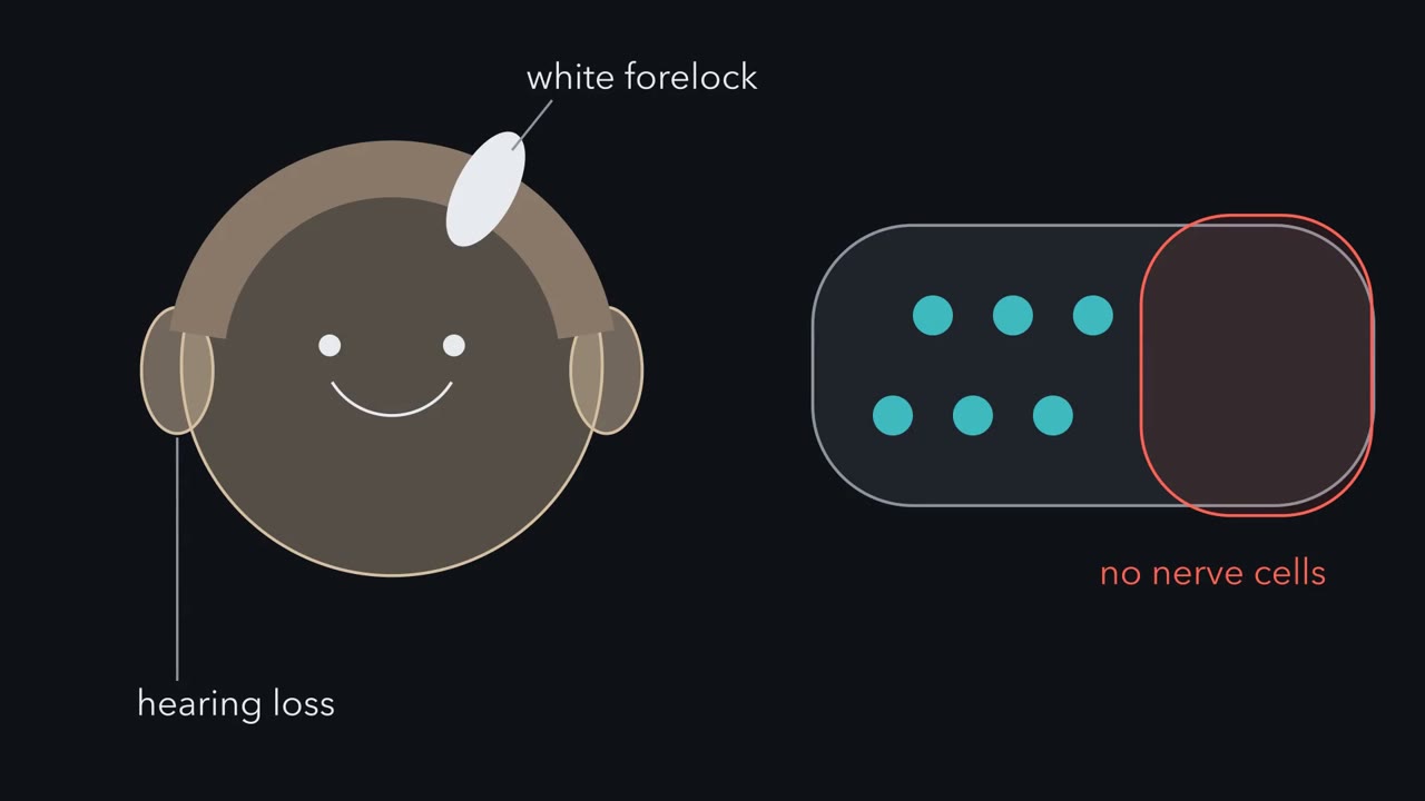
Why can one mutation leave a child with part of the colon without gut neurons, a white forelock and deafness?
Pause when the reading reaches a film, and offer to play it. Saved on this device.
Watch the film now, or keep listening
Builds on:Neural Induction: The Default ModelNeurulation and Neural Tube DefectsTools for Following Cells and GenesWhy these?
 ▶ Watch One Border, Many Destinations · 2:11
Read the transcript of “One Border, Many Destinations”
Film transcript: One Border, Many Destinations
Hearing loss, a white forelock, and a stretch of gut with no nerve cells. Why would one gene cause all three? Some patients have Hirschsprung disease and Waardenburg syndrome together, from one mutant gene. Hold that puzzle. Early on, the ectoderm splits into neural plate and future skin. The neural crest forms at the border between them. When the plate rolls up into the neural tube, the border cells end up along its top. In the chick trunk, they then let go of their neighbours, break through the basement membrane, and crawl away. In the mouse head, many leave before the folds have closed. This change, from a cell in a sheet to a free cell, is an epithelial-to-mesenchymal transition, What they become differs along the body.
Cranial crest makes cartilage and bone of the face and much of the skull. Vagal and sacral crest make the neurons and glia of the gut wall. Trunk crest makes sensory and sympathetic neurons, adrenal medulla, Schwann cells and melanocytes, the pigment cells. Here is the one idea to carry through. One border, many destinations. Where along the body a crest population starts biases what it can become. The signals on its route, and where it stops, help decide which fates appear. This holds for populations. Some single cells are already biased before they leave. Many tissues share that start, and many need the same genes. So one failed gene can strike tissues that look unrelated.
Predict. If gut neurons and pigment cells share a start, what could one broken gene do? It could fail both lineages at once, if both need that gene. Chapter three names the genes. The takeaway. The start biases the options, and the journey helps choose.
From the film
- Where along the body a crest population starts biases what it can become; the signals on its route, and where it stops, help decide which fates appear.
- Crest cells form at the border between the neural plate and future skin, then change from epithelial cells into free mesenchymal cells in an epithelial-to-mesenchymal transition.
- Because many tissues share that start and many need the same genes, one failed gene can strike tissues that look unrelated.
A child with Hirschsprung disease has a stretch of colon with no nerve cells in its wall. A person with Waardenburg syndrome may have a white forelock, eyes of two colours and hearing loss. Some patients have both conditions, from a mutation in a single gene.
Why would gut neurons, hair pigment and hearing fail together? Part of the answer is ancestry. The gut’s neurons and the pigment cells, called melanocytes, of skin, hair and inner ear share an ancestor: the neural crest. But a shared ancestor is only half an answer. It does not say why one gene can break both lineages, or why it is almost always the far end of the gut that is left empty. Hold that puzzle; the lesson follows crest cells from the neural plate to the clinic and solves it at the end.
A border population that leaves home
In the lesson on neural induction you saw the ectoderm, the embryo’s outer cell layer, split into neural plate and future skin. The neural crest forms at the border between the two. Bone morphogenetic protein (BMP) signaling helps define that border: in the standard gradient model, the middle of the plate gets little BMP, the future skin a lot, and the border, in between, an intermediate dose (see the lesson on TGF-β and Smads). Wnt signals are needed to make crest there (see the lesson on Wnt signaling). When the plate rolls up into the neural tube, described in the lesson on neurulation and tube defects, the border cells end up along the dorsal midline, the top of the tube.
Then they do what almost no other cells in the tube do. They loosen their grip on their neighbours, lose the top-to-bottom polarity of an epithelial cell, break through the basement membrane, a mat of extracellular matrix under the epithelium, and crawl away. That change, from a cell in an epithelium to a free mesenchymal cell, is an epithelial-to-mesenchymal transition, or EMT.
Where they end up, and what they become, differs along the body:
| Axial level | Main derivatives |
|---|---|
| Cranial | Cartilage and bone of the face and much of the skull, connective tissue, some cranial sensory and parasympathetic neurons, glia, melanocytes |
| Vagal and sacral | Neurons and glia of the gut wall (the enteric nervous system) |
| Cardiac (part of the vagal level) | Tissue that divides the heart’s outflow into aorta and pulmonary trunk |
| Trunk | Dorsal root ganglion sensory neurons, sympathetic neurons, adrenal medulla, Schwann cells, melanocytes |
In birds and mammals, trunk crest does not normally make skeleton; crest bone and cartilage are a head speciality.
The one idea to carry through this lesson. One border, many destinations. Where along the body a crest population starts biases what it can become; the signals it meets on its route and where it stops help decide which of those fates appear. Because many tissues share that start, and many need the same genes, one failed gene can strike tissues that look unrelated.
How do you follow a cell that leaves?
Read the transcript of “Mark It, Move It”
Film transcript: Mark It, Move It
How do you follow a cell that leaves home? You have to mark it before it goes. And the mark has to last. Julia Platt opened the question in 1893, working on the mudpuppy, a salamander. In fixed sections, she followed cells from the crest into the arches, where head cartilage forms. Cartilage was supposed to be mesoderm, and her evidence was indirect. The claim was long doubted. The lasting mark came from Nicole Le Douarin. Quail nuclei carry a dense block of chromatin. Chick nuclei lack it. The mark is built into the cell. It neither fades nor dilutes as cells divide. Graft quail neural tube into a chick at the same level. Then map where the quail cells go.
But a same-level graft cannot say why. Origin and route are tangled together. To separate them, move the cells. Predict. Take trunk crest from the adrenal level. At home it makes cells that release noradrenaline and adrenaline. Move it into the neck. Where will it go, and what transmitter will it use?
They go where neck crest goes, into the gut wall. There they form enteric ganglia. Some donor neurons are cholinergic: they use acetylcholine. The new site changed what the population made. Swap the other way, and neck crest placed at the adrenal level makes adrenal medulla and sympathetic derivatives. The skeleton behaves differently. In most early grafts, trunk crest in the head made little or no cartilage. Under some conditions, trunk crest can make cartilage. So the start biases the options, and the surroundings help choose. Now predict. One cell in a dish makes neurons and pigment cells. Would it make both in the embryo?
Not necessarily. A dish shows what a cell can make. Tracing in the embryo shows what it did make. In 1988, both methods found some single cells that made several types. Whether the typical cell does is still debated. The takeaway. Move the cells, and the pattern appears. The start biases the options, and the journey helps choose.
From the film
- Quail nuclei carry a built-in mark that neither fades nor dilutes as cells divide, so quail-into-chick grafts show where each level's crest goes.
- Moving cells to a different level changes what the population makes: adrenal-level trunk crest at the vagal level formed enteric ganglia, some with cholinergic neurons.
- A dish shows what a cell can make, tracing in the embryo shows what it did make, and whether the typical crest cell is multipotent is still debated.
To know where a migrating cell came from, you have to mark it before it goes, and the mark has to last.
The question of what crest makes was opened by Julia Platt, working on the mudpuppy Necturus, a salamander with large, yolky embryos. Its ectoderm cells carry less yolk than the deeper layers, and that difference helped her tell them apart in a section, a natural marker of sorts. Reading serial sections of embryos fixed at successive stages, she followed cells from the neural crest and thickened head ectoderm into the branchial arches. In 1893 she concluded that ectoderm contributes to the cartilages of the head. That broke a rule, since cartilage was supposed to be mesoderm, and her evidence was indirect, from fixed sections with no living cell labelled or moved. The conclusion was long doubted.
Classic experiment: quail-chick chimeras
The lasting mark came from Nicole Le Douarin. She noticed that quail nuclei, stained for DNA, show a large dense mass of condensed chromatin associated with the nucleolus, which chick nuclei lack. Graft a short piece of quail neural tube into a chick embryo at the same level, and the quail cells that emigrate can be recognised in sections long afterwards. The mark neither fades nor dilutes as cells divide.
With Marie-Aimée Teillet and others, Le Douarin used these same-level grafts, called isotopic, to map the crest origin of sensory, sympathetic and enteric ganglia, the adrenal medulla and the skeleton of the face. The neurons of the bird gut, for example, come from crest at the vagal level of the neck, with a contribution from the lumbosacral level to the hindgut.
A same-level graft shows where each level’s cells go. It cannot show why they become what they do, because origin and route are confounded: the cells start and travel as they would have anyway. To separate the two, move the cells.
Moving the cells: heterotopic grafts
A heterotopic graft puts cells at a different level from the one they came from.
Predict. Take quail neural tube from the adrenomedullary trunk region, opposite somites 18 to 24. At home its crest makes sympathetic neurons and adrenal chromaffin cells, which release noradrenaline and adrenaline. Graft it into a chick at the neck, the vagal level. Where will the quail cells go, and what transmitter will their neurons use?
Check your prediction
They go where vagal crest goes, into the gut wall, and form enteric ganglia. Some donor-derived neurons are cholinergic: they use acetylcholine, as many gut neurons do (Le Douarin and Teillet, 1974; Le Douarin, Renaud, Teillet and Le Douarin, 1975). The new site changed what the population made. The graft cannot say whether it instructed cells that would have become noradrenergic or favoured a subset already able to become cholinergic. Run the swap the other way, and vagal crest grafted to the adrenomedullary level makes adrenal medulla and sympathetic derivatives.
The skeleton behaves differently. In most early reports, trunk crest grafted to the head joins the arches but makes little or no cartilage. So the start biases what the cells can make, and the surroundings help choose.
| Graft (quail into chick) | Result | What it shows |
|---|---|---|
| Same level | cells reach that level’s usual targets | where each level’s crest goes |
| Adrenomedullary trunk crest to vagal level | enteric ganglia, some cholinergic neurons | the new site changes the population’s outcome |
| Vagal crest to adrenomedullary level | adrenal medulla, sympathetic derivatives | the same, in reverse |
| Trunk crest to head | little or no cartilage in most early grafts | origin biases what the cells make |
Neither simple story survives the table. “Origin fixes fate” fails on the second and third rows. “The surroundings alone decide” fits the last row poorly.
One cell, or a mixed population?
A graft carries many cells, so it shows what a population can do, not whether each cell could make everything the population makes. Two kinds of single-cell experiment asked that, and they ask different questions.
Predict. One crest cell, alone in a dish, grows into a colony containing both neurons and pigment cells. Does that show the same cell would have made both in the embryo?
Check your prediction
Not necessarily. Culture shows what a cell can make under one condition; in the embryo its route and surroundings might have chosen only one of the two. To learn what a cell did make, label it in place and find its descendants later.
In a dish, Anne Baroffio, Elisabeth Dupin and Le Douarin plated single migrating crest cells from the quail midbrain and anterior hindbrain region onto a feeder layer of 3T3 cells in 1988. They analysed 243 clones, colonies each grown from one cell, after 7 to 10 days. Some clones contained a single neuronal type. Others contained several neuron types, non-neuronal cells and melanocytes. Many fell in between.
In the embryo, Marianne Bronner-Fraser and Scott Fraser injected a fluorescent dextran into single premigratory cells in the chick trunk neural tube, also in 1988. Some single clones spread into more than one derivative, and some included cells that stayed in the neural tube.
So some crest cells can make several cell types. Whether the typical premigratory crest cell is broadly multipotent in the living embryo is still debated: later single-cell tracing in chick and mouse has produced both multipotent and restricted clones. The grafts speak for populations, not for every cell.
What the rule covers.
- Which cells. Crest derivatives at each axial level. The graft tests are in birds; in mice, genetic lineage tracing gives the fate map and also shows differences between levels.
- Biases rather than fixed menus. Because some cells are already biased or restricted before they leave, a cell’s start and its route are not fully separate causes.
- Context. The trunk-cartilage limit is not absolute. Cultured under the right conditions, trunk crest can make cartilage, and trunk crest placed directly into developing head tissue has contributed to head skeleton (McGonnell and Graham, 2002). So the limit is partly what the cell carries and partly what its surroundings and stage allow.
Leaving home: an identity first, then a release
Read the transcript of “Leave, Steer, Arrive: The Puzzle Solved”
Film transcript: Leave, Steer, Arrive: The Puzzle Solved
How does a crest cell let go and find its way? And what happens when a step fails? To leave, a crest cell must loosen its grip. In chick, a cadherin called Cadherin-6B helps hold it to its neighbours. A transcription factor, Snail2, is needed. Block it, and fewer cells leave. Suppose Snail2 works by switching off Cadherin-6B. Lower Snail2. What happens to Cadherin-6B?
Cadherin-6B RNA rises within about half an hour, just as predicted. The authors concluded Snail2 represses it directly. Lower Cadherin-6B instead, and cells leave early. It is a brake. Snail2 probably lifts it, at least partly. Now steering. In the chick trunk, crest cells pass only through the front half of each sclerotome. The back half repels them on contact. In chick, ephrin-B is one such signal. In mouse, semaphorin 3F also repels them. Neither is known to be the only cue. Crest cells also steer each other. When two touch, they collapse protrusions at the contact and move apart. Edge cells tend to move outward. Mutual attraction holds the group together, and a chemokine gives it direction.
In frogs, a cluster follows that chemokine better than a lone cell. For trunk crest, this is less clear. Now the puzzle. The patient has Waardenburg syndrome type 4. The genes are SOX10, the endothelin receptor B and EDN3. Enteric precursors need them, and so do melanocytes, including those the inner ear needs for hearing. Try a toy model. One front moves down the gut from the mouth end, within a fixed time. Slow it. Which part stays empty?
The end the front reaches last, the anal end. In the model, that segment is left without neurons. That is a toy model. In the clinic, too, the empty segment usually starts at the anal end. Rare cases skip a segment. An earlier failed step need not mean wider damage. Treacher Collins syndrome cuts crest numbers early. Yet it mainly affects the face, ears and eyelids. Tissues that fail together often share a requirement. Which tissues fail depends on where the gene is needed, the variant, and each tissue's sensitivity. One border, many destinations. The start biases the options, and the journey helps choose.
From the film
- In chick, Snail2 is needed for crest cells to leave and probably lifts the Cadherin-6B brake, at least partly.
- In the chick trunk, crest cells pass only through the front half of each sclerotome because the back half repels them; ephrin-B in chick and semaphorin 3F in mouse are repellents, and neither is known to be the only one.
- Tissues that fail together often share a requirement, and which tissues fail depends on where the gene is needed, the variant and each tissue's sensitivity.
A crest cell has two jobs before it can travel. It has to become a crest cell, and it has to let go.
Becoming crest. The border signals, bone morphogenetic protein (BMP) and Wnt from earlier, along with fibroblast growth factor (FGF), switch on a first tier of genes, the border genes. These switch on a second tier, the crest genes, such as Snail2, FoxD3 and Sox10. The crest genes in turn control the genes that change adhesion and movement and, later, influence fate. Daniel Meulemans and Marianne Bronner-Fraser drew this in 2004 as a proposed gene-regulatory network, a diagram of which genes switch on which. Read each arrow as a hypothesis until it has been tested; some since have been.
Letting go. In the neural tube, premigratory chick crest cells are held to their neighbours partly by a cadherin, Cadherin-6B. To leave, they must loosen that grip. In 1994 a group including Angela Nieto showed that blocking the transcription factor Slug, now called Snail2, reduced crest emigration in chick embryos. Snail2 was needed for departure. How it acted was still open.
Predict. Suppose Snail2 promotes departure by switching off Cadherin-6B. Lower Snail2 in premigratory crest with a morpholino, a synthetic molecule that blocks the making of one protein. What should happen to Cadherin-6B RNA soon afterwards, and to the number of cells that leave?
Check your prediction
Cadherin-6B RNA should rise, and fewer cells should leave. In 2007 Lisa Taneyhill, Edward Coles and Bronner-Fraser did this experiment, and Cadherin-6B RNA rose within about half an hour. Snail2 bound clusters of E boxes, short DNA motifs, in the Cadherin-6B regulatory region, and the authors concluded that it represses the gene directly.
That makes Cadherin-6B a candidate brake. A brake needs its own test: release it without touching Snail2, and the cells should leave early. In a companion study, Coles, Taneyhill and Bronner-Fraser lowered Cadherin-6B, and crest cells did emigrate early.
So Snail2 probably promotes departure at least partly by lifting the Cadherin-6B brake, and EMT involves more than one adhesion gene. “Partly” matters. If lowering Cadherin-6B along with Snail2 restored only half of the lost departure, that would be consistent with Cadherin-6B explaining part of the defect; other Snail2 targets, or incomplete knockdown, could explain the rest. That is the reasoning in the third MCAT question below, a hypothetical experiment, not a published result.
Steering: a forbidden half and cells that back away
In the chick trunk, crest cells take two main routes. The ventromedial route runs through the somites, the blocks of mesoderm beside the neural tube, and gives dorsal root ganglia, sympathetic ganglia and adrenal medulla. The dorsolateral route, taken later, runs under the skin and gives mostly melanocytes.
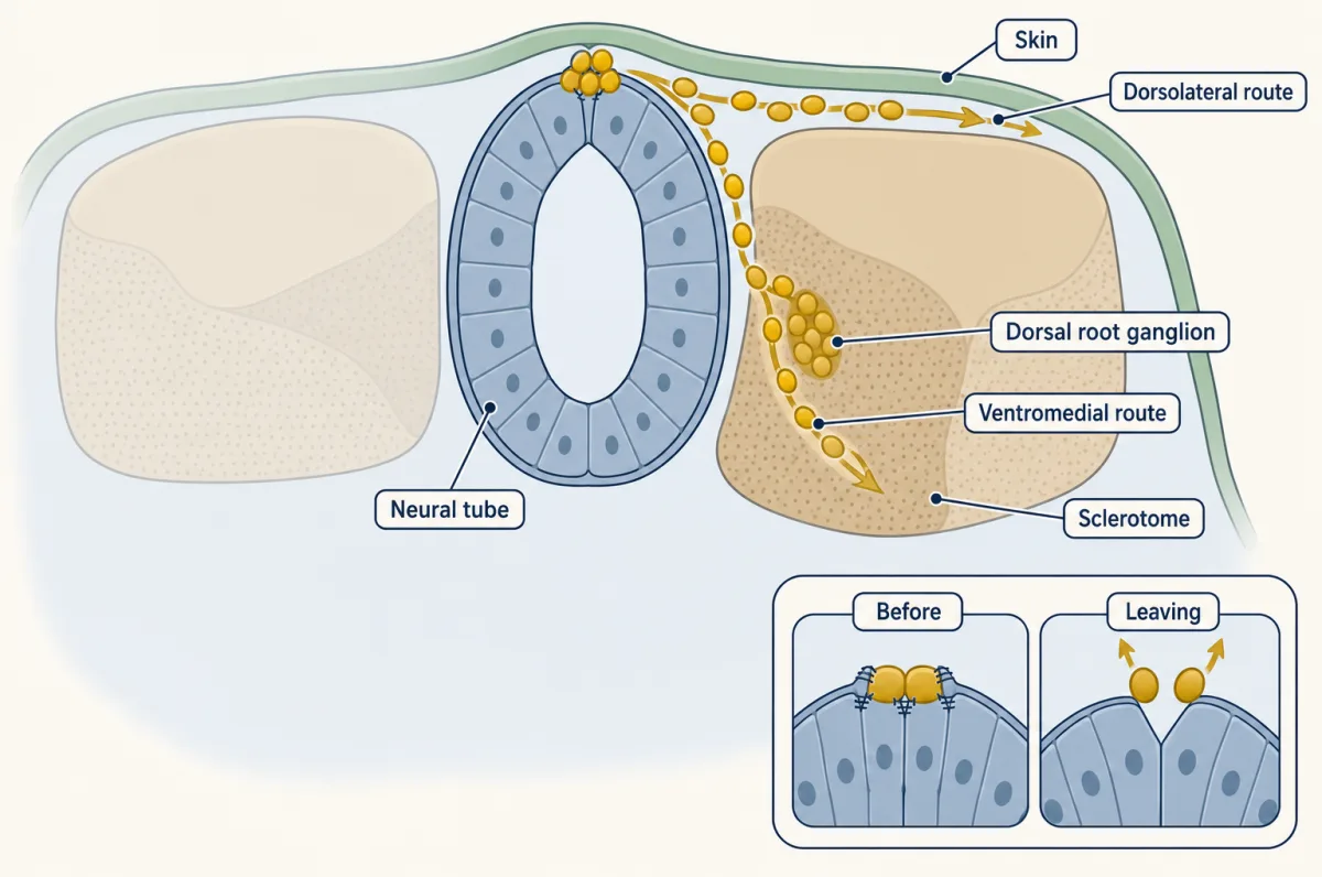
On the ventral route the cells obey a rule. They pass only through the rostral (front) half of each sclerotome, the part of the somite that will form vertebrae. The caudal (back) half keeps them out.
What keeps them out? In chick, crest cells carry EphB receptors, and caudal-half sclerotome cells carry ephrin-B, a membrane-bound ligand. Because both sit in membranes, the signal passes only where a crest cell touches a caudal-half cell. In 1997 two groups showed that this repels crest. Catherine Krull and colleagues, working with chick, found that trunk crest plated on alternating stripes avoids ephrin-B1 stripes. Hai Wang and David Anderson showed that clustered ephrin-B ligands repel crest cells and motor axons. Ephrin-B can also signal back into its own cell (see the lesson on reverse signaling), but here the forward signal into the crest cell repels.
In mouse the caudal half also makes semaphorin 3F, which repels crest through its receptor neuropilin-2 (Laura Gammill and colleagues, 2006). Mice lacking either protein lose segmental crest migration. So the caudal half uses more than one repellent: ephrin-B is the best-studied in chick, semaphorin 3F in mouse, and neither is known to be the only one.
Design problem
Trunk crest cells in culture avoid stripes coated with ephrin-B. A sceptic says the stripe is simply a poor surface to stick to, and the cells drift onto the better one. How would you show that the avoidance is active repulsion through Eph receptors?
Saved in this browser only.
One way to do it
Add soluble ephrin-B to the medium, or a reagent that blocks Eph receptors. Soluble ligand (or a blocking reagent) occupies or saturates the receptors everywhere, so the cell can no longer read the stripe as different. If cells now cross the stripes and spread evenly, the avoidance needed Eph-ephrin signaling. That is what the 1997 chick study showed: soluble ephrin-B1 blocked avoidance of ephrin-B1 stripes. A passive adhesion difference would not be relieved by adding ligand, because the stripe surface itself is unchanged. Watching cells turn away at the stripe edge, rather than slip off it, adds further evidence. As a control, add a soluble protein that does not bind Eph receptors; avoidance should persist.
The same study took the logic into tissue: soluble ephrin-B1 added to trunk explants disrupted segmental migration, and crest cells entered caudal as well as rostral halves. One trap: clustered soluble ephrin can activate the receptors, while unclustered ligand can block them, so say which form and dose you used, and check that the cells still move.
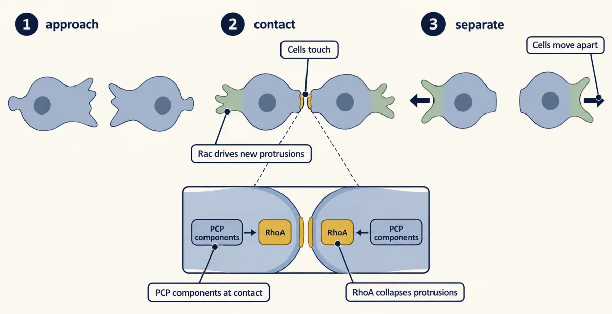
Crest cells also steer each other. Contact inhibition of locomotion was first described by Michael Abercrombie in cultured fibroblasts in the 1950s: two cells that touch stop moving towards each other and head off in new directions. In 2008 Roberto Mayor’s laboratory showed that frog and zebrafish crest cells do this. Collective migration of frog cranial crest is thought to depend on three ingredients.
- Contact inhibition pushes cells apart and gives each a front. At the contact, planar cell polarity signaling (a Wnt branch that acts without β-catenin) activates RhoA, a small GTPase. RhoA collapses protrusions there. Another GTPase, Rac, drives new protrusions at the free edge (see the lesson on integrins and Rho GTPases). So a cell at the edge of the group tends to protrude from its free side and move outward.
- Mutual attraction keeps the group from scattering. In Xenopus, crest cells attract one another chemically.
- A chemoattractant gives the group a direction. The cells move up a gradient of the chemokine Sdf1.
The payoff: in Xenopus a cluster follows Sdf1 better than a single cell does. How far this recipe applies to trunk crest in birds and mammals is less clear.
Stopping and gathering
Then the travellers have to stop and gather. This is the coalescence problem: no central planner tells cells where to form a ganglion, and how they manage it is still poorly understood. Three kinds of signal have some support: adhesion molecules such as N-cadherin, which help cells sort and hold together; chemoattractants such as SDF1, which draw cells to some sites; and boundaries such as Eph-ephrin and semaphorin signals, which divide the embryo into zones. Their roles and timing differ between ganglia. In chick sympathetic ganglia, crest cells first spread out along the aorta and only then sort into separate clusters, with N-cadherin contacts helping to shape them.
Predict. Remove N-cadherin from trunk crest cells. What happens to dorsal root ganglia if adhesion only holds clusters together once they have begun? What if it is needed to start them?
Check your prediction
If N-cadherin only holds clusters together, the cells should still arrive and begin to gather, but the ganglia would be loose or poorly separated from their neighbours. If it is needed to start clusters, the cells would stay scattered along the route. The evidence above is from chick sympathetic ganglia; for dorsal root ganglia, treat both outcomes as predictions to test, not results.
Arriving is not the end of the test. Once these sensory and sympathetic neurons send axons to their targets, many die, as the lesson on the neurotrophic hypothesis shows.
Neurocristopathies: tissues that fail together often share a requirement
A disorder of neural crest cells is a neurocristopathy, a name Robert Bolande gave in 1974. Back to the opening patient. First, is it a coincidence? Using commonly cited rough rates, Hirschsprung disease affects about 1 in 5,000 births and Waardenburg syndrome about 1 in 42,000. If the two were independent, both would turn up together in about 1 in 210 million births. Yet the combination is a recognised syndrome that runs in families, with variants in the same few genes. That points to a shared cause, not chance.
That patient has Waardenburg syndrome type 4, also called Shah-Waardenburg syndrome. The genes are SOX10, EDNRB (the endothelin receptor B) and EDN3. Each is needed by both enteric precursors and melanocytes, including the melanocytes the inner ear needs for hearing. One broken gene, two lineages that need it.
Hirschsprung disease has a second feature to explain. In typical cases the segment without neurons begins at the far, anal end of the gut and extends back for a variable length. Rare skip segments break this pattern.
Predict. Take a toy model. Vagal crest colonises the gut as a single front, moving from the mouth end towards the anus, within a limited time window. Suppose the front moves more slowly than normal. Which part of the gut is left without neurons?
Check your prediction
The end the front reaches last: a segment at the anal end. Put numbers on it. Give the gut a fixed length, and let the front move at constant speed and normally reach the anal end just as the window closes. Then the empty fraction equals the slowing: a front at 90 percent of normal speed leaves the last 10 percent empty, and one at 80 percent leaves the last 20 percent. If the gut grows unevenly, the numbers shift. This is an illustration, not a measured law, and it uses speed alone. Real front advance also depends on cell division and survival, which is why the table below says crest fails to colonise the distal gut rather than fails to migrate. Real colonisation is more complex still: in mice some vagal cells take a shortcut across the mesentery to the colon, sacral crest adds a small contribution, and the gut wall becomes less welcoming as it ages.
A shared crest start does not explain every pattern; a shared requirement explains several rows below. The step labels are entry points, not exclusive mechanisms: one disorder can touch several steps. Read the table by how widely each disorder spreads, and test a common guess against it: the earlier the failed step, the more tissues are hit.
| Disorder | Gene | Step that fails | Tissues hit |
|---|---|---|---|
| Treacher Collins syndrome | TCOF1, a ribosome biogenesis gene | survival of early crest; in mice, cell death reduces crest | mainly the face: small jaw and cheekbones, ears, lower eyelids |
| Waardenburg syndrome type 1 | PAX3, needed later to switch on MITF in melanocyte precursors | melanocyte development | pigment changes and deafness |
| Waardenburg syndrome type 3 | PAX3 | as type 1 | as type 1, plus limb muscle (not crest) |
| Waardenburg syndrome type 4 | SOX10, EDNRB, EDN3 | development of enteric and melanocyte precursors | pigment, hearing, distal gut |
| PCWH | particular SOX10 variants | as type 4, plus glial development and myelination | as type 4, plus Schwann cells (crest) and central oligodendrocytes (not crest) |
| Hirschsprung disease | RET, EDNRB, EDN3, SOX10 | crest fails to colonise the distal gut | distal gut; RET is also used in the developing kidney (not crest) |
| Piebaldism | KIT | melanoblast survival or migration | patches that lack melanocytes altogether |
| Oculocutaneous albinism type 1 (OCA1) | tyrosinase | differentiation: melanocytes arrive but make little or no melanin | skin and hair; vision, mainly through the retinal pigment epithelium (not crest) |
PCWH stands for peripheral demyelinating neuropathy, central dysmyelinating leukodystrophy, Waardenburg syndrome and Hirschsprung disease.
Two lessons sit in the table. First, Treacher Collins syndrome is a counter-example to the guess: it cuts crest numbers early, yet its effects stay mainly in the head and face. Which tissues are hit depends on where the failed gene is needed at vulnerable stages, and on the variant, its dose and each tissue’s sensitivity: different SOX10 variants give type 4 or the wider PCWH. Second, a shared requirement is not always a shared lineage: it reaches outside the crest, so a disorder can hit crest and non-crest tissues together. PAX3 also works in limb muscle precursors, SOX10 in central oligodendrocytes, and tyrosinase in the retinal pigment epithelium, which comes from the optic cup. Albinism sits at the very end of the chain for crest-derived pigment cells.
Two steps are missing from the table. Border genes such as PAX3 act in specification at the border in animal embryos, but no disorder in this table traces to that step alone. Few human disorders trace to EMT alone either; the clearest evidence on EMT is from chick.
Putting it together
| Experiment | Animal | Result | What it supports | What it does not establish |
|---|---|---|---|---|
| Serial sections (Platt, 1893) | mudpuppy | ectoderm-derived cells reach the arches, where cartilage forms | ectoderm, including crest, contributes to head cartilage | the lineage of any one living cell |
| Same-level quail-chick grafts | quail into chick | each level’s crest reaches its usual derivatives | the fate map by level | why the cells take those fates |
| Heterotopic grafts | quail into chick | adrenomedullary trunk crest at the vagal level makes enteric neurons, some cholinergic; in most early grafts, trunk crest in the head made little or no cartilage | the new site changes the outcome; origin biases what cells make | what each single cell could do, or whether the site instructs or selects |
| Clonal culture (1988) | quail cranial crest | single neuronal types, mixed clones and clones in between | some cells can make several types | what those cells did make in the embryo |
| Single-cell dye injection (1988) | chick trunk | some clones in several derivatives | some premigratory cells are multipotent in vivo | whether the typical cell is |
| Slug/Snail2 block (1994) and knockdown (2007) | chick | less emigration; Cadherin-6B RNA rises | Snail2 is needed and, the authors concluded, represses Cadherin-6B directly | that Cadherin-6B is its only target |
| Cadherin-6B knockdown | chick | early emigration | Cadherin-6B is a brake on leaving | the other adhesion changes of EMT |
| Stripe assay (1997) | chick trunk crest | avoids ephrin-B1 stripes; soluble ephrin-B1 blocks the avoidance | ephrin-B repels crest | that ephrin-B is the only caudal-half cue |
| Sema3F and neuropilin-2 mutants | mouse | segmental crest migration lost | a second caudal-half repellent | that segmented migration builds segmented ganglia |
| Contact inhibition | frog, zebrafish | cells collapse protrusions at contact and move apart; in frog, clusters follow Sdf1 better | a recipe for collective migration | that trunk crest of birds and mammals uses it |
Going deeper
History of the crest idea
Grafting and removal experiments in amphibians gave Platt’s conclusion experimental support in later decades, notably the work of Sven Hörstadius and Sven Sellman in the 1940s. Some authors call the crest a “fourth germ layer”. That is a popular label, not a formal term: the crest arises from ectoderm.
Genetic lineage tracing
The modern equivalent of the quail mark is genetic: a Cre line is a mouse strain that makes the enzyme Cre recombinase in a chosen cell type, and Cre switches on a reporter gene permanently in those cells and all their descendants.
A second, later source of trunk derivatives
In mice, Schwann cell precursors, crest-derived cells that travel along growing nerves, give rise to many adrenal chromaffin cells and skin melanocytes. They reach their targets along nerves, a later route than the two main streams.
The gene-regulatory network, arrow by arrow
Border genes include Msx, Pax3/7 and Zic; Sox9 is another crest gene. In chick cranial crest, for example, the Bronner laboratory later showed that Sox9, Ets1 and cMyb act directly on an enhancer of Sox10.
Timing and a cadherin switch
Timing of EMT differs: chick trunk crest leaves after the tube has closed, while in the mouse head many cells leave while the folds are still open. A subset of migrating chick crest cells switches from Cadherin-6B to Cadherin-7.
Segmented migration and segmented ganglia
Segmented migration is thought to help segment the dorsal root ganglia, but the two can be separated. In mice lacking one caudal-half repellent, crest enters both halves of the sclerotome and the ganglia still form in segments; the ganglia lose their segments only when two guidance pathways are lost together.
Cleft palate
Cleft palate is harder to place in the table. The palatal shelves are built largely of crest-derived mesenchyme that must grow, lift and fuse, and many genes and environmental factors can make that fail.
Your notes
Select any text in the lesson to highlight it or add a note to it. Your highlights and notes are saved in this browser and collected in your notebook.
This browser is not letting the site save anything (a private window or strict privacy settings), so highlights and notes will be lost when you leave the page.
How we measure it
Quail-chick chimeras
A piece of quail embryo is grafted into the same place in a chick embryo. Quail nuclei carry a dense chromatin mass that chick nuclei lack, so every donor descendant can be identified later in sections. The method maps where a population goes, not what a single cell can do.
Single-cell lineage tracing by dye injection
A large fluorescent tracer is injected into one cell; it is passed to that cell's descendants and not to neighbours, though it dilutes as the cells divide. Finding the labelled cells later shows what one cell actually made in the embryo.
Clonal culture
Single cells are placed one per well and grown under fixed conditions, and the cell types in each colony are scored with markers. It measures what a cell can make in that dish, which may differ from what it would make in the embryo.
Stripe assay
Alternating stripes of a test protein and a control protein are laid on a dish, and cells are plated over them. If cells pile up on the control stripes, the test protein repels them or fails to support them, and further controls tell which.
Practice questions on this lesson (3) → MCAT-style, original, each with an explanation
Where this lesson fits
Before thisReview first
- Neural Induction: The Default ModelThis course · Lesson 6
The border between neural plate and future skin, where crest forms, comes from the BMP gradient set up there.
- Neurulation and Neural Tube DefectsThis course · Lesson 7
You need the rolling up of the plate, which puts border cells at the dorsal tube, before they can leave.
- Tools for Following Cells and GenesThis course · Lesson 4
The lesson uses grafts, lineage tracing and clonal logic for fate mapping, which that lesson introduces.
After thisStudy next
- Pioneers, Guideposts and the Logic of Guidance CuesThis course · Lesson 22
Repellent cues like ephrin-B that fence off the caudal sclerotome are developed there as a general logic of guidance.
- Semaphorins and RepulsionThis course · Lesson 25
Semaphorin 3F and neuropilin-2, the mouse caudal-half repellent for crest, are treated there as repulsive cues.
- Too Many Neurons: Naturally Occurring Cell DeathThis course · Lesson 28
Crest-derived sensory and sympathetic neurons that arrive at their targets then face heavy cell death, as covered there.
Elsewhere on the siteFilms, simulations and method films in other courses
- Cre-lox and binary expression systemsMethod film
Genetic lineage tracing of crest derivatives in the lesson's deeper section uses recombinase-based marking of crest cells.
- Wnt Plugs the DrainFilm · Cell Signaling
Wnt signaling helps specify the crest at the plate border, so this film shows that signal in detail.
Suggested by an AI model from the lessons themselves, then checked. How we use AI
Finish line
Test yourself
9 questions
Practice and MCAT-style, with explained answers.
Next · Lesson 21
One Axon, Many Dendrites: Neuronal Polarity
A neuron takes in signals on dendrites and sends them out along one axon. Cut that axon close to the cell body in a dish and one of its dendrites can become a new axon. How does a neuron choose its axon, and keep it?
Continue →← Lesson 19 · Tangential Migration: Interneurons and the Long Way Round