Developmental Neuroscience · Unit 2: Becoming neural
The Organizer: Spemann and Mangold
Rewritten on : new text, flashcards and film. Your notes and highlights stay, but the text around them may have moved.
How can a small graft make its host build a second spinal cord from cells meant for skin?
Pause when the reading reaches a film, and offer to play it. Saved on this device.
Watch the film now, or keep listening
Builds on:The Wiring Problem: How a Brain Gets BuiltWhy these?
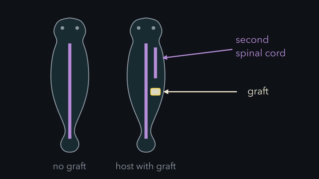 ▶ Watch One Sheet, Two Fates · 2:28
Read the transcript of “One Sheet, Two Fates”
Film transcript: One Sheet, Two Fates
How can a small graft make its host build a second spinal cord, from cells meant for skin? First, a little embryology. The egg divides into a ball of smaller cells. In amphibians, the ball becomes a blastula, a sphere of cells around a fluid-filled cavity. Then, in gastrulation, cells move inside, and the embryo sorts into three layers. The outer layer, the ectoderm, makes the outer skin and the nervous system. The middle layer, the mesoderm, makes muscle, bone and the notochord, a stiff rod along the back. The inner layer lines the gut.
Cells move inside through a slit, the blastopore. Its edge on the future back is the dorsal lip. Now watch the ectoderm. It looks like one sheet. Along the back, a strip becomes the neural plate. It rolls up into the neural tube, the future brain and spinal cord. On the belly, the same sheet stays flat and becomes skin. So why does the back strip become neural, while the belly strip becomes skin?
There are two kinds of answer. Perhaps the belly cells are already fixed as skin. Or they can still change, and something nearby tells the back strip what to become. Moving tissue tests the first answer. Put dorsal-lip tissue next to belly ectoderm, and watch. The graft worked. The belly cells built a second neural tube. Chapter two shows whose cells they were. That experiment gives this lesson its one idea. A cell's fate can be set by its neighbours. Move the organizer somewhere new, and host cells there build much of a second back around it. The graft itself becomes notochord. But only while they are still competent, that is, able to answer.
Notice one limit. The idea says who changed fate, and where. Not what the signal is. One sheet, two fates, and neighbours can decide which. Next: whose cells built the second back?
From the film
- A cell's fate can be set by its neighbours.
- In newts, an organizer moved to a new place makes host cells build much of a second back around it.
- This works only while the host cells are still competent, that is, able to answer.
- The idea says who changed fate and where, not what the signal is.
Move a small piece of tissue from one newt embryo into the side of another, and the host can build a second neural tube, mostly out of its own cells that were on their way to becoming skin.
That result came from grafts Hilde Mangold made in Hans Spemann’s laboratory in Freiburg in 1921 and 1922, published with Spemann in 1924. The graft did more than add nervous system: the donor tissue, the organizer, organized a whole second axis. This lesson follows that graft as an argument, from the puzzle it was built to answer to the question it left open: what is the signal?
One sheet, two fates
After fertilization the egg divides without growing, a process called cleavage, into a ball of smaller cells. In amphibians the ball becomes a blastula, a sphere of cells around a fluid-filled cavity. Then comes gastrulation, the stage at which cells move from the surface to the inside and the embryo sorts itself into three germ layers:
- The ectoderm, the outer layer, makes the epidermis, the outer layer of the skin, and the nervous system.
- The mesoderm, the middle layer, makes muscle, bone, blood, kidney and the notochord, a stiff rod of tissue along the back.
- The endoderm, the inner layer, lines the gut and forms organs such as the liver.
In an amphibian, cells move inside through a slit in the surface called the blastopore. It first appears as a short groove on one side of the embryo, and the edge of that groove is the dorsal lip, on what will become the back (dorsal means toward the back; ventral, toward the belly). Cells that roll inward over the dorsal lip become the mesoderm under the back, including the notochord.
Now the puzzle. The brain and spinal cord come from a strip of ectoderm along the back, the neural plate, which later rolls up into the neural tube. Ectoderm on the belly becomes skin. Both start as the same kind of surface tissue.
Predict. Give two different kinds of explanation for why the back strip becomes neural and the belly strip does not. For each, say what should happen if you put dorsal-lip tissue next to belly ectoderm.
Check your prediction
One kind of answer: belly ectoderm is already fixed as skin, and stays skin whatever lies next to it. The other: belly ectoderm can still change, and something nearby tells the back strip what to become; then dorsal-lip tissue placed beside belly ectoderm could make it neural. Moving tissue tests the first answer in its strong form. If belly ectoderm becomes neural beside a dorsal lip, it was not fixed as skin. That would not show that back and belly cells started identical; both kinds of answer can be partly true.
Spemann’s hair-loop experiments, in the reading he developed over the following years and that entered the textbooks, had already pointed to the future dorsal side, where gastrulation will begin: a half embryo made a whole one only if it contained material from that side (Going deeper). The loop could not say what that material did. The graft was built to find out.
The one idea to carry through this lesson. A cell’s fate can be set by its neighbours. Move the organizer somewhere new, and host cells there build much of a second back around it (the graft itself becomes notochord), but only while they are still competent, that is, able to answer.
The graft: whose cells built the second back?
Read the transcript of “Whose Cells Built It?”
Film transcript: Whose Cells Built It?
A dorsal lip moved to a new place can give a second body axis. But whose cells built it? Grafts within one species had given extra axial structures. But donor and host cells look alike. So, two readings for the new neural tube. Self-differentiation: the graft's cells develop by their own origin, and make the tube themselves. Or induction: the graft changes the fate of its neighbours, and host cells build the new tube. The solution was colour. Hilde Mangold did the test in Hans Spemann's laboratory, for her doctoral thesis. She used two newt species. Cells of one carry little pigment. Cells of the other carry pigment granules. The granules stay visible in sections, so colour shows which embryo a cell came from.
In 1921 and 1922, she cut a piece of dorsal lip from a pale gastrula. She placed it in the belly of a pigmented gastrula, in tissue that would normally become skin. Most embryos died. In the survivors, a second neural plate rose, folded, and closed into a second neural tube. Predict. If the graft simply grew, what colour is the new tube? If it changed its neighbours?
If the graft grew, the tube should be pale. If it changed its neighbours, the tube should be pigmented. In one embryo, most of the second tube was pigmented, with a narrow pale strip along its floor. In another, the whole tube was host tissue. The pale graft lay beneath it, as a notochord and nearby mesoderm.
Host ectoderm that would have made skin had made a neural tube. For the tube, induction explained it. Induction is a change in the fate of one tissue, caused by a signal from another. The graft did three things. Its own cells became notochord. It recruited host mesoderm into somites, blocks beside the tube that form vertebrae and muscle. And it induced host ectoderm to become a neural tube. Hence the name, organizer. The second axis formed at the graft, not along the host's own back. The pattern followed the source. So the organizer decides where neural tissue forms. It does not yet say how: by a go signal, or by releasing a brake.
The responder must also be able to answer. That ability is competence. Wrap older and older ectoderm around early organizer. The response falls, and largely stops by the end of gastrulation. The control: late organizer with young ectoderm. If that still works, the loss is in the responder.
So host cells built the second neural tube, at the graft, while they could still answer.
From the film
- A pale graft in a pigmented host showed that most of the second neural tube was built by host cells, so the graft induced it.
- The graft's own cells became notochord, it recruited host mesoderm into somites, and it induced host ectoderm to become neural tube.
- Older ectoderm responds less and largely stops responding by the end of gastrulation, so the responder must be competent.
Earlier grafts within a single species had shown that a dorsal lip moved to a new place could give extra axial structures. But in a same-species graft the donor’s cells look like the host’s, so you cannot count whose cells are in the new neural tube. Its origin has two possible readings:
- Self-differentiation. The graft’s cells develop according to their own origin and supply the new neural tissue themselves, so the second neural tube is made of donor cells. (In its own embryo the dorsal lip mainly makes notochord and other dorsal mesoderm.)
- Induction. The graft changes the fate of its neighbours. Then host cells build the second neural tube.
The solution was colour. Mangold mainly used two newt species, then called Triton cristatus and Triton taeniatus. Cells of cristatus carry little pigment. Cells of taeniatus carry pigment granules that stay visible after the embryo is fixed and cut into thin sections. Natural colour became a lineage marker, a visible trait that shows which embryo a cell came from.
She cut a small piece of the dorsal lip region from a pale cristatus gastrula and placed it in the flank or belly of a pigmented taeniatus gastrula (host stages in the series varied), in tissue that would normally become belly skin. The operations were hard, and most operated embryos died. In the survivors, a second neural plate rose at the graft site, separate from the host’s own in the embryos described in detail, folded, and closed into a second neural tube.
Predict. In thin sections of that second neural tube, what colour should most of the cells be if the graft simply grew? What colour if it changed its neighbours?
Check your prediction
| If the second tube is built by… | Pale (cristatus) cells in the tube | Pigmented (taeniatus) cells in the tube | What it would mean |
|---|---|---|---|
| donor cells, the graft developing by its own origin (self-differentiation) | nearly all | few or none | no induction shown |
| host cells whose fate was changed (induction) | few or none | nearly all | the graft changed its neighbours |
| both | a mix | a mix | both lineages contribute; the host cells still show induction |
The sections settled the question. In one embryo, most of the second neural tube was pigmented, and so made of host cells, with a narrow pale strip from the graft along its floor. In another, the second neural tube was entirely host tissue, while the pale graft lay beneath it and formed a second notochord and much of the nearby mesoderm. The graft had changed the fate of host ectoderm that would otherwise have made skin: for the neural tube, induction explained the sections. The graft’s own cells self-differentiated into that notochord and mesoderm, so both happened at once. Spemann and Mangold called the dorsal lip region the organizer.
Induction needs two parties
Induction is a change in the fate of one tissue caused by a signal from another, the event the pigment sections show. Every induction has two parties. The inducer sends the signal. The responder has to be able to answer it, and that ability is called competence.
Competence belongs to the responder, and it does not last. You can test this by wrapping ectoderm around organizer tissue in a dish, the explant method Holtfreter used, in the salt solution he developed.
Predict. Cut ectoderm from newt embryos at four stages: early, mid and late gastrula, and early neurula (the stage after gastrulation, when the neural plate forms). Wrap each piece around organizer tissue from an early gastrula and score how often it becomes neural. If the falling response is in the ectoderm, what pattern do you expect? What one extra combination would show that the organizer had not simply stopped signalling?
Check your prediction
The response falls with the age of the ectoderm: in amphibians, by the end of gastrulation it largely stops forming neural tissue in such tests. The extra combination is late organizer wrapped in early ectoderm. If that still gives neural tissue, the organizer is still signalling, and the loss is in the responder.
Without the inducer, or without competence, that induction does not happen, although some ectoderm can become neural by other routes, as the end of this lesson shows.
More than one induction: the organizer builds an axis
The second axis in the grafts could have a neural tube, a notochord and somites, blocks of mesoderm beside the neural tube that form vertebrae and muscle, aligned head to tail and back to belly. Three things happened at once:
- The organizer’s own cells became notochord and other dorsal mesoderm (pale in the sections).
- It recruited neighbouring host mesoderm into dorsal fates such as somites.
- It induced host ectoderm to become a neural tube.
That is why the name fits. An organizer is a signalling region that can make its neighbours form a coordinated set of tissues, here a whole axis, in the right order.
Now look at where the second axis formed. A permissive signal allows an outcome that something else chooses; an instructive signal chooses the outcome itself. One test asks who chooses the place: keep the signal present but move its source. If the pattern stays where it was, something else is choosing the place. If the pattern follows the source, the signal is instructive for place: it decides where the pattern forms.
Predict. The graft sat in tissue that would have made belly skin. If the organizer only allowed neural tissue where the host had already marked out a back, where should neural tissue form?
Check your prediction
Along the host’s own back, and the graft site would stay skin. Instead a second neural plate rose at the graft, so the organizer decides where neural tissue forms. You will ask the same question about nerve growth factor in the neurotrophic hypothesis lesson: when the source or delivery moves, does the pattern move with it?
What the test cannot say is how. Suppose ectoderm becomes neural unless something blocks it. An organizer that removed the block locally would also make a plate appear wherever it sat, so supplying a neural signal and releasing a brake both move the plate with the source. If it releases a brake, the neural fate itself was the ectoderm’s own, and the organizer would be permissive for neural fate yet still instructive for place. The next lesson takes up which it is.
In the frog Xenopus, where this has been worked out best, the place where the dorsal lip forms is set soon after fertilization: β-catenin, the Wnt pathway’s transcriptional co-activator, is stabilized on the future dorsal side and enters nuclei there, and with signals from the yolky cells below it switches on organizer genes in the tissue that will form the dorsal lip (the lesson on Wnt signalling).
Dead tissue that still induces
Read the transcript of “Dead Tissue That Still Induces”
Film transcript: Dead Tissue That Still Induces
If the organizer sends a signal, the signal should be a substance. So kill the cells. Does it still work? From 1933, Johannes Holtfreter froze, dried and heated pieces of newt embryos, some to boiling. He placed them against early-gastrula ectoderm. On its own, that ectoderm became skin. Next to dead organizer, does the ectoderm become neural? And does a second axis form?
The ectoderm often became neural. A living organizer was not needed for the neural response. But in these tests, organizing did not survive. Killed tissue made ectoderm neural without building an organized second axis. A factor is sufficient if it is enough, by itself, to produce an effect. It is necessary if that effect does not happen without it. Dead organizer was sufficient to make ectoderm neural. But sufficient does not mean natural. Then the controls were run. Many other killed tissues induced too. So did ether extracts of amphibian tissue, in the 1930s. Unrelated materials, from adult tissues to synthetic chemicals, induced in some cases.
In the 1940s, salamander ectoderm became neural with no organizer at all, or after brief damage. When almost everything scores yes, a yes cannot name the signal. Waddington described the trap. Perhaps competent ectoderm already holds a hidden tendency to become neural. Then a substance might be the natural signal. Or it might merely injure the tissue, and release that tendency. The salamander result is one explanation for why the chemical hunt stalled for decades. Suppose neural is what ectoderm becomes when a brake is released. Then many injuries and chemicals that weaken it would be expected to induce too.
The next lesson shows frog ectoderm, its cells separated, becoming neural with no inducer at all. So neighbours can set a cell's fate, while it can still answer. What the signal is comes next.
From the film
- Killed and even boiled organizer tissue could still make ectoderm neural, though it did not build an organized second axis.
- Many other killed tissues and unrelated materials induced too, so a yes could not name the natural signal.
- That result left the natural signal unidentified and stalled the search.
If the organizer acts through a signal, the signal should be a substance. Johannes Holtfreter tested this from 1933. He froze, dried and heated pieces of newt embryos, in some series to boiling, then placed them against ectoderm from early gastrulae. On its own such ectoderm became skin.
Predict. Next to dead organizer, does the ectoderm become neural? And does dead organizer still build an organized second axis?
Check your prediction
Next to dead organizer the ectoderm often became neural tissue: neural-inducing activity survived the death of the cells that carried it. Organizing did not: killed tissue made ectoderm neural but did not build an organized second axis in these tests.
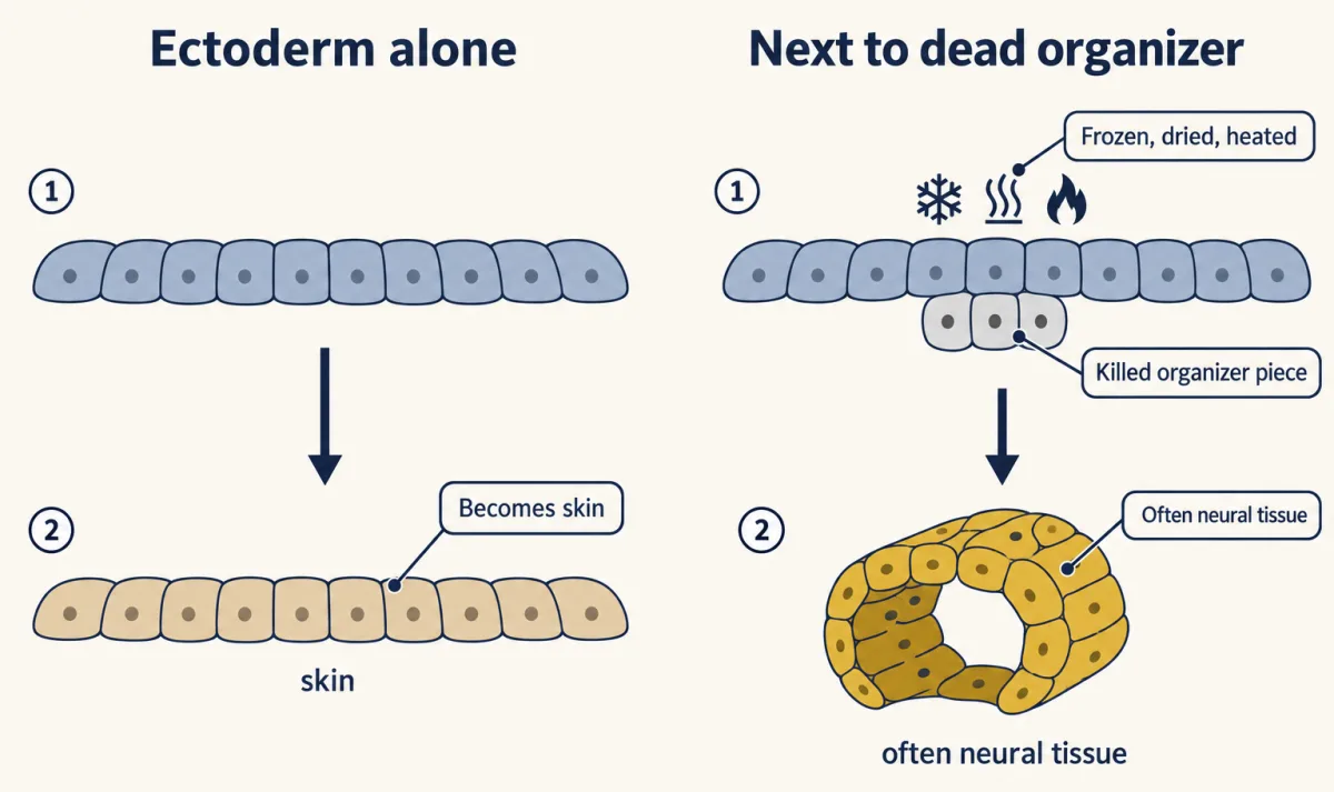
Two words help here, and you will meet them again in the neurotrophic hypothesis lesson. A factor is sufficient if it is enough, by itself, to produce an effect. It is necessary if the effect does not happen without it. Holtfreter’s result is a sufficiency result: dead organizer was enough to make competent ectoderm neural. But sufficient does not mean natural: a factor can be enough to produce an effect without being the signal the embryo uses. The controls showed why that mattered. In the 1930s many other killed tissues, ether extracts of amphibian tissue (ether dissolves fats, so the extract holds the tissue’s fat-soluble substances) and more and more unrelated materials were found to “induce”, at least in some cases.
| Condition | Scores neural? |
|---|---|
| Early-gastrula ectoderm cultured alone (the control in these assays) | no; it becomes skin |
| Ectoderm with living organizer | yes, and in the embryo the organizer builds an axis |
| Ectoderm with organizer killed by freezing, drying or heat (Holtfreter, from 1933) | often; neural tissue, but no organized axis |
| Ectoderm with other tissues, once killed | yes, many |
| Ectoderm with ether extracts of amphibian tissue (Waddington, Needham and Needham, by 1934) | yes, in some tests |
| Ectoderm with unrelated materials, from adult tissues to synthetic chemicals | in some cases |
| Salamander (Ambystoma) ectoderm with no organizer, or after brief damage that did not kill it (Barth, then Holtfreter, 1940s) | yes, in some cultures |
The rows come from different labs, species and culture conditions, so they are examples, not seven matched trials; the salamander row is a different ectoderm and treatment from the control in the first row. Still, almost every row gives some neural tissue, one with no organizer at all. When almost everything scores yes, a yes cannot name the signal.
C. H. Waddington described the trap. If competent ectoderm already holds a hidden tendency to become neural, an implanted substance might be the natural signal, or it might merely injure the tissue and release that tendency. The salamander result in the table’s last row adds a second blow: for that ectoderm, an organizer was not even necessary for neural tissue to form. That finding is one explanation for why the chemical hunt stalled for decades.
The confusion also pointed toward the answer. The next lesson shows that frog ectoderm whose cells are separated from one another becomes neural tissue with no inducer at all, and that the organizer’s products, Noggin, Chordin and Follistatin, act by blocking a signal of the TGF-beta family (see that lesson). If neural is what ectoderm becomes when a brake is released, then many injuries and chemicals that weaken the brake would be expected to “induce” as well.
Putting it together
| Experiment | Animal and stage | Observed result | What it supports | What it does not establish |
|---|---|---|---|---|
| Hair-loop constriction | newt eggs, from 1901 | some halves lived and divided but formed only a belly piece, with no neural plate or notochord | a half needs material from the future dorsal side (the textbook reading) | which tissue supplies it, or what it does |
| Dorsal lip grafted within one species | amphibian gastrula | extra axial structures | a moved dorsal lip can give a second axis | whose cells built it |
| Pale cristatus dorsal lip into the belly of pigmented taeniatus | newt gastrulae, 1921 and 1922 (host stages varied) | second neural tube mostly or entirely host cells; graft forms a second notochord and much of the nearby mesoderm | induction: the graft changed the fate of host cells | what the signal is; that the underlying mesoderm is the normal source |
| Same graft, read for position | newt gastrula | a second neural plate forms at the graft, from host ectoderm that would have made belly skin; the axis also holds donor notochord and recruited host mesoderm | the organizer decides where neural tissue forms (the move-the-source test) | whether it supplies a neural signal or removes a brake |
| Organizer with ectoderm from older and older embryos | amphibian gastrula to neurula | neural response falls and largely stops by the end of gastrulation | competence belongs to the responder and fades | what ends competence |
| Killed organizer against ectoderm | amphibian early-gastrula ectoderm, from 1933 | often neural tissue; no organized axis | neural-inducing activity survives cell death, so a substance can carry it | which substance |
| Other killed tissues, extracts, unrelated materials | amphibian ectoderm, 1930s | many induce in some cases | the yes-or-no neural test is not specific | the natural signal |
| Ambystoma ectoderm alone or briefly damaged | salamander, 1940s | becomes neural in some cultures | an organizer is not necessary for neural tissue in this ectoderm | why; see the next lesson |
The guiding idea holds within its limits. Neighbours can set a cell’s fate, and the host’s own cells answer only while they are still competent. The experiments also mark what the idea does not cover:
- Which tissue. Amphibian ectoderm and mesoderm during gastrulation. After that the ectoderm’s competence for the organizer’s neural induction has largely gone, though it still answers other signals later in development.
- “Can be set”, not “must be set”. Signals set fate in part; the responder’s own state, including competence, also counts. Nor does neural tissue always need an organizer: salamander ectoderm (Barth and Holtfreter), and frog ectoderm whose cells are separated (the next lesson), can become neural with no organizer at all, though signalling inside those cells still takes part.
- Which question. The idea says who changed fate and where. What the signal is, the graft could not say, and the dead-tissue era showed why the neural test could not say either.
Design problem
You have a dorsal lip from a pale newt species and host gastrulae from a pigmented species. Design experiments to show (a) whether the induced neural tube is made by host or donor cells, (b) whether the signal needs the two tissues to touch, and (c) when the responding ectoderm stops being able to respond.
Saved in this browser only.
One way to do it
(a) Graft the pale dorsal lip into the belly of the pigmented host, let a second axis form, then section it. Pigmented cells in the second neural tube are induced host cells; pale ones came from the donor. A dye injected into the donor before grafting is a more reliable lineage label than natural pigment (how far to trust a pigment marker).
(b) Take competent ectoderm and organizer tissue out of the embryo and culture them on opposite sides of a thin porous filter. The pores should let molecules through but be too small for cells or their processes to cross. If the ectoderm still becomes neural, a diffusible signal is enough. A solid sheet in place of the filter, and ectoderm on the filter with no organizer, are the controls.
(c) Run the competence test above: early organizer with ectoderm from later and later stages, from early gastrula to neurula, plus late organizer with early ectoderm to show that the drop is in the responder and not in the signal.
Going deeper
Roux, Driesch and the loop of hair
The question began as an argument about the first cell divisions. In 1888 Wilhelm Roux reported experiments on the two-cell embryo of a frog. He killed one of the two cells with a hot needle and left the dead cell in place. The living cell could go on to make something like half an embryo, with structures on one side and not the other. Roux read this as a mosaic: each early cell carries its own share of the future body.
A few years later Hans Driesch shook two-cell sea-urchin embryos until the cells came apart. A separated cell, instead of making half a larva, could make a smaller whole one. The two results are less opposed than they look. The experiments differed in species, and in one detail that mattered: Roux’s dead cell stayed attached to its living sister. In later experiments that separated frog cells completely, the survivor often formed a whole embryo.
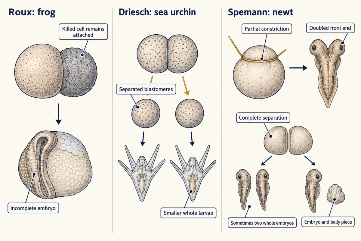
Spemann wanted a gentler tool. By 1901 he was tying a loop of very fine hair around newt eggs and tightening it slowly, dividing the embryo partly or completely without an open wound. Partial constriction gave embryos with doubled front ends. Complete separation sometimes gave two whole embryos. In other cases one half made a normal embryo while its partner stayed alive and kept dividing but formed only a “belly piece”, with no neural plate and no notochord. Cells could live and divide without building an axis.
Where the organizer comes from
Among the organizer genes that dorsal β-catenin switches on in Xenopus, an early example was goosecoid, a homeobox gene expressed in the organizer, cloned by Eddy De Robertis’s laboratory in 1991.
The classical picture of what happens next is called primary induction: mesoderm that has moved inside signals to the ectoderm above it, and that ectoderm becomes neural. The 1924 graft showed that dorsal-lip tissue can induce; it did not itself show that the mesoderm underneath is the normal source, which came from later work. The classical picture has since been refined in two ways. In Xenopus, evidence indicates that neural induction starts earlier, at the blastula stage, in dorsal cells that already make bone morphogenetic protein (BMP) antagonists. And signals from the organizer also pass sideways within the ectoderm itself, not only up from the mesoderm below.
How far to trust a pigment marker
Pigment granules are not shared evenly by every descendant. A host cell that inherits few granules can look pale and be counted as donor, which would understate the host’s share. Errors can also run the other way, because cristatus cells carry a little pigment rather than none. Without measuring how often each mistake happens, neither the size nor the direction of the net error is known. The main result, a second tube made mostly of host cells, is a pattern across sections rather than a single count, but without validated error rates a better lineage label would make it firmer.
Operations, priority and credit
Spemann proposed the pigment test in print early in 1921, before Mangold began, and she carried it out for her doctoral thesis. By one historical reconstruction she performed a few hundred operations over two breeding seasons, and the paper described six embryos in detail. The paper appeared in 1924 with Spemann’s name first; Mangold died the same year. In 1935 Spemann alone received the Nobel Prize in Physiology or Medicine, for the organizer effect, and in his Nobel lecture he said the experiment was first carried out at his instigation by her.
The chemical search, year by year
In 1932, before Holtfreter’s tests, a note from Spemann’s circle reported that organizer activity could survive destructive treatment. C. H. Waddington, Joseph Needham and Dorothy Needham began physico-chemical tests of the organizer in 1933; when their ether extracts induced, they suspected a fat-soluble substance, and the Cambridge group went on to test sterols. Lester Barth’s salamander report came first in the 1940s, and Holtfreter’s followed.
Your notes
Select any text in the lesson to highlight it or add a note to it. Your highlights and notes are saved in this browser and collected in your notebook.
This browser is not letting the site save anything (a private window or strict privacy settings), so highlights and notes will be lost when you leave the page.
How we measure it
Interspecies grafting with a pigment marker
Tissue is moved between embryos of two species whose cells differ in a visible trait, here pigment granules. After the embryo is sectioned, each cell's species shows which embryo it came from, so you can tell cells that were induced from cells that were transplanted.
Hair-loop constriction
A loop of very fine hair is tied around an amphibian egg and tightened by degrees, partly or completely dividing it without an open wound. Comparing what each side makes shows how much an early fragment can form on its own and what material it needs.
Explant culture of competent ectoderm
Ectoderm is cut from an early gastrula and cultured in a salt solution, alone or wrapped around a test tissue. Alone it usually becomes skin; if it becomes neural tissue when ectoderm cultured alone under the same conditions does not, the test tissue induced it. The method let Holtfreter test killed and heated tissues.
Serial histological sectioning
The embryo is fixed, embedded and cut into a series of thin slices that are stained and examined in order. The series shows the internal anatomy, such as a second neural tube and notochord, and the identity of the cells that built it.
Practice questions on this lesson (3) → MCAT-style, original, each with an explanation
Where this lesson fits
Before thisReview first
- The Wiring Problem: How a Brain Gets BuiltThis course · Lesson 1
It sets out how a brain is built from signals between cells, the question this graft experiment answers.
After thisStudy next
- Neural Induction: The Default ModelThis course · Lesson 6
It asks whether the organizer supplies a neural signal or removes a brake, the question left open here.
- Neurulation and Neural Tube DefectsThis course · Lesson 7
It follows the induced neural plate as it folds and closes into the neural tube.
- Head to Tail: Hox Genes, Retinoic Acid and RhombomeresThis course · Lesson 8
It shows how the neural tube the organizer induces is given head-to-tail identity.
Suggested by an AI model from the lessons themselves, then checked. How we use AI
Finish line
Test yourself
9 questions
Practice and MCAT-style, with explained answers.
Next · Lesson 6
Neural Induction: The Default Model
Pull a square of frog skin-to-be apart into single cells, and it becomes neural. Why? Because BMP signaling, which makes skin, falls in those cells. In the embryo, the organizer's secreted proteins make it fall.
Continue →