Cell Signaling · Unit 3: G-protein-coupled receptors and second messengers
Calcium: The Messenger Kept at Arm's Length
Rewritten on : new text, flashcards and film. Your notes and highlights stay, but the text around them may have moved.
How can the same calcium ion trigger contraction, secretion or gene expression without making the cell respond everywhere at once?
Pause when the reading reaches a film, and offer to play it. Saved on this device.
Watch the film now, or keep listening
Builds on:Binding: Affinity, Occupancy and Dose–ResponsePLC, IP3, DAG and Protein Kinase CPhosphorylation: Kinases and PhosphatasesWhy these?
 ▶ Watch How a Cell Makes a Calcium Signal · 3:28
Read the transcript of “How a Cell Makes a Calcium Signal”
Film transcript: How a Cell Makes a Calcium Signal
How can a cell signal with an ion it cannot make or destroy? Cyclic AMP can appear, carry information, then disappear. Calcium atoms remain calcium atoms throughout a response. The puzzle is therefore not how to build the messenger. It is how to change the calcium that proteins can reach.
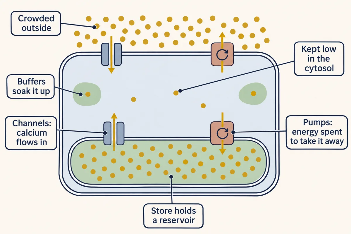
A resting cell keeps free calcium scarce in its cytosol. Calcium is crowded outside and stored inside the endoplasmic reticulum. Coral pumps maintain that separation by moving calcium away from the cytosol. The result is stored potential: many ions are waiting across membranes, while few remain free beside cytosolic proteins. A channel does not push calcium or spend fuel. It opens a path, and the existing gradient drives calcium through. Gold channel doors separate, and teal ions enter the cytosol. The free-calcium gauge rises immediately. A protein near that opening now encounters calcium that was unavailable a moment earlier. Now predict. Which machine can raise free cytosolic calcium immediately: opening a channel, or speeding a pump?
Follow one teal ion. Decide whether each machine brings it toward cytosolic proteins or carries it away. Ask which direction the calcium moves, not which machine is larger. Opening the channel raises the cytosolic pool. Speeding the pump lowers it. That gives two functional teams. Channels are on mechanisms because they admit calcium to the cytosol. Pumps, exchangers, and buffers are off mechanisms because they remove calcium from the free pool. These names describe direction, not molecular shape or energy source. Calcium signals begin and end by moving calcium, not changing it. During a rise, entry outruns removal. When channels close, the off machinery wins again.
Buffers bind some ions quickly. Pumps return others to the store or outside, restoring the low free concentration that makes another rise noticeable. The source can be outside the cell or inside its store. Either route changes the same measured quantity: free calcium in the cytosol. Which channel opens determines where the rise begins. How long it stays open helps determine the rise's timing. Movement creates a pattern that downstream proteins can read. So the signal is not a newly manufactured substance. It is a controlled redistribution of an existing ion. The gradient makes opening fast. The off machinery makes recovery possible.
Together they turn calcium movement into a brief, repeatable change in what cytosolic proteins can encounter.
From the film
- Calcium signals begin and end by moving calcium, not changing it.
- Opening a channel lets an existing gradient raise free cytosolic calcium.
- Pumps, exchangers and buffers lower free cytosolic calcium again.
- Restoring the low resting level makes the next rise noticeable.
In 1883 a frog heart kept beating in salt water mixed with London tap water, and stopped within about twenty minutes when the same recipe was mixed with distilled water. Sydney Ringer traced the difference to calcium in the hard pipe water. Sixty-four years later Lewis Heilbrunn and Floyd Wiercinski injected a calcium salt into a frog muscle fibre. With no nerve impulse at all, the fibre shortened: calcium alone can trigger contraction. How can the same ion control other responses without switching on the whole cell at once?
Build the rise: gradient, channels and removal
As the first film showed, calcium is an element that no enzyme can make or destroy, so it signals only by being moved. Free cytosolic calcium means calcium ions dissolved in the cytosol and not bound to proteins or other molecules. A resting cell holds it near 100 nanomolar; outside, calcium is 1 to 2 millimolar, ten to twenty thousand times higher. In the endoplasmic reticulum (ER), the cell’s folded internal calcium store, free calcium is several hundred micromolar; muscle’s version is the sarcoplasmic reticulum.
A signal may first make another messenger, such as inositol trisphosphate (IP3) from the previous lesson, which opens its receptor channel on the ER. Ions begin crossing within milliseconds of a channel opening. Because the resting level is so low, a modest number of ions raises bulk cytosolic calcium about tenfold, to roughly 1 micromolar, and much higher right beside the pore.
Calcium is an element: the cell cannot make or break it, only move it, and an open channel only opens a path while the gradient does the work. Time is in relative units and the picture is a sketch of the logic, not a measurement. The store bar's height is drawn at 500 µM only for the picture; the lesson says several hundred micromolar, and the outside bar spans 1 to 2 mM.
| Role | Where | Immediate action |
|---|---|---|
| On | Plasma membrane | Voltage-, ligand- or store-operated channels admit calcium from outside |
| On | Store membrane | IP3 and ryanodine receptor channels release stored calcium |
| Off | Store membrane | SERCA moves 2 calcium ions into the store per ATP |
| Off | Plasma membrane | PMCA expels 1 calcium ion per ATP; the sodium/calcium exchanger uses the sodium gradient, trading 3 sodium ions in for 1 calcium ion out |
| Off | Cytosol | Buffers such as parvalbumin bind calcium and remove it from the free pool |
Predict 1. A cell in calcium-free fluid receives two identical signals that open IP3 receptors. Calcium that leaves the cell between them cannot be replaced. How should the second cytosolic rise compare with the first?
Check your prediction
It should be smaller: SERCA could refill the ER only partly, because calcium pumped out of the cell was not replaced from outside. The exact change depends on how much calcium remained and how completely the first signal emptied the store.
The proof that a quiet cytosol is paid for came from muscle. By late 1961 Wilhelm Hasselbach and Madoka Makinose, in Heidelberg, had isolated sarcoplasmic-reticulum vesicles, added ATP and watched calcium leave the surrounding fluid. Setsuro Ebashi and Fritz Lipmann, in New York, independently reported ATP-driven uptake in 1962. Within two years the Heidelberg measurements put the cost at about two calcium ions per ATP split: relaxed muscle spends ATP to stay relaxed. Back in Tokyo, Ebashi and Ayako Kodama identified the separate protein through which calcium acts on the filaments of skeletal muscle in 1965, and named it troponin in 1966.
An empty store asks for a refill
A store that never refills supports only a limited release. James Putney, reassessing earlier findings on calcium stores, proposed in 1986 that depletion is itself an instruction to open plasma-membrane channels: store-operated entry. STIM1 crosses the ER membrane once, with a calcium-binding site facing the lumen. When luminal calcium falls, STIM1 clusters beneath the plasma membrane and opens Orai1. Orai1 admits calcium to the cytosol; SERCA can then pump some into the ER.
Predict 2. A drug empties the ER while no calcium is outside. Calcium is then restored outside. What happens in a cell lacking STIM1?
Check your prediction
The sharp store-operated rise should be largely missing. Orai1 cannot sense ER calcium by itself; without STIM1, the main store sensor, an empty store cannot efficiently open that route.
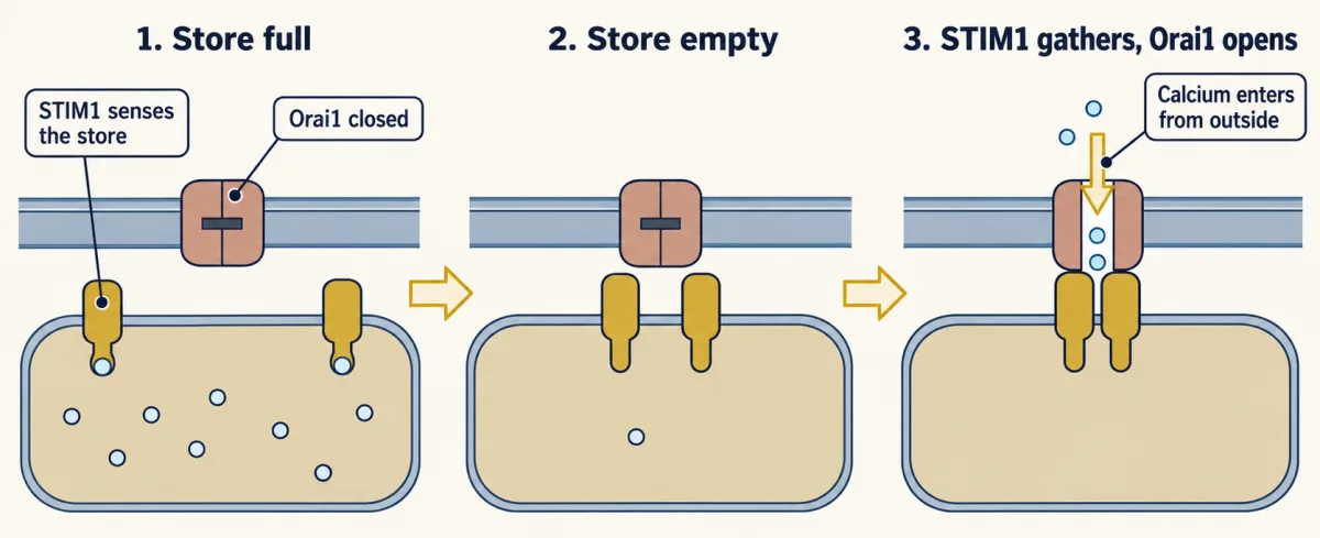
The parts took twenty years to name. Two groups, including laboratories led by Michael Cahalan and Tobias Meyer, independently reported STIM1 as the sensor in 2005. Stefan Feske and colleagues identified Orai1 in 2006 through a family whose immune cells lacked store-operated calcium entry.
How a protein reads the rise
Calcium offers a double positive charge and a preference for oxygen atoms. A protein can therefore make an oxygen-lined pocket that is mostly empty at 100 nanomolar and fills around a micromolar: the Kd idea from the lesson on binding and dose-response.
A common calcium-binding motif is the EF hand. In the parvalbumin structure published by Robert Kretsinger and C. E. Nockolds in 1973, calcium sits in a twelve-residue loop between helices E and F. Aspartate and glutamate side chains supply oxygens around it. The same helix-loop-helix occurs in a large protein family, including troponin C and the myosin light chains, although a few EF hands have lost calcium binding.
Predict 3. Two proteins bind the same amount of calcium. Only one exposes a target-binding surface. Which is a sensor, and which could be only a buffer?
Check your prediction
The one that exposes a surface is a sensor: binding changes what it can do. The other may still matter as a buffer, but binding alone sends no instruction.
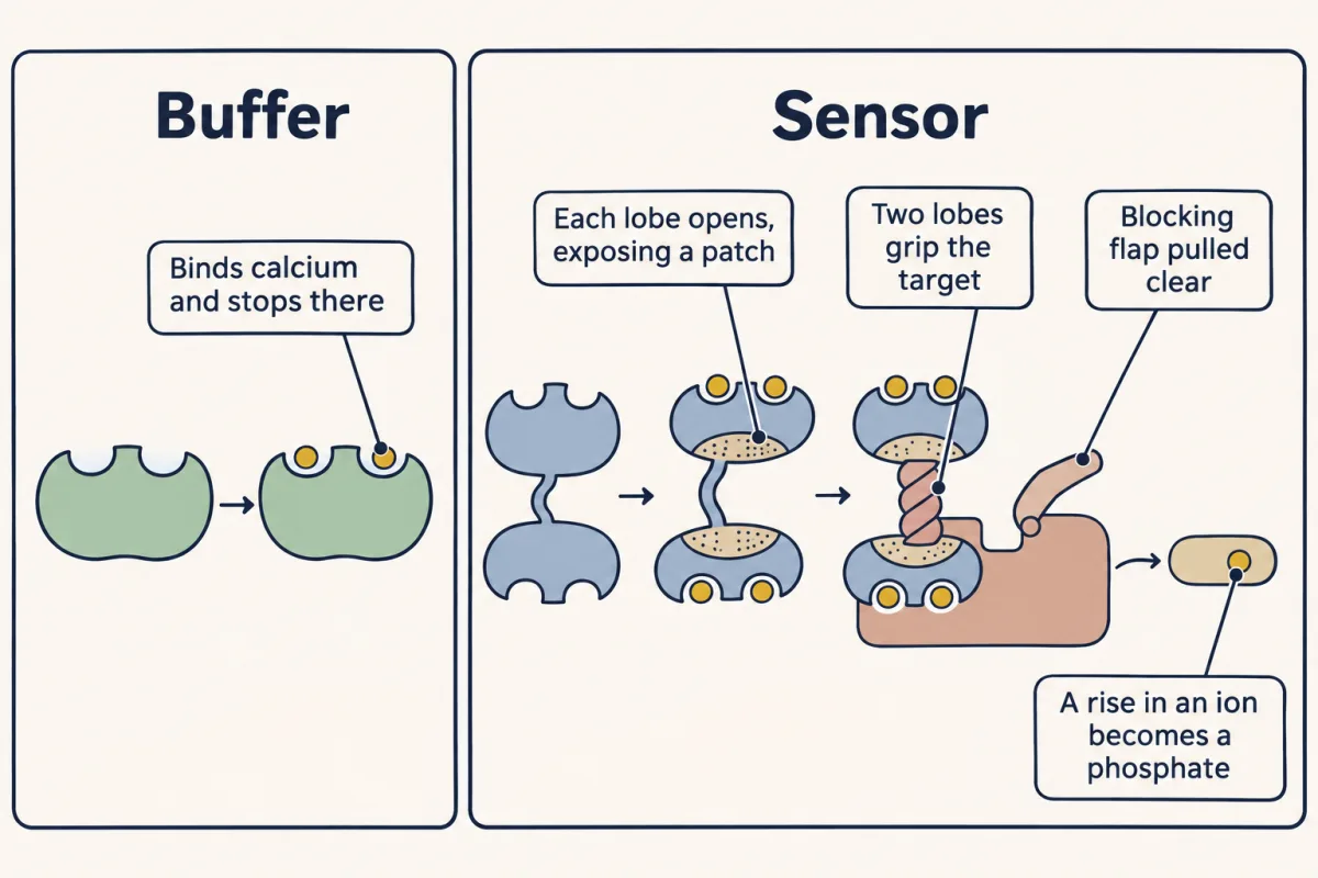
Parvalbumin binds calcium and acts as a buffer. Calmodulin binds calcium and changes shape. It has 148 amino acids and four EF hands, two in each lobe, joined by a flexible linker. Calcium exposes a hydrophobic, water-avoiding patch on each lobe. With many targets, the lobes close around a target helix; other targets use one lobe or separate sites.
Calmodulin has no catalytic activity, so one molecule can serve many targets: each target carries its own calmodulin-binding site. In CaM kinase II (CaMKII), a segment blocks the active site at rest. Calcium-loaded calmodulin grips that segment and pulls it clear, letting the kinase phosphorylate substrates. A rise in an ion has become a phosphate on a protein, read as in the lesson on the phosphorylation switch.
Which readers a rise reaches depends on where and when it happens; the second film builds that picture.
Read the transcript of “How One Ion Carries Different Messages”
Film transcript: How One Ion Carries Different Messages
The first film showed how moving calcium creates a cytosolic rise. How can that same ion trigger different responses? A cell cannot label one calcium ion for contraction and another for secretion. Instead, it controls where the rise happens, and when and how often.
Open one gold channel. Calcium is highest near its pore before buffers capture and pumps remove it. The teal plume fades with distance. A sensor anchored beside the channel samples that local domain. An identical sensor across the cytosol meets much less free calcium. Predict before either sensor changes. Both sensors switch on only above the same calcium threshold. The channel opens briefly. Which sensor responds: the one beside the pore, the one across the cell, or both? Follow the fading plume.
Only the nearby sensor responds. Calcium beside the pore crosses the threshold; the lower level far away does not. The near sensor binds calcium, changes shape and exposes an orange output surface. Location has selected a reader without changing the calcium or the reader's binding properties. A local event can also recruit more release. Calcium from one site may help open a neighbouring release channel. Then the next site can recruit another. The moving front becomes a wave, and many sensors across the cell can respond. This spreading route is positive feedback. Now transform distance into time. One sensor sees isolated calcium spikes, each followed by full recovery.
Another sees closely spaced spikes. Both trains can contain similar individual rises, yet their intervals differ. A reader with a slowly fading active state can distinguish those intervals. With long gaps, the reader returns toward rest before the next spike.
Close spikes arrive while some activity remains. The responses can build. Calcium has not acquired a new identity. Timing matters because the reader keeps a biochemical memory that outlasts each brief rise. Cells give calcium meaning by controlling where and when free calcium rises. The size of a rise matters too; this film concentrates on place and timing.
Local domains favour nearby readers. Coordinated release can spread the response. Repeated spikes favour readers that retain activity between rises. Cells combine these patterns with different sensors; there is no universal decoder. The cell therefore writes information without changing the ion itself.
Channel position sets the starting place. Feedback can enlarge the territory. Opening and closing set the rhythm. Sensors read only the calcium pattern that reaches them, on the time scale their own chemistry can remember.
From the film
- Cells give calcium meaning by controlling where and when free calcium rises.
- Buffers can keep a rise local, so a sensor beside the channel can respond while an identical one farther away does not.
- Calcium-dependent release can recruit more sites and spread a wave.
- Closely spaced spikes can build activity in a reader that keeps some activity from one spike to the next.
Where and how often: the evidence and its limits
In heart muscle, a local release through ryanodine receptors is a spark. Each electrical impulse sets off thousands of sparks at once, and together they make the whole-cell rise behind a beat. In eggs and liver cells, release travels instead: calcium from one cluster stimulates the release channels of the next, by positive feedback, and a wave crosses the cell. Not every global rise, however, is built only from sequential local events.
Predict 4. A fast calcium buffer is added, and the whole-cell calcium peak falls sharply. Should a sensor anchored beside an open channel lose its response just as sharply?
Check your prediction
Probably not. It sits where calcium is highest, before the buffer can bind much of it; the whole-cell average reflects calcium that has spread, so a distant sensor loses more. The result depends on the buffer’s concentration and binding kinetics.
In 1986 Woods, Cuthbertson and Cobbold used a light-emitting reporter in single rat liver cells. More hormone produced more frequent calcium spikes: the dose was written in frequency, not in a higher calcium level.
To respond to frequency, a reader must keep some activity from one spike to the next. Mitochondrial enzymes respond to spike rate in liver cells, and transcription factors such as nuclear factor of activated T cells (NFAT) respond best to frequent spikes in immune cells. For faster neuronal pulses, calcium/calmodulin-dependent protein kinase II (CaMKII) is one counter. In its hub of twelve or fourteen subunits, when two neighbours hold calmodulin together, one can phosphorylate the other, and activity can persist after calcium falls. In 1998 De Koninck and Schulman showed with purified enzyme that lasting activity depends on pulse frequency. Which molecules decode slower cellular oscillations, and how much they read frequency rather than summed calcium, remain open questions.
Seeing calcium without confusing the signal
None of this could be studied until calcium could be watched in a living cell. Aequorin, purified from the jellyfish Aequorea by Osamu Shimomura with Frank Johnson and Saiga and described in 1962, emits blue light when it binds calcium. Injected into one cell, as in the liver cells above, it reports rises as flashes. Fura-2, from Roger Tsien’s laboratory in 1985, shifts its best excitation wavelength on binding calcium. The ratio at two wavelengths depends little on dye loading and can be calibrated to concentration.
Design problem
How could calmodulin’s shape change make a calcium sensor? Design a protein that reports calcium by light. State the control that shows the response depends on calcium.
Saved in this browser only.
One way to do it
Build one chain: cyan fluorescent protein, calmodulin, a calmodulin-binding peptide and yellow fluorescent protein. At low calcium the fluorescent proteins are apart. When calcium rises, calmodulin grips its peptide and brings the ends within a few nanometres. Energy can then pass from excited cyan protein to yellow protein by FRET, raising the yellow-to-cyan ratio. Miyawaki, Tsien and colleagues published this “cameleon” design in 1997.
Make a calcium-blind twin by changing the glutamate at the end of each EF-hand loop to glutamine, so calmodulin can no longer hold calcium at cellular concentrations. The ratio could also move with pH or bleaching; any change the twin shows is not calcium. The twin shows that the response requires calcium binding, but not where the calcium came from. Calibrate the working sensor inside cells: an ionophore, which carries calcium across membranes, gives the maximum ratio in high calcium, and a chelator, which grips calcium tightly, gives the minimum. The reporter is itself a calcium buffer, so use as little as can be read.
Going deeper
Kretsinger and Nockolds lettered parvalbumin’s six helices A to F, hence the name. Kretsinger’s right-hand analogy (forefinger as helix E, thumb as F, curled middle finger as the loop) captures the topology, not a molecular structure.
Your notes
Select any text in the lesson to highlight it or add a note to it. Your highlights and notes are saved in this browser and collected in your notebook.
This browser is not letting the site save anything (a private window or strict privacy settings), so highlights and notes will be lost when you leave the page.
How we measure it
Aequorin luminescence
Aequorin is a jellyfish protein that gives off blue light when it binds calcium. Injected into a single cell, it reports a calcium rise as a flash that a light detector can count. It needs no excitation light, which keeps the background dark.
Ratiometric calcium dyes (fura-2)
Fura-2 is a small fluorescent molecule that binds calcium, and binding shifts its excitation spectrum toward shorter wavelengths. The dye is excited in turn at 340 nm and at 380 nm. Dividing the two emission signals largely cancels out how much dye the cell holds, so the ratio can be calibrated to a calcium concentration.
FRET protein sensors (cameleons)
A cameleon is one polypeptide: a cyan fluorescent protein, calmodulin, a calmodulin-binding peptide and a yellow fluorescent protein. Calcium makes calmodulin wrap around the peptide, which brings the two fluorescent proteins together and raises energy transfer from cyan to yellow. Because the cell makes the sensor from a gene, it can be sent to one cell type or one compartment.
Calcium uptake by membrane vesicles
Break open muscle, collect the sealed vesicles that form from sarcoplasmic reticulum, add ATP, and measure how much calcium disappears from the surrounding fluid. Comparing calcium taken up with ATP split gives the stoichiometry of the pump, about two calcium ions per ATP.
Practice questions on this lesson (3) → MCAT-style, original, each with an explanation
Where this lesson fits
Before thisReview first
- Binding: Affinity, Occupancy and Dose–ResponseThis course · Lesson 3
You need Kd and occupancy from there to see why an EF hand fills at about a micromolar.
- PLC, IP3, DAG and Protein Kinase CThis course · Lesson 14
The IP3 made there is what opens the IP3 receptor on the store here.
- Phosphorylation: Kinases and PhosphatasesThis course · Lesson 6
CaMKII turns a calcium rise into a phosphate, which you read using kinase and phosphatase logic from there.
After thisStudy next
- Turning the Signal Off: Arrestins, RGS Proteins and PhosphodiesterasesThis course · Lesson 16
It takes the idea of switching a signal off further, with arrestins, RGS proteins and phosphodiesterases.
- The Shape of a Signal: Size, Duration, Frequency and PlaceThis course · Lesson 35
It uses calcium spike size, duration, frequency and place as examples of how a cell reads signal shape.
- Signaling to the Nucleus: CREB, AP-1 and Immediate Early GenesThis course · Lesson 32
Calcium and CaMKII signals reaching the nucleus switch on CREB and immediate early genes.
Elsewhere on the siteFilms, simulations and method films in other courses
- The length constantSimulation · Neuroscience
Resting leak and pump balance show how a steep ion gradient is held by pumps.
- Two Pushes, One HillFilm · Cell Biology
Concentration and voltage together set which way calcium flows when a channel opens.
- Blips from a Silent NerveFilm · Neuroscience
Calcium entry triggers vesicle release at the terminal, a key calcium-triggered response.
- A Tap, a Hole, a LevelFilm · Cell Biology
A pump keeps a store's gradient against leak, as in the calcium store.
Suggested by an AI model from the lessons themselves, then checked. How we use AI
Finish line
Test yourself
8 questions
Practice and MCAT-style, with explained answers.
Next · Lesson 16
Turning the Signal Off: Arrestins, RGS Proteins and Phosphodiesterases
The hormone is still bound and the response has faded. What switched the receptor off?
Continue →