Developmental Neuroscience · Unit 2: Becoming neural
Neural Induction: The Default Model
Rewritten on : new text, flashcards and film. Your notes and highlights stay, but the text around them may have moved.
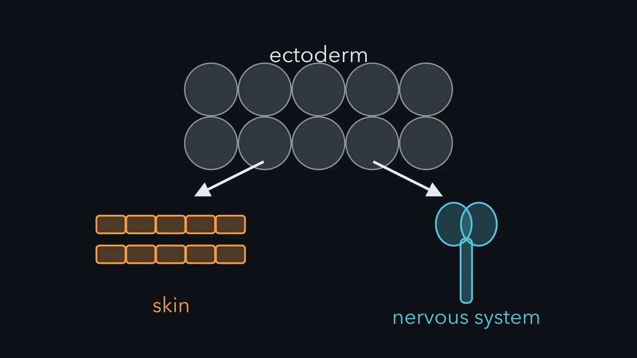
Pull a square of frog skin-to-be apart into single cells, and it becomes neural. Why? Because BMP signaling, which makes skin, falls in those cells. In the embryo, the organizer's secreted proteins make it fall.
Pause when the reading reaches a film, and offer to play it. Saved on this device.
Watch the film now, or keep listening
Builds on:The Organizer: Spemann and MangoldWhy these?
 ▶ Watch The Map and the Question · 2:54
Read the transcript of “The Map and the Question”
Film transcript: The Map and the Question
Ectoderm can become skin or nervous system. What decides which one it becomes? Start with the map. A blastula is the early frog embryo, a hollow ball of cells. Inside is a fluid-filled cavity, the blastocoel. The roof of that cavity is the animal cap. The cap is ectoderm, the outer germ layer. It gives rise to both the skin and the nervous system.
The outer layer of skin is the epidermis. Keep that word; it is one of the two answers. Gastrulation is the set of cell movements that form the germ layers. It begins at the blastopore, a groove on the lower half of the embryo, where cells start to move inside. The tissue at the blastopore's dorsal lip is the organizer. Dorsal is the side where the back and the neural plate will form. Ventral is the belly side. In the 1920s, Hilde Mangold and Hans Spemann grafted an organizer into another embryo. The host formed a second body axis. The organizer could change what nearby ectoderm became. For decades, the question was what positive signal the organizer sends to make ectoderm neural.
Now the dish. Cut out the animal cap and culture it alone in salt solution. Left alone, what should a piece of skin-to-be become? Make a guess. It becomes epidermis. No organizer tissue is attached to it. One more pair of names. BMPs, bone morphogenetic proteins, are secreted signals of the TGF-beta family. Inside a cell, their signal is carried to the nucleus by Smad1, Smad5 and Smad8. The map raises three puzzles. First, what holds an intact cap as skin? Second, what does the organizer send, and how does it change the ectoderm's fate? Third, does the answer hold outside the frog cap: in the whole embryo, in a fly, in a chick?
Here is the idea to carry through every chapter. BMP says skin. In early frog animal caps, turn its signal down far enough, and the cells become neural, as long as they can still respond and their other needed inputs, such as FGF, are intact. BMP's effect comes down to one dial, Smad1 activity in the nucleus. The next chapters show three hands that turn it down.
From the film
- Ectoderm can become skin or nervous system, and an animal cap left intact becomes epidermis.
- BMP says skin: in early frog animal caps, turning its signal down far enough lets competent cells become neural, provided other needed inputs such as FGF are intact.
- BMP's effect comes down to one dial, the activity of Smad1 in the nucleus.
Cut a small square from the top of a frog blastula, culture it in salt water, and it becomes skin. Pull the same square apart into single cells, keep them apart for a few hours, then let them clump again, and they become neural tissue.
Both results come from the frog Xenopus laevis, and together they turned an old question around. For decades the question had been what positive signal makes ectoderm neural. The square suggested another answer. Perhaps the signal in the ectoderm is the one that makes skin, and the job of the embryo’s organizer is to stop it. This lesson follows how that idea was tested, which molecules carry it, and where it falls short.
The layout
Every experiment in this lesson happens on one map, which the opening film draws. (For how the organizer was discovered, see the lesson on the organizer.)
- The blastula is the early embryo, a hollow ball of cells around a fluid-filled cavity, the blastocoel.
- The animal cap is the roof of that cavity, at the top of the ball. It is made of ectoderm, the outer germ layer (mesoderm and endoderm are the other two), which gives rise to both the skin and the nervous system. The outer layer of skin is the epidermis.
- Gastrulation is the set of cell movements that form the germ layers. It begins at the blastopore, a groove on the lower half of the embryo where cells start to move inside.
- The organizer is the tissue at the blastopore’s dorsal lip. Dorsal is the side where the back and the neural plate will form; ventral is the belly side.
- The graft that started it. In the 1920s Hilde Mangold and Hans Spemann grafted an organizer into another embryo and got a second body axis. The organizer could change what nearby ectoderm became.
- The animal-cap explant. Cut out the cap and culture it alone in salt solution, and it becomes epidermis, making epidermal keratin, a skin protein scored as the readout. Any treatment that makes it switch on neural genes reveals what that treatment can induce in this tissue.
- BMPs, bone morphogenetic proteins, are secreted signals of the TGF-beta family. Inside a cell, their signal is carried to the nucleus by the proteins Smad1, Smad5 and Smad8.
The map raises three puzzles.
- What holds an intact cap as skin? In the dish, no organizer tissue is attached to it.
- What does the organizer send, and how does it change the ectoderm’s fate?
- Does the answer hold outside the frog cap: in the whole embryo, in a fly, in a chick?
The one idea to carry through this lesson. BMP says “skin.” In early frog animal caps, turn its signal down far enough, by any of the routes in this lesson, and the cells become neural, provided they are still competent and their other required inputs, including early fibroblast growth factor (FGF) signaling, are intact. BMP’s effect comes down to one dial: the activity of Smad1 (with its close relatives Smad5 and Smad8) in the nucleus. The organizer turns the dial down from outside the cell, by catching BMP. A broken receptor turns it down at the membrane. Mitogen-activated protein kinase (MAPK) signaling turns it down inside the cell.
What the rule covers. The BMP side of the choice between epidermis and neural tissue in standard Xenopus animal-cap cultures, while the ectoderm is competent, able to respond, early in development. Even there the dial is not the whole decision: early FGF signaling is needed too, in caps and in the whole embryo. The rule says nothing about where along the head-to-tail axis the neural tissue will lie. And in chick, turning the dial down is not enough on its own.
Three cap experiments
Read the transcript of “Break the Receptor, Add the Ligand”
Film transcript: Break the Receptor, Add the Ligand
Break one signaling receptor in a frog cell, without adding anything new. What will an animal cap become? Every experiment here uses the same tissue, the animal cap, cultured in salt solution. Left intact, it becomes epidermis and makes epidermal keratin. That is the control. BMPs signal through pairs of receptor kinases, a type II and a type I. The type I receptor phosphorylates Smad1, 5 or 8. It moves to the nucleus with Smad4. Now remove one receptor's kinase domain. It still binds ligand and still pairs with normal partners. But it cannot pass the signal on, so it jams the complexes it joins. This is a dominant negative: a broken copy that blocks the working copies.
In 1994, Ali Hemmati-Brivanlou and Douglas Melton injected eggs with RNA for such a receptor. It was built from an activin receptor. But it also blocks signaling by related ligands. Cut caps from the injected embryos and culture them alone. What do they become?
Neural tissue, with no mesoderm detected that could have induced it. Blocking a receptor, not adding a signal, made neural tissue. Which blocked signal normally keeps ectoderm as skin? The receptor blocks activin, and related signals too. But activin added to caps makes mesoderm, not skin. A relative fit better: BMP4. Its RNA is in the ectoderm early. And it is cleared from the dorsal side as the neural plate appears. To test BMP4 directly, you need cap cells that would otherwise turn neural. In 1989, Horst Grunz and Lothar Tacke had found them. They separated cap cells in medium lacking the calcium and magnesium the cells need to stick together.
They kept the cells apart for hours, then let them clump again. The cells made neural tissue. In 1995, Paul Wilson and Hemmati-Brivanlou separated cap cells the same way and added BMP4 protein. What do these cells become?
Epidermis. Their neural genes stayed off. The ligand alone was enough to restore skin. The takeaway: jam the receptor and a cap turns neural. Add BMP4 back and separated cells return to skin.
From the film
- In frog animal caps, jamming a receptor with a dominant-negative copy makes neural tissue, with no mesoderm detected that could have induced it.
- Blocking a receptor, not adding a signal, made neural tissue.
- Adding BMP4 protein back to separated cap cells restores epidermis and keeps neural genes off.
Each experiment starts from the intact cap, which becomes epidermis. The question is what holds it there.
Break the receptor
Bone morphogenetic proteins (BMPs) signal through pairs of receptor kinases. A BMP dimer brings together a type II and a type I receptor; the type I receptor then phosphorylates Smad1, 5 or 8, which moves to the nucleus with Smad4 and turns on target genes (see the lesson on TGF-beta and Smads). A receptor missing its kinase domain still binds ligand and still pairs with normal partners, but it cannot pass the signal on, so it jams the complexes it joins. This is a dominant negative: a broken copy that blocks the working copies.
In 1994 Ali Hemmati-Brivanlou and Douglas Melton injected eggs with RNA for such a receptor. It had been built from an activin receptor, but it also blocks signaling by related ligands.
Predict. Cut animal caps from the injected embryos and culture them alone, with no other tissue. What do they become?
Check your prediction
Neural tissue, with no mesoderm detected that could have induced it. Blocking a receptor, not adding a signal, made neural tissue.
Which blocked signal normally holds ectoderm as skin? The result was first read as a block of activin. But activin added to caps makes mesoderm, not skin, so it is a poor candidate for the skin signal. A relative fit better: BMP4, whose RNA is in the ectoderm early and is cleared from the dorsal side as the neural plate appears.
Add the ligand back
To test BMP4 directly you need cap cells that would otherwise turn neural. In 1989 Horst Grunz and Lothar Tacke had found them. They separated animal-cap cells in medium lacking the calcium and magnesium the cells need to stick together, kept them apart for hours, then let them clump again. Instead of epidermis, the cells made neural tissue. Assume for now that separation lowers BMP signaling; the next section shows how.
Predict. In 1995 Paul Wilson and Hemmati-Brivanlou separated cap cells in the same way and added BMP4 protein to the medium. What do the cells become?
Check your prediction
Epidermis, and their neural genes stayed off. The ligand alone was enough to restore skin.
One dial
Read the transcript of “One Dial”
Film transcript: One Dial
Separated cells turn neural, yet their BMP receptors keep signaling. How can both be true? Start by putting the four results in one table. The intact cap: receptors working, Smad1 output high, epidermis. The dominant negative cap: receptors jammed, output low, neural. Separated cells: neural. Leave their output for a moment. Separated cells with BMP4: output high enough, epidermis. The output column is the model's reading. It was not measured directly in every row. Read down the last two columns. In these four conditions, where the dial was high, the cells made skin. Where it was low, they made neural tissue. In a 1997 review, Hemmati-Brivanlou and Melton called this the default model.
In these early cap cultures, neural is the fate frog ectoderm takes when BMP signaling is kept low, while the cells can still respond and their other inputs, such as FGF, are intact. BMP promotes epidermis. Default is an operational word. It names the outcome in these cultures, with competent cells and their other inputs intact, not a fate reached with no signals. Now the third row. The first explanation was dilution. Separation spreads the cells through a large volume of fluid. The BMP they pass to one another would be diluted away. It was a fair guess. If BMP spreads freely, the physics of diffusion supports it. But later work tested it. BMP receptors keep signaling in separated cells.
So how could the dial fall? Separation switches on lasting signaling by MAPK. It is a kinase cascade, usually started by growth factors such as FGF. MAPK phosphorylates Smad1 in its middle, or linker, region. That can keep Smad1 out of the nucleus and mark it for destruction. The cells need this activity to turn neural. The dial still goes down, by a different hand. the fourth row? The damping is partial. So enough added BMP4 can push the output back up. That is why the rule names the dial, Smad1 in the nucleus. It does not name BMP in the fluid. The takeaway: three hands can turn the dial down. Outside the cell, at the membrane, or inside it.
From the film
- In these four cap conditions, where Smad1 output was high the cells made skin, and where it was low they made neural tissue.
- Separated cells still have signaling BMP receptors, but MAPK activity can damp Smad1, so dilution is probably not the reason they turn neural.
- The rule names the dial, Smad1 in the nucleus, and not BMP in the fluid.
Put the results in one table. The fourth column is the model’s reading of each row; it was not measured directly in every case.
| Condition | What was changed | BMP receptors | Smad1 output (the dial) | Fate |
|---|---|---|---|---|
| Intact cap | nothing (the control) | working | high | epidermis |
| Intact cap, dominant-negative receptor | broken activin receptor, which also blocks BMP | jammed | low | neural |
| Separated cells | kept apart for hours | still signaling | low (MAPK; next section) | neural |
| Separated cells + BMP4 | ligand added | working | high enough | epidermis |
Rule to keep: in this frog tissue neural is the default fate, and cell-to-cell BMP signaling suppresses it. The picture is a sketch of the logic, with dots standing for relative amounts of BMP, not for measured ones.
Read down the last two columns. In these four conditions, where the dial was high the cells made skin; where it was low, they made neural tissue. In a 1997 review Hemmati-Brivanlou and Melton called this the default model: neural is the fate frog ectoderm takes when bone morphogenetic protein (BMP) signaling is kept low, and epidermis the fate BMP instructs.
Default is an operational word. It names the outcome in these cultures, with the cells competent and their other inputs intact. It does not mean that no signals are acting, and the third row shows why that matters.
Why separated cells turn neural
The first explanation of the third row was dilution. Separation spreads the cells through a large volume of fluid, so on this reading the BMP they normally pass to one another is diluted away. It was a fair guess, and the physics of diffusion supports it if BMP spreads freely (Going deeper works it). But later work found that BMP receptors keep signaling in separated cells. Something else had changed.
Separation switches on lasting signaling by MAPK (mitogen-activated protein kinase), a kinase cascade usually started by growth factors such as fibroblast growth factor (FGF). MAPK phosphorylates Smad1 in its middle, or linker, region. That can keep Smad1 out of the nucleus and mark it for destruction. The cells need this MAPK activity to turn neural. So the dial still goes down in separated cells, by a different hand, and dilution is probably not the reason.
That is why the rule names the dial, Smad1 in the nucleus, and not BMP in the fluid. It also reads the fourth row: the damping is partial, so enough added BMP4 can push the pathway’s output back up. Keep MAPK in mind; it returns in the frog embryo and in the chick.
The organizer’s catchers
Read the transcript of “The Organizer's Catchers”
Film transcript: The Organizer's Catchers
If BMP makes skin, what should the organizer make to turn ectoderm neural? Something that blocks BMP. It makes at least three such proteins. First, where they come from. Chordin and Noggin are made early, at the blastula stage, in dorsal animal cells. So neural induction starts before gastrulation. Then the organizer and dorsal mesoderm turn inward and spread beneath the ectoderm. This is involution.
The involuting mesoderm secretes all three. The ectoderm above it becomes the neural plate. Noggin was found in 1992 by William Smith and Richard Harland. It is a secreted protein made in the organizer. In 1993, Teresa Lamb, Harland and colleagues added Noggin protein to intact caps. What should the caps become?
Neural tissue, without dorsal mesoderm. Noggin acted on the ectoderm directly. The markers were all anterior. Forebrain is neural. Cement gland, a mucus gland at the head end, is not. Chordin was cloned in 1994 by Yoshiki Sasai and colleagues, in Eddy De Robertis's laboratory. Both Noggin and Chordin bind BMP4 and keep it off its receptors. Follistatin was known as a protein that binds activin. It also neuralizes caps and binds some BMPs. It works differently. It can stay bound to BMP as BMP docks on its receptor. That complex does not signal. None of the three needs to enter a cell. Each grabs BMP outside the cell. In the crystal structure, Noggin clasps a BMP dimer and covers the faces the receptors would bind.
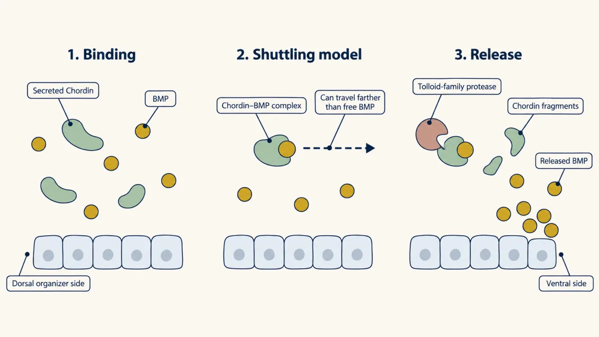
Chordin has a further twist. In the shuttling model, BMP bound to Chordin travels toward the ventral side. There, Tolloid proteases cut Chordin and release the BMP. Caps show what the catchers can do. Does the embryo need them? In 2005, Khokha and colleagues depleted Chordin, Noggin and Follistatin together from frog embryos. What happens to the neural plate?
The embryos lost their dorsal structures, the neural plate among them. With all three catchers gone, BMP signaling stays high. The reverse test agreed. Reversade's group removed three BMPs from embryos whose organizer signals were blocked. The embryos formed massive amounts of brain. Early FGF signaling is also needed in the frog, in addition to blocking BMP. It can turn the dial down through MAPK. It also does more.
The takeaway: the frog embryo needs the catchers to keep the dial down. And FGF adds a second input.
From the film
- The organizer makes at least three proteins that catch BMP outside the cell: Noggin, Chordin and Follistatin.
- Noggin protein turns intact caps neural, with anterior markers, without dorsal mesoderm.
- In Xenopus tropicalis embryos, depleting all three catchers lost the neural plate, so the frog embryo needs them; early FGF signaling is needed too.
If bone morphogenetic protein (BMP) makes skin, the organizer should make something that blocks BMP. It makes at least three such proteins.
Chordin and Noggin are made early, at the blastula stage, in dorsal animal cells, so neural induction starts before gastrulation. Then, during gastrulation, the organizer and the dorsal mesoderm turn inward and spread beneath the ectoderm. This is involution. The involuting mesoderm secretes Chordin, Noggin and Follistatin, and the ectoderm above it, where BMP signaling is kept low, becomes the neural plate.
Noggin. Noggin was found in 1992 by William Smith and Richard Harland. It encodes a secreted protein expressed in the organizer, and it binds BMP4 and keeps it off its receptors (Zimmerman, De Jesús-Escobar and Harland, 1996).
Predict. In 1993 Teresa Lamb, Harland and colleagues added Noggin protein to intact animal caps. Using the dial, what should the caps become? Should they need to make dorsal mesoderm first?
Check your prediction
Neural tissue, without dorsal mesoderm, so Noggin was acting on the ectoderm directly. The markers were all anterior: forebrain, which is neural, and cement gland, a mucus gland at the head end that forms from non-neural ectoderm. There were no hindbrain or spinal cord markers.
Chordin. Chordin was cloned in 1994 by Yoshiki Sasai and colleagues in Eddy De Robertis’s laboratory as a gene switched on by organizer transcription factors. It neuralizes animal caps and BMP4 opposes the effect (Sasai, Lu, Steinbeisser and De Robertis, 1995), and it binds BMP4 tightly and blocks its receptor binding (Piccolo, Sasai, Lu and De Robertis, 1996).
Follistatin. Known as a protein that binds activin, follistatin is made in the organizer, and in 1994 Hemmati-Brivanlou, O. G. Kelly and Melton showed that it neuralizes caps. It was later found to bind some BMPs as well. It works differently from the other two: instead of blocking receptor binding, it can stay bound to BMP as BMP docks on its receptor, forming a three-part complex that does not signal.
None of the three needs to enter a cell; each grabs BMP outside it. The structure viewer shows how Noggin does it: in the crystal, a Noggin dimer clasps a BMP-7 dimer and covers the faces the receptors would bind.
Chordin does one more thing, which the course calls the shuttling mechanism. In the model, BMP held by Chordin travels farther than free BMP, so it is carried away from the organizer; Tolloid-family proteases (Xolloid in the frog) then cut Chordin and release the BMP toward the ventral side.
The embryo test
Caps show what the antagonists can do. Does the embryo need them? Two groups asked by depleting proteins from whole frog embryos.
Predict. In 2005 Khokha and colleagues depleted Chordin, Noggin and Follistatin together from embryos of Xenopus tropicalis, a related frog. What happens to the neural plate?
Check your prediction
The embryos lost their dorsal structures, the neural plate among them. With all three catchers gone, BMP signaling stays high, which fits the ectoderm not becoming neural.
The reverse test points the same way. Reversade and colleagues depleted BMP2, BMP4 and BMP7 in embryos whose organizer signals had been prevented, by ultraviolet treatment or by knocking down beta-catenin. The embryos formed massive amounts of brain, even without normal organizer signals. So in the frog, low BMP signaling is needed for the neural plate, not only able to make neural tissue in a dish.
The dial is not the whole story. Early fibroblast growth factor (FGF) signaling is also needed for neural induction, in the embryo and in caps, in addition to BMP inhibition (Delaune and colleagues, 2005). FGF can turn the dial down through mitogen-activated protein kinase (MAPK), the hand you met in separated cells, but it also does things that lowering BMP does not explain.
The same contest in a fly, upside down
Read the transcript of “A Fly Turned Over”
Film transcript: A Fly Turned Over
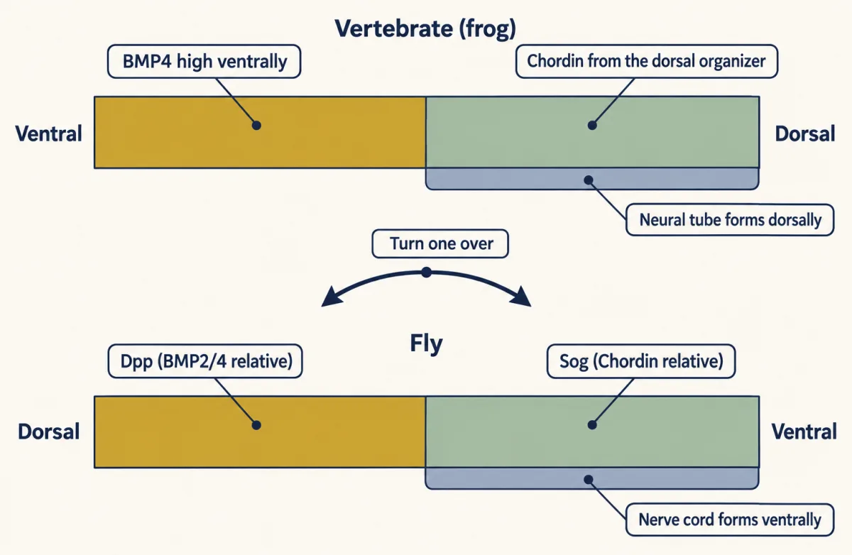
A fruit fly and a frog look nothing alike. Do they build a nervous system the same way? The fly has the same pair. Its gene decapentaplegic encodes a relative of BMP2 and 4. And short gastrulation, or Sog, encodes a relative of Chordin. So Dpp plays the part of the frog's BMP4. Sog plays the part of its Chordin. In 1995, Holley and colleagues swapped the two antagonists between species. Frog Chordin works in the fly. And fly Sog works in the frog. Tolloid was first named as a fly gene, and it cuts Sog. Now the map. In the fly, Dpp is made on the dorsal side. Sog is made in broad lateral stripes of ectoderm on the ventral half. Use the rule. Where should the fly's nerve cord form?
Ventrally, from that ectoderm, where Sog keeps Dpp signaling low. The picture is simplified. The ventral-most fly cells are mesoderm, not nerve cord. In a vertebrate, BMP4 signaling is high on the ventral side. Chordin comes from the dorsal organizer, and the neural tube forms dorsally. In both animals, nervous system forms where the antagonist keeps the BMP signal low. What happens if you turn the fly over?
Its BMP and its antagonist lie where frog's do. The two maps line up. In 1996, De Robertis and Sasai argued the common ancestor already patterned its body this way. One lineage then inverted the plan. On this view, the fly's map and the frog's map are one ancestral map, read from opposite sides. Long before, in the 1820s, Étienne Geoffroy Saint-Hilaire had proposed this inversion from anatomy alone. The takeaway: fly and frog run the same contest, BMP against its catcher. Only the body
From the film
- The fly's Dpp and Sog are the counterparts of the frog's BMP4 and Chordin, and frog Chordin works in the fly and fly Sog in the frog.
- In both animals, nervous system forms where the antagonist keeps the BMP signal low.
- The fly's map is inverted relative to the frog's: Dpp is dorsal and the nerve cord forms ventrally.
The fruit fly has the same pair. Its gene decapentaplegic (dpp) encodes a BMP2/4 relative, and short gastrulation (sog) encodes a relative of Chordin. In 1995 Holley and colleagues, a collaboration of fly and frog laboratories including De Robertis’s, reported that the two antagonists act alike when swapped between species: frog Chordin works in the fly and fly Sog works in the frog. Tolloid itself was first named as a fly gene, and it cuts Sog.
The map is inverted. In the fly, Dpp is made on the dorsal side and Sog in broad lateral stripes of ectoderm on the ventral half, and the nerve cord forms from that ectoderm ventrally, where Sog keeps Dpp signaling low. In a vertebrate, BMP4 signaling is high on the ventral side, Chordin comes from the dorsal organizer, and the neural tube forms dorsally. In both animals, nervous system forms where the antagonist keeps the bone morphogenetic protein (BMP) signal low.
In 1996 De Robertis and Sasai argued that the common ancestor of flies and vertebrates already patterned its body this way, and that one lineage inverted the plan: turn a fly over, and its BMP and antagonist lie where a frog’s do.
Where the rule stops
Read the transcript of “Where the Rule Stops”
Film transcript: Where the Rule Stops
The dial is the BMP side of the choice between skin and neural. In frog caps and in the whole embryo, FGF is needed too. Where else does the rule stop? First limit: it says neural, not where. Noggin caps made forebrain and cement gland markers. They made no hindbrain or spinal cord markers. Neural tissue made by Noggin in caps is anterior. This fits an older idea from Nieuwkoop: two steps, activation and transformation. Activation makes ectoderm neural and anterior. Transformation shifts part of it toward hindbrain and spinal cord. In molecular terms, the BMP antagonists look like activators. Wnt signals, FGFs and retinoic acid are leading candidates for transformation.
The spinal cord may follow a different order. Much of it may come from progenitors made posterior before they become neural. Second limit: the chick. Read blindly, the rule says blocking BMP anywhere should make neural tissue. In 1998, Claudio Stern's laboratory made Chordin in chick epiblast outside the future neural plate. Do those cells turn neural?
No. They made no early neural markers. Blocking BMP there was not sufficient. Yet Chordin did stabilize early neural markers in cells that had already received neural-inducing signals. In 2000, Andrea Streit and colleagues looked earlier. FGF signaling before gastrulation is needed to begin neural induction in chick. In 2001, Sara Wilson and colleagues added Wnt. Its state decides how epiblast cells respond to FGF. So in chick, two more inputs join the dial. One is early FGF. The other is a Wnt state that lets cells answer it. Third limit: the mouse. Embryos lacking both chordin and noggin have severe forebrain defects. Yet they still form other neural tissue.
So the two are needed for a normal forebrain, but not for all neural tissue. A fair summary: BMP inhibition is needed for the frog neural plate and for mouse forebrain. In chick epiblast, it is not sufficient on its own. None of this shows the frog results were wrong. So, back to the one idea. In early frog caps, BMP says skin. Turn its signal down far enough, and competent cells become neural, as long as other inputs such as FGF are intact. The takeaway: the dial is the BMP branch of the choice between skin and neural. FGF and competence are part of that choice too, in caps and embryos. Where the tissue lies along the body, and how chick and mouse differ, are further questions.
From the film
- The rule says neural, not where: Noggin-treated caps made only anterior markers.
- In chick, Chordin outside the future neural plate did not induce early neural markers, so lowering BMP is not enough on its own there; early FGF signaling and Wnt state matter.
- In mouse, embryos lacking both chordin and noggin still form neural tissue but have severe forebrain defects.
Beyond fibroblast growth factor (FGF), three more findings mark the rule’s edges.
It says neural, not where. The Noggin-treated caps made only anterior markers. This fits an older idea from Nieuwkoop: activation, which makes ectoderm neural and anterior, and transformation, which shifts part of it toward hindbrain and spinal cord. In molecular terms the bone morphogenetic protein (BMP) antagonists look like activators. Wnt signals (see that lesson), FGFs and retinoic acid are leading candidates for transformation. The two-step frame still serves for the brain. The spinal cord may follow a different order: lineage tracing in mouse embryos and stem-cell work suggest that much of it comes from progenitors that take on a posterior identity before they become neural.
In chick, turning the dial down is not enough. Read blindly, the rule predicts that blocking BMP anywhere in competent ectoderm should make it neural.
Predict. In 1998 Claudio Stern’s laboratory misexpressed Chordin in chick epiblast, the top cell layer of the early chick embryo, outside the future neural plate. Do those cells switch on early neural markers?
Check your prediction
No. Chordin did not induce early neural markers there, although it did stabilize early neural markers in cells that had already received neural-inducing signals.
What else is needed? In 2000 Andrea Streit and colleagues reported that FGF signaling before gastrulation is needed to begin neural induction in chick. In 2001 Sara Wilson and colleagues in Thomas Edlund’s laboratory reported that in chick epiblast the state of Wnt signaling decides whether cells respond to FGF by becoming neural or epidermal. FGF can turn the dial through mitogen-activated protein kinase (MAPK), but here, as in the frog, it does more than that.
In mouse, the antagonists matter for the forebrain. Embryos lacking both chordin and noggin show severe defects in forebrain development, yet they still form neural tissue. That shows the two antagonists are needed for the forebrain, not that they are needed for neural tissue as such.
None of this shows the frog results were wrong. All the animal-cap work is in Xenopus laevis, and the rule was written for that tissue.
Design problem
You have Xenopus animal caps and five conditions. Predict the fate of each: (1) an intact cap; (2) an intact cap given Noggin protein; (3) cells separated for three hours, then reaggregated; (4) separated cells given BMP4; (5) an intact cap from an embryo injected with RNA for a dominant-negative BMP receptor. Then say which single result would have falsified the default model.
Saved in this browser only.
One way to do it
(1) Epidermis: the cells keep exchanging BMP, so the dial stays high. (2) Neural, and anterior in character, since Noggin catches the BMP before it reaches the receptors and supplies no posteriorizing signal. (3) Neural: separation switches on MAPK, which damps the BMP pathway (the classic reading was dilution). (4) Epidermis: the damping is partial, so enough BMP4 pushes the pathway’s output back up. (5) Neural, if enough of the broken receptor is made: the pathway is jammed, so BMP in the fluid has little effect.
The default model says that competent cap ectoderm with BMP signaling blocked becomes neural. What could break it is a cap with BMP signaling measured and shown to be blocked, for example by the dominant-negative receptor, that still fails to make neural markers, or makes keratin, while its competence, timing and other required inputs such as FGF are preserved. Then something other than BMP would be holding it as skin, or neural fate would need a further positive signal. A cap that was too old or lacked FGF signaling does not test the model, and nor does separation alone, since separated cells also switch on MAPK.
Putting it together
In one line: neural induction involves inhibition of bone morphogenetic protein (BMP) signals, with fibroblast growth factor (FGF) and competence alongside. That answers the three puzzles. An intact cap stays skin because its own BMP keeps the dial up. The organizer sends catchers, Noggin, Chordin and Follistatin, that hold BMP outside the cell, and the frog embryo needs them for its neural plate. Outside the frog cap, the fly runs the same contest upside down, the chick shows that turning the dial down is not always enough, and none of it says where along the head-to-tail axis the new tissue will lie.
The general lesson is that a cell’s fate is set in part by the signals it is exposed to, and in part by how the cell reads them. You will meet the second half again in netrin and DCC, where a growth cone’s internal state helps decide its answer to a cue, as mitogen-activated protein kinase (MAPK) does here.
Going deeper
The shuttling mechanism, step by step
While bound to Chordin, bone morphogenetic protein (BMP) cannot reach its receptors, so it is silent as it travels. Where Chordin is cut, the freed BMP piles up on the ventral side. The antagonist thus both lowers BMP signaling dorsally and helps shape a gradient that peaks ventrally, which can sharpen and scale it. The fly’s Sog, with its partner Tsg, concentrates Dpp at the dorsal midline in a comparable way.
Why dilution was a good guess, and what would make it fail
The numbers here are illustrative, not measured for frog BMP4. Picture a cap as a ball of cells about 200 micrometres in radius, each cell secreting BMP at the same rate, and compare it with one isolated cell 15 to 25 micrometres in radius, both in a large bath kept near zero BMP, with no removal. For steady, free diffusion, the concentration a cell inside the ball sees is roughly (R/a)² times what an isolated cell sees, where R is the ball’s radius and a the cell’s: about 60 to 180-fold here. So if BMP diffused freely, dispersal should dilute it strongly. A real cap is a sheet a few cells thick, not a ball, so the true effect would be smaller.
Now suppose instead that cells remove BMP by uptake or breakdown, idealized as a uniform first-order sink with rate constant k, so each molecule lasts on average 1/k. The concentration then decays over a length √(D/k), where D is the diffusion coefficient: a characteristic scale, not a hard limit, and only an approximation to uptake at cell surfaces. Reversible binding alone would not do this. With D near 2 µm²/s and an average lifetime of 5 to 30 minutes, the decay length is about 25 to 60 micrometres, roughly half a cell diameter to two. Then much of each cell’s BMP signal is its own or its near neighbors’, and dispersal changes it far less. That is one physical reading consistent with BMP receptors still signaling after separation, not a measured explanation; the signal also depends on secretion, where removal happens and how many receptors are occupied.
The truncated receptor and the whole axis
Noggin-treated caps are anterior, but caps neuralized by the truncated receptor expressed markers along the whole head-to-tail axis. This lesson does not explain the difference. It is a reminder that the broken receptor blocks more than one ligand and acts in every cell that inherits it, from the egg onward.
Stem cells: between frog and chick
In mammals the closest test of the default idea is in embryonic stem cells: mouse cells grown alone at low density, and human cells given blockers of BMP and related signals, mostly become neural. These cells make some fibroblast growth factor (FGF) of their own, so they sit between the frog and chick results.
An inversion guessed from anatomy
Étienne Geoffroy Saint-Hilaire had proposed an inversion of the body plan between arthropods and vertebrates in the 1820s on anatomy alone, long before Dpp and Sog.
How noggin and chordin were found
Noggin was found by expression cloning: RNA from pools of cloned genes was injected into ultraviolet-treated embryos that lack dorsal structures, and the pools were narrowed to the one gene that rescued them. Chordin’s 1995 neuralizing paper appeared in the same issue of Nature as Wilson and Hemmati-Brivanlou’s BMP4 result; the 1994 cloning paper was published separately, in Cell. The 1996 papers showing that Noggin and Chordin bind BMP4 appeared side by side.
The Utrecht fold experiments of 1952
In 1952 Pieter Nieuwkoop and coauthors at the Hubrecht Laboratory in Utrecht reported experiments in which a fold of responsive amphibian ectoderm, planted with one end in a host’s neural plate, could form posterior structures near the attachment and forebrain farther along it. These folds were the basis of his two steps.
Your notes
Select any text in the lesson to highlight it or add a note to it. Your highlights and notes are saved in this browser and collected in your notebook.
This browser is not letting the site save anything (a private window or strict privacy settings), so highlights and notes will be lost when you leave the page.
How we measure it
Animal-cap explant
The roof of the blastocoel is cut from a Xenopus blastula and cultured in salt solution. Left alone it becomes epidermis, so any treatment that makes it express neural or mesoderm genes reveals what that treatment can induce in competent ectoderm.
Dissociation and reaggregation
Cap cells are separated in medium lacking calcium and magnesium, kept apart for hours, then allowed to clump again. On the classic reading, signals the cells normally pass to one another are diluted away while they are separate, so a change in fate points to a signal among the cells themselves. Later work found that separation also switches on MAPK signaling, which damps the BMP pathway inside the cell.
Dominant-negative receptor
RNA for a receptor missing its kinase domain is injected into the egg. The broken receptor binds ligand and pairs with normal partners but cannot signal, so it blocks the pathway in cells that inherit enough of it. Its weakness is that it may block more than one ligand.
Expression cloning
RNA made from pools of cloned genes is injected into embryos that lack a structure, and pools that restore it are divided again and again until one gene remains. Noggin was found this way by its ability to rescue dorsal development.
Method films · the methods library
- Binding assays: immunoprecipitation, pull-down and gel shift 3:43
- Cell, primary and explant culture 3:58
- Engineered mutants: site-directed, dominant-negative and constitutively active 4:25
- Gene targeting: knockout and knock-in mice 4:13
- Targeted cell ablation 3:58
- Tissue grafting and transplantation 3:53
Practice questions on this lesson (3) → MCAT-style, original, each with an explanation
Where this lesson fits
Before thisReview first
- The Organizer: Spemann and MangoldThis course · Lesson 5
The organizer graft and its power to change nearby ectoderm is the starting point for asking what it sends.
After thisStudy next
- Head to Tail: Hox Genes, Retinoic Acid and RhombomeresThis course · Lesson 8
Neural identity is separate from head-to-tail position, and this lesson takes up how posterior fates are set.
- Ventral to Dorsal: Sonic Hedgehog and BMP in the Neural TubeThis course · Lesson 10
It uses BMP as a graded signal once the neural tube exists, extending the BMP dial to dorsal patterning.
- Proneural Genes and Lateral InhibitionThis course · Lesson 14
It continues the neural-versus-other choice inside neural ectoderm, now between neuron and neighbor cell.
Elsewhere on the siteFilms, simulations and method films in other courses
- RNAi and antisense knockdownMethod film
The lesson's depletion of Noggin, Chordin and Follistatin relies on knockdown and checking specificity.
- β-catenin levelSimulation · Cell Signaling
Not available
Suggested by an AI model from the lessons themselves, then checked. How we use AI
Finish line
Test yourself
8 questions
Practice and MCAT-style, with explained answers.
Next · Lesson 7
Neurulation and Neural Tube Defects
In a human embryo the neural tube has closed by the end of about the fourth week after fertilization. What happens if it doesn't? Part of the brain or spinal cord stays open, and folic acid taken before conception lowers the risk.
Continue →