Ecology and Evolution · Unit 2: Genomes, development and diversification
Evolution of Development
Rewritten on : new text, flashcards and film. Your notes and highlights stay, but the text around them may have moved.
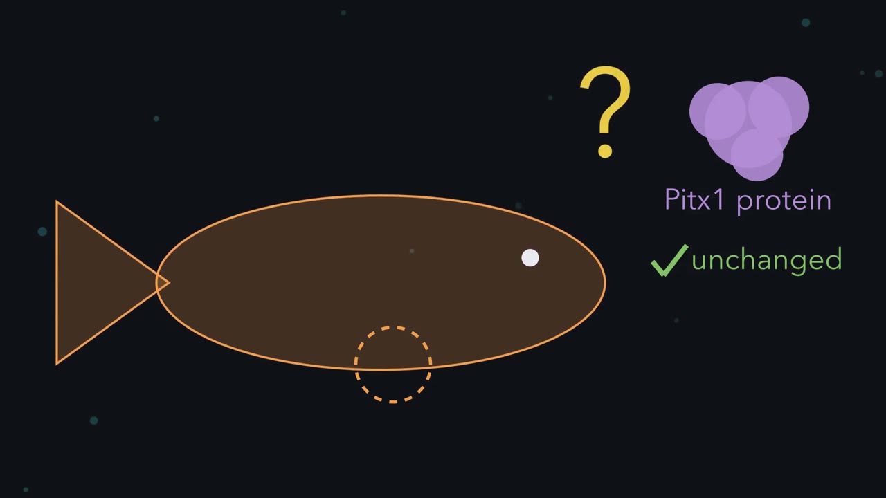
Some lake sticklebacks have almost no pelvis, yet their Pitx1 protein is unchanged. If the protein did not break, what did?
Pause when the reading reaches a film, and offer to play it. Saved on this device.
Watch the film now, or keep listening
Builds on:Reading Phylogenetic TreesDrift, Gene Flow and Competing ExplanationsHow to Test an Evolutionary ExplanationWhy these?
 ▶ Watch Same Protein, Different Switch · 3:00
Read the transcript of “Same Protein, Different Switch”
Film transcript: Same Protein, Different Switch
This lake stickleback has almost no pelvis, yet its Pitx1 protein is unchanged. So what broke, if the protein did not? The likely answer is a kind of switch. Marine sticklebacks carry a pelvis: a bony girdle with a pair of long spines. It is the fish's version of your hips and legs. In Paxton Lake, British Columbia, the bottom-feeding fish have a greatly reduced pelvis. Most have no pelvic bones at all. The gene most strongly tied to that difference is Pitx1. Its protein is a transcription factor. It binds DNA and switches other genes on or off. In the lake fish examined, that protein was unchanged. Pitx1 is not only a pelvis gene. It is also used in the jaw.
Mice with no working Pitx1 die, and they have malformed hind limbs among other defects. Suppose the lake fish had broken the protein itself. Where would you expect the damage to show? Everywhere the gene works. A protein with no function would fail in the jaw and the pelvis at once. The mouse shows how costly that can be. The lake fish do not look like that.
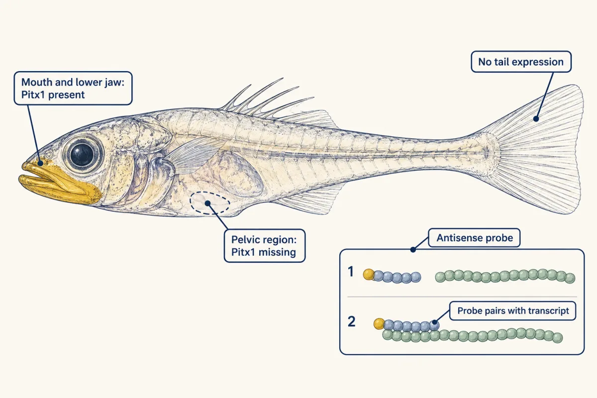
The way out is an enhancer. An enhancer is a short stretch of noncoding DNA that switches a gene on in particular tissues. One gene can have several. One can serve the jaw, another the pelvis. Their jobs can overlap. So where is Pitx1 RNA in a lake fish larva? A labelled probe pairs with the RNA and leaves a gold mark wherever it is. In Paxton larvae, gold sits in the mouth and lower jaw. Where the pelvis forms, there is none. So suppose the jaw switch still works, but the pelvic switch does not. Then that body part shrinks, while the gene keeps its other jobs. Compare the two fish once more. Both make the same Pitx1 protein. Both use it in the jaw.
Of the tissues compared, only the pelvic use differs. The pelvic use changed, and the jaw use was spared. Other tissues were not all checked. This is the best-studied route to a reduced pelvis in sticklebacks. It is not the only one. A missing gold mark also leaves a question open. Is the break in this gene's own DNA, or in another gene?
From the film
- In the pelvic-reduced Paxton Lake sticklebacks examined, the Pitx1 protein is unchanged.
- An enhancer is a stretch of noncoding DNA that switches a gene on in particular tissues, and one gene can have several.
- Paxton larvae still have Pitx1 RNA in the mouth and lower jaw, but none where the pelvis forms.
- Losing the protein outright would remove Pitx1 wherever it works; losing pelvic enhancer activity can shrink one body part and spare the gene's other jobs.
The first film set the puzzle: Paxton Lake (British Columbia) benthic sticklebacks, most with no pelvic bones at all, make an unchanged Pitx1 protein. How was a lost switch proved, and where does the proof stop?
The one idea. A gene’s protein and its pattern of use can change separately. Pitx1’s expression is set by several enhancers (stretches of noncoding DNA that switch a gene on in particular tissues). In Paxton fish, a deletion in the pelvic enhancer PelA, with changes in a second enhancer, PelB, goes with the loss of Pitx1 from the developing pelvis, while the protein and its jaw expression are kept. Losing the protein outright would remove Pitx1 wherever it works; losing pelvic enhancer activity can shrink one body part and spare the gene’s other jobs.
Scope: “Switch” is shorthand: several enhancers can serve one tissue, and one can act in more than one. The case is Pitx1 in threespine sticklebacks: the best-studied route, found in many pelvic-reduced populations but not all, and explaining much of the trait, not all. It is not a claim that form mostly evolves this way.
The puzzle: a lost pelvis with a working gene
The threespine stickleback, Gasterosteus aculeatus, lives in the sea along northern coasts and in many lakes colonised after the last ice sheets retreated. Its pelvis is homologous to the tetrapod hip girdle and hind limb: inherited, modified, from a common ancestor (see reading phylogenetic trees).
The first film showed why a broken protein is a poor route: Pitx1, a transcription factor, also works in the jaw, and mice with no working Pitx1 have malformed hind limbs among other defects and die at or shortly after birth. Enhancers are taught in eukaryotic gene regulation; the question here is how to show evolution used one.
Mapping: one major region, four minor
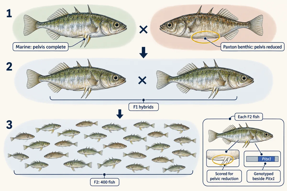
Shapiro, Kingsley and colleagues reported in 2004 that pelvic reduction in their stickleback crosses mapped to one major and four minor chromosome regions, the major one near Pitx1. The method is linkage mapping: cross the two forms to make F1 hybrids, breed them together to make an F2 generation in which the parents’ chromosomes are shuffled, and find regions whose genotype travels with the trait.
Worked problem, part 1. These data are invented. A marine fish (pelvis complete) is crossed with a Paxton benthic fish (pelvis reduced). The F1 fish are intercrossed, giving 400 F2 fish. Each is scored for pelvic reduction and genotyped at a marker beside Pitx1: M is the marine allele, P the Paxton allele. All fish are raised together and scored on the same scale. Of 100 MM fish, 2 are reduced; of 200 MP fish, 6; of 100 PP fish, 32.
Predict. Does one Paxton copy do much? (Dominant means one copy shows the effect; recessive means it takes two.) And if you knew only a fish’s marker genotype, how well could you guess whether its pelvis is reduced?
Check your prediction
- Frequency of reduction in each genotype. MM: 2 of 100, or 2%. MP: 6 of 200, or 3%. PP: 32 of 100, or 32%.
- Read dominance. One Paxton copy (MP) barely changes the frequency; two raise it from 2% to 32%. The region acts close to recessively.
- How well does the marker predict reduction? Of the 40 reduced fish, 32 (80%) are PP. But 68 of the 100 PP fish are not reduced, so other loci or the environment matter too. And 8 reduced fish carry a marine allele; those other factors, or a crossover between the marker and the causal change, could explain them. (How much variation the region explains needs a statistical model, not a head count.)
In the real 2004 cross the link was tighter: all 70 F2 fish with no pelvic spines on either side carried two Paxton alleles at Pitx1.
A mapped region tells you where to look, not what changed: it can hold many genes. No coding mutation changed the encoded Pitx1 protein sequence in the pelvic-reduced fish they examined. In situ hybridization, shown in the first film, gave the pattern in the figure (the first published pictures used the wrong probe; see the anecdote “The probe that was the wrong way round”).
Two possibilities remain. The Paxton copy’s own pelvic control could be broken: a cis change, travelling with that copy of the DNA, like a broken switch wired to one lamp. Or another gene that supplies Pitx1’s regulators could be broken: a trans change, acting through a diffusible product on every Pitx1 copy in the cell, like a power cut that dims every lamp in the house. Mapping cannot tell these apart, since the regulator’s gene could sit in the same region. One kind of fish can.
Cis or trans: the F1 test
An F1 hybrid cell holds one marine and one Paxton allele in one nucleus, bathed by the same regulators. Measure how much Pitx1 RNA comes from each allele (allele-specific expression), tissue by tissue.
Predict. Suppose the Paxton fish’s problem were trans: a shortage of some regulator in the pelvis. In an F1 pelvic bud, what share of Pitx1 RNA would come from each allele?
Check your prediction
About half each. The F1 cell’s regulators act on both alleles alike, so a trans change cannot make one allele lower than the other. Only a difference in one allele’s own DNA can, so an unequal share in one tissue only points to a tissue-specific cis change.
| If the Paxton change is… | It sits… | F1 pelvis, marine : Paxton RNA | F1 jaw, marine : Paxton RNA |
|---|---|---|---|
| cis, pelvis-specific | on the Paxton copy of the DNA (linked to Pitx1) | unequal, Paxton low | about 50 : 50 |
| trans | in a shared regulator | about 50 : 50 | about 50 : 50 |
Worked problem, part 2. Same invented cross. In F1 pelvic buds, 90% of Pitx1 transcripts come from the marine allele and 10% from the Paxton allele. In jaw tissue the split is 50:50.
- Cis or trans? The alleles share one trans environment, so a 90:10 ratio means the Paxton allele itself is less active: a cis change.
- Tissue-specific? The 50:50 jaw ratio shows no cis defect there; of the two tissues tested, the defect is in the pelvis.
- What has improved since part 1? Unlike mapping, the result places the difference on the Paxton copy of Pitx1. It could lie in an enhancer, the promoter (where transcription starts) or the RNA itself; the reporter tests ask which sequence.
In this sketch: invented data from the lesson, one nucleus shared by both alleles, shares of transcripts in relative units, no other source of difference between the alleles. The lesson predicts only that a cis change gives unequal shares in the pelvic bud, so no size is predicted.
Both alleles share one nucleus, so a difference built into one allele’s DNA lies in cis (that allele alone is down), while a change in a regulator would affect both alleles alike (trans). This is a sketch of the logic, not a measurement.
Predict. In part 1, one Paxton copy barely raised the chance of reduction. In the F1 pelvis, a Paxton copy makes 10 RNA molecules for every 90 from a marine copy. Roughly how much pelvic Pitx1 RNA does an MP fish make, compared with a marine MM fish?
Check your prediction
Assume the change is purely cis, so each allele’s output does not depend on its partner. A Paxton copy then makes 10/90, about 0.11, of a marine copy’s output. An MP fish, like the F1, makes (1 + 0.11) / 2, about 56% of the marine level; a PP fish about 11%. Over half the marine dose would fit the near-recessive pattern if one marine copy is nearly enough: an inference from invented numbers, not something they show.
Chan and colleagues ran the real version in 2010: in F1 hybrids, the Paxton benthic allele was expressed less in pelvic tissue but not in head tissue.
The hybrid test puts the fault in the Paxton copy’s own DNA, but not which stretch, or whether it matters for bone. The second film puts one candidate on trial: an upstream enhancer first called Pel, now PelA.
Read the transcript of “Putting the Switch on Trial”
Film transcript: Putting the Switch on Trial
The hybrid test pinned the fault on the lake copy's own DNA. Pitx1 RNA is missing where the pelvis forms. So which stretch of DNA is the broken pelvic switch? And how would you prove it? The colours are the same: blue for marine, orange for Paxton, gold for Pitx1 RNA. Here is the suspect, a stretch of noncoding DNA just upstream of the gene. It was first called Pel. A second switch was found later, so this one became PelA. Three tests put it on trial. First test: a reporter. Cut out the suspect DNA. Join it to a short start signal and a gene for a visible marker. Put that into fish embryos, and watch where the marker turns on.
The marine version of PelA drove the marker in the developing pelvis. The Paxton version carries a deletion. It gave pelvic expression in none of the fish tested, as the counter shows. So the Paxton switch drives no pelvic glow. Does that prove the fish's own pelvis needs PelA?
Not yet. A reporter shows what a sequence can drive, inside a construct. It does not show that the body depends on it. For that, you change the fish itself. Each test answers its own question. A reporter asks what a sequence can drive. Adding the switch back asks whether that can restore form. Removing it from the animal asks whether form depends on it. Rescue comes closer.
Marine PelA, driving Pitx1, was put into fertilized eggs from another pelvic-reduced lake. Those fish normally grow no pelvic spine. Compared with uninjected siblings, pelvic spine growth was variably enhanced. Now the wild fish. Many pelvic-reduced populations carry deletions at PelA. The deletions differ in where they start and stop. So the same switch was lost more than once. In one survey, deletions turned up in most pelvic-reduced populations, as the tally shows. None of the pelvis-complete populations had them. Common, then, but not universal. Most of the others showed signs of another route. Different deletions, same switch. Pelvic reduction evolved again and again by losing one enhancer, in many lakes but not all. The deletions hit the switch, not the Pitx1 protein.
From the film
- In a reporter test, marine PelA drove expression in the developing pelvis, and the Paxton version with a deletion drove none in 8 fish.
- A reporter shows what a sequence can drive in a construct, not that the body depends on it.
- Marine PelA driving Pitx1, added to eggs from another pelvic-reduced lake, variably enhanced pelvic spines.
- Deletions with different endpoints remove PelA in many pelvic-reduced populations, so the same switch was lost more than once.
- Not every pelvic-reduced population carries a PelA deletion.
Enhancers on trial: a second switch, and why the first keeps breaking
The second film’s tests came from Chan’s 2010 study. It left out a second enhancer, and why PelA is lost so often.
In 2018 Thompson, Capellini, Kingsley and colleagues found a second hind-limb enhancer, PelB, downstream of Pitx1. Deleting it in mice trimmed hind-limb Pitx1 expression rather than abolishing it, so PelB supplies part of the limb’s Pitx1 expression and other enhancers the rest; it is not an on-off switch. Paxton PelB also drove weaker pelvic reporter expression, so Paxton fish carry changes in both enhancers (Going deeper).
Predict. The second film found the same switch lost in lake after lake. Why that one? What evidence would point to selection, what to mutation, and could both be true?
Check your prediction
Both can contribute: mutation may explain why Pel deletions arise again and again, selection why they spread (with the limits under “The agent of selection” below).
- Selection. Chan’s 2010 study found signs of positive selection (a variant spreading because it helps its carriers) around Pel in pelvic-reduced populations: an excess of derived alleles (newer variants, not the ancestral ones) and reduced genetic diversity not seen in pelvis-complete lake populations.
- Mutation. Xie and colleagues (2019) found that the Pel sequence breaks easily in a yeast chromosome assay, a clue to why deletions recur. That is a yeast result, not a fish mutation rate (anecdote: “A fragile stretch of DNA, measured in yeast”).
What stays open
Each method asks its own question: mapping localises, in situ hybridization shows where transcript is, the F1 test detects a cis difference, a reporter shows what a sequence can drive, rescue asks whether adding it back restores form, and a native-locus edit tests whether form depends on the sequence (done for mouse PelB; in sticklebacks, Xie and colleagues reported that CRISPR breaks at Pel could give local deletions and pelvic loss in crosses). Together they support one claim, a lost pelvic switch rather than a lost protein. The open questions ask how far it reaches:
- PelA versus PelB. Swapping the natural Paxton PelA and PelB variants into fish, separately and together, remains to be done; reporters cannot split their contributions (see the Design problem).
- Other loci. The four minor regions, and background, are not explained.
- The agent of selection. The signals support positive selection on Pel-region variants in pelvic-reduced populations, consistent with a smaller pelvis being favoured. Because a sweep marks a linked region, they do not show which variant or trait was the direct target, or which ecological pressure acted in particular lakes; this lesson names no cause.
- Generality. Pel deletions are common but not universal among pelvic-reduced populations, and a 2023 study of two small Norwegian lakes found no obvious association (details under Going deeper). For gene-level change across lineages and reused developmental toolkits, see molecular evolution, how signaling evolved and the Hox genes.
Design problem
Paxton fish carry changes in both PelA (upstream) and PelB (downstream). Design an experiment to measure each one’s contribution to pelvic size.
Saved in this browser only.
One way to do it
Work at the native locus, not with reporters. In marine (pelvis-complete) fish, use genome editing to install the Paxton-type change at PelA alone, PelB alone, and both. Make several independent lines of each (so one off-target edit cannot explain the result), with unedited siblings as controls.
Raise all genotypes together and, blind to genotype, measure spine length and girdle length on each side, with the left-right summary, sample size and statistical model chosen in advance. Measure pelvic Pitx1 RNA in the same lines to link expression to form.
Measure each edit as its loss relative to controls. If effects add on that length scale, the double edit’s loss is about the sum of the single losses (double = PelA alone + PelB alone − control); if they interact, larger or smaller (each alone may do little if the other compensates). Ordered categories cannot be added like this; for them, fit an ordinal model chosen in advance and define interaction on its scale. A fish with no pelvis left cannot lose more, and that floor can mimic an interaction. One marine background may not represent others, so repeat in a second marine population and keep the result to the backgrounds tested.
Going deeper
What the F1 test does and does not separate
The F1 logic assumes regulators act alike on both copies; one that reads one allele’s sequence differently is itself evidence of a cis difference. To ask whether trans factors add to it, compare the parents (invented numbers, every ratio Paxton over marine, like tissues compared). The F1 allele ratio, 0.11, estimates the cis effect. If cis and trans effects multiply, parents’ ratio = cis × trans, so a parents’ ratio of 0.11 gives trans = 0.11 / 0.11 = 1.0 (none), and 0.05 gives 0.05 / 0.11, about 0.45.
PelB in mice and in sticklebacks
Because PelA is not conserved in mammals, Thompson and colleagues tested PelB in mice, deleting 9,425 base pairs containing it at its native mouse locus using CRISPR/Cas9 (methods in CRISPR and functional genomics). Homozygous mice were viable and fertile, and several hind-limb bones were shorter. Their hind-limb Pitx1 RNA, measured at embryonic day 12.5, kept about 85% of the control level, a 15% drop. Patellae were absent only in mice carrying the PelB deletion on one chromosome and a Pitx1 null allele on the other, a sensitized background. PelB’s reporter is active mainly in the posterior part of the developing foot (the posterior autopod), so its share may be regional rather than an even trim everywhere.
In sticklebacks, the Paxton benthic PelB carried a 125-base-pair deletion and a 341-base-pair insertion, and its reporter drove weaker, more restricted pelvic expression than the marine one while keeping strong jaw expression.
The 2023 Norwegian lakes
A 2023 study of sticklebacks from two small subarctic Norwegian lakes and a nearby marine site found no obvious association between PelA variants and pelvic status in the lake where spineless fish occur, and found no variation in PelB.
Your notes
Select any text in the lesson to highlight it or add a note to it. Your highlights and notes are saved in this browser and collected in your notebook.
This browser is not letting the site save anything (a private window or strict privacy settings), so highlights and notes will be lost when you leave the page.
How we measure it
Linkage mapping in experimental crosses
Two populations that differ in a trait are crossed, the hybrids are bred together, and hundreds of offspring are scored for the trait and genotyped at markers across the genome. A region whose genotype co-segregates with the trait is linked to a cause, but it can hold many genes.
Whole-mount in situ hybridization
A labelled antisense RNA probe that pairs with a gene's transcript is applied to a whole embryo or larva, showing where the transcript is present. A sense probe is the negative control; mixing the two up was the error the 2006 stickleback correction fixed.
Allele-specific expression in F1 hybrids
In a hybrid, both parental alleles share one nucleus and one set of transcription factors. Measuring how much RNA comes from each allele, tissue by tissue, tells you whether an expression difference is built into the allele's own DNA (cis). Comparing that ratio with the difference between the parent fish shows whether the cell's other factors (trans) add to it.
Reporter assays and engineered deletions
A candidate enhancer is placed beside a minimal promoter and a reporter such as GFP or lacZ to ask where it can drive expression. Deleting the enhancer at its native locus with CRISPR/Cas9 asks a different question: does the organism's form depend on it?
Practice questions on this lesson (3) → MCAT-style, original, each with an explanation
Where this lesson fits
Before thisReview first
- Reading Phylogenetic TreesThis course · Lesson 2
The lesson treats the pelvis as a homologous structure, and you need tree reading to follow that inherited-and-modified idea.
- Drift, Gene Flow and Competing ExplanationsThis course · Lesson 4
Judging whether repeated pelvic loss came from selection or chance needs the competing explanations for genetic change taught there.
- How to Test an Evolutionary ExplanationThis course · Lesson 1
Deciding what the mapping, F1 and reporter evidence does and does not show uses the evidence-testing habits taught there.
After thisStudy next
- Speciation: How Barriers to Gene Flow EvolveThis course · Lesson 7
It takes up how genetic differences like these between populations can build barriers to gene flow.
Elsewhere on the siteFilms, simulations and method films in other courses
- Gene targeting: knockout and knock-in miceMethod film
It shows how to delete a chosen DNA stretch in a mouse to test what it does, like the enhancer deletion tests.
- Enhancer logic board: combinations and open chromatinSimulation · Genetics and Molecular Biology
Its enhancer logic board shows how separate enhancers switch one gene on in different tissues.
- Access, Then AssemblyFilm · Genetics and Molecular Biology
It shows how deleting pieces of regulatory DNA reveals which enhancer controls a gene's pattern of use.
Suggested by an AI model from the lessons themselves, then checked. How we use AI
Finish line
Test yourself
8 questions
Practice and MCAT-style, with explained answers.
Next · Lesson 7
Speciation: How Barriers to Gene Flow Evolve
Two monkeyflowers can be crossed by hand, yet they almost never exchange genes in the wild. What barriers hold them apart, and how did adapting to different places and pollinators build those barriers?
Continue →