Biology · Unit 2: Cells
The Cell Cycle and Mitosis
Rewritten on : new text, flashcards and film. Your notes and highlights stay, but the text around them may have moved.
When one cell becomes two, what keeps either new cell from receiving only half the chromosomes?
Pause when the reading reaches a film, and offer to play it. Saved on this device.
Watch the film now, or keep listening
Builds on:The Cell Theory and Its EvidenceOrganelles as a SystemCrossing the MembraneWhy these?
Where this lesson sits
- Virginia SOL BIO.3c
- Practices BIO.1c, BIO.1d, BIO.1e, BIO.1f
Essential question: Why do organisms make more cells instead of just bigger ones, and how does a cell divide so each new cell gets a full copy?
-
Life Science (grade 7) LS.2 d
Review it at grade 7 level
In Life Science you learned that all living things are made of cells and that new cells come from cells that already exist. You saw that cell division makes new cells, which lets living things grow and reproduce. Under a microscope one cell can become two. Grade 7 named interphase, mitosis and the four stages. Biology goes further and asks what a cell does to get ready, or how it shares its chromosomes so that both new cells get a full set. It adds the phases G1, S, G2 and M, cytokinesis, and the reasons cells divide and stay small.
-
Biology BIO.3c You are here
This lesson
-
Biology II Biology II: Advanced Survey of Biology Topics, BIOII.2 e; Genetics, GEN.4 a and c
Preview where it goes next
Virginia's Biology II courses, including Advanced Survey of Biology Topics and Genetics, come back to cell division through signaling that regulates growth and division, and through mitosis compared with meiosis. The standards do not name checkpoints, which this lesson left out: points in the cycle where a cell stops unless conditions are right, for example until its DNA is fully copied or every chromosome is attached to the spindle. Proteins called cyclins rise and fall through the cycle and switch on partner enzymes that push the cell to the next stage. When genes for these controls are damaged, cells can divide when they should not, which is part of how cancers develop. You also compare mitosis with meiosis and, in Genetics, trace what happens when chromosomes are shared out unevenly.
-
College
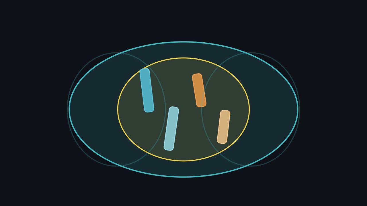 ▶ Watch How One Set Becomes Two · 3:15
Read the transcript of “How One Set Becomes Two”
Film transcript: How One Set Becomes Two
How can one cell become two without leaving either new cell with half a chromosome set? Watch one blue chromosome through the whole answer. Its partners will stay visible, but blue is our guide. A new cell enters G1, where it grows and does its everyday work. This busy stretch is interphase, not a rest. Next comes S phase, when the cell copies its DNA. G2 prepares the cell to divide. During S phase, every chromosome is copied. The blue rod becomes two matching blue sister chromatids joined at one centromere. Its partners copy too. The DNA amount doubles, but each joined pair still counts as one copied chromosome. Soon the copied chromosomes will line up across the cell's middle.
Where must the two blue sister chromatids go so both new nuclei receive a complete set? Hold your answer. The blue copies must move to opposite poles. The same is true for every copied chromosome. Copy each chromosome first; separate the copies so each nucleus gets one. That rule preserves the chromosome set through a typical mitotic division. M phase begins with mitosis, division of the nucleus. In prophase, the copied chromosomes coil into thick visible shapes. Green spindle fibers form. Near the end, the yellow nuclear envelope breaks down, opening access to the chromosomes. In metaphase, spindle fibers line the copied chromosomes across the middle.
Each sister chromatid faces a different pole. The row is not the goal. It is the setup that lets matching copies move in opposite directions. In anaphase, the sister chromatids separate at their centromeres. Spindle fibers pull one copy of every chromosome toward each pole.
Follow blue. One blue copy moves left, while its match moves right. Each separated chromatid now counts as a chromosome. In telophase, a complete set reaches each end. Yellow nuclear envelopes form, and the chromosomes loosen.
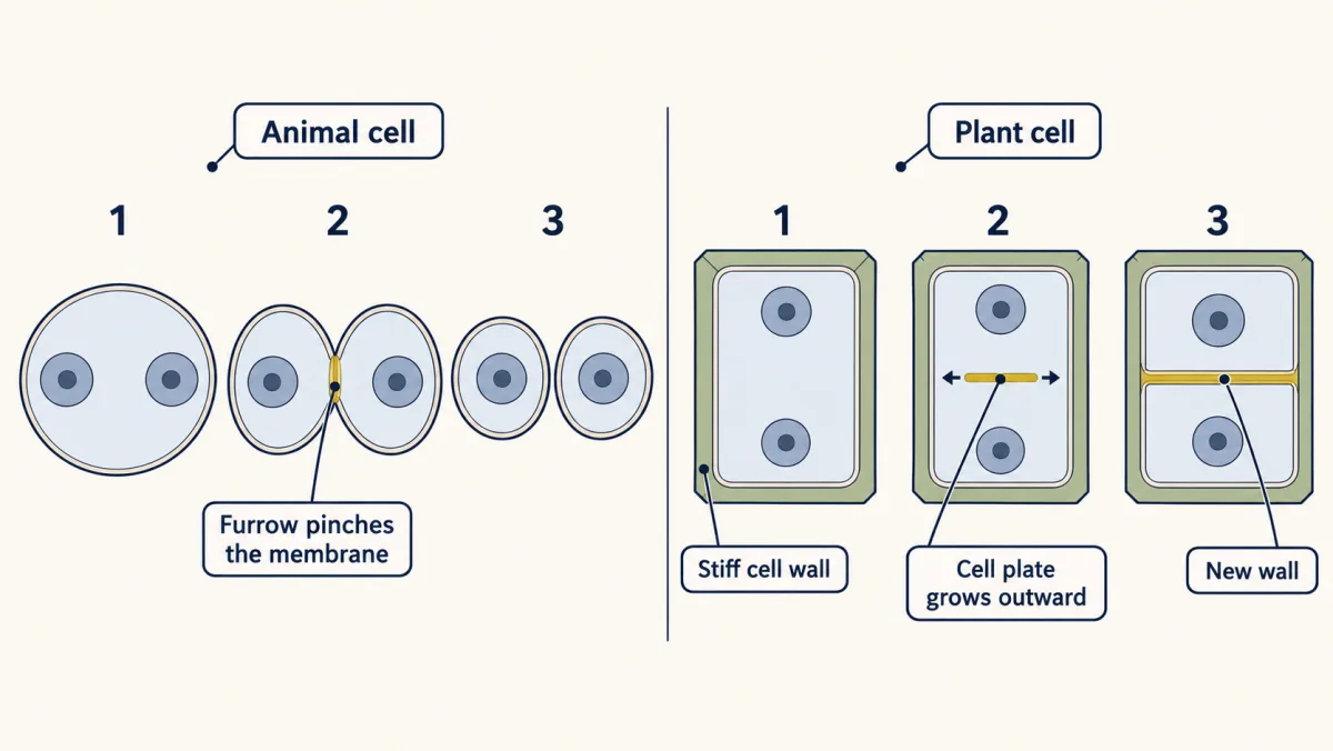
Mitosis has made two nuclei. Cytokinesis often begins in late anaphase or telophase. It divides the cytoplasm. An animal cell pinches inward; a plant cell builds a plate outward into a new wall. Notice what did not happen. The cell did not split its original chromosomes in half. It copied every chromosome before separation. That is why each finished nucleus can receive a complete set, not a random half.
From the film
- Copy each chromosome first; separate the copies so each nucleus gets one.
- Interphase includes growth, DNA copying and preparation; it is not a rest.
- After S phase, two joined sister chromatids still count as one copied chromosome.
- Mitosis divides the nucleus; cytokinesis divides the cytoplasm, often starting in late anaphase or telophase.
The first film followed one chromosome’s copies into two nuclei, but it never counted them. The count is where most students slip. Check three common ideas first, then count with the model below.
What do you think? Decide before you read.
-
Scientists disagree: Mitosis divides the nucleus. A second step, cytokinesis, divides the rest of the cell. Biologists know these are separate because they can happen apart. In the early embryo of a fruit fly, for example, the nucleus divides many times before membranes split the embryo into separate cells.
What scientists think
Scientists disagree: Mitosis divides the nucleus. A second step, cytokinesis, divides the rest of the cell. Biologists know these are separate because they can happen apart. In the early embryo of a fruit fly, for example, the nucleus divides many times before membranes split the embryo into separate cells.
-
Scientists disagree: Some single-celled organisms do reproduce by dividing. In a plant or an animal, though, most divisions build and repair the body. Studies of healing skin in mice found many cells copying their DNA near a wound, and cutting down division with a drug made the wound cover over less well.
What scientists think
Scientists disagree: Some single-celled organisms do reproduce by dividing. In a plant or an animal, though, most divisions build and repair the body. Studies of healing skin in mice found many cells copying their DNA near a wound, and cutting down division with a drug made the wound cover over less well.
-
Scientists disagree: A cell between divisions looks quiet under a light microscope, because its chromosomes are spread out and hard to see. Tests that mark new DNA show that it is busy. It grows, makes proteins and copies every chromosome before it divides again.
What scientists think
Scientists disagree: A cell between divisions looks quiet under a light microscope, because its chromosomes are spread out and hard to see. Tests that mark new DNA show that it is busy. It grows, makes proteins and copies every chromosome before it divides again.
Explore: step a cell through division
The stepper carries a model cell with two color-coded chromosome pairs through interphase and mitosis. Its second panel turns root-tip cell counts into phase-time estimates.
1. Step one cell through division
- Stage
- G1
- Chromosomes
- 4
- Copies in each chromosome
- 1
- Cells
- 1
orange = the long pair blue = the short pair (2 chromosomes in each pair) dark dot = centromere thin lines = spindle fibers
In this sketch: one cell with two pairs of chromosomes, 4 in all. Shapes are schematic, not to scale, and each stage is drawn as a still picture, not a timing.
2. Count a field of onion root-tip cells
What to look for: round nucleus = interphase; two small X shapes = prophase; one bar across the middle = metaphase; two bars = anaphase; two dots = telophase.
| Phase | Counted | From lesson counts | Minutes | Minutes from lesson counts |
|---|
In this sketch the field is drawn by chance from the lesson’s counts of 36 onion root-tip cells (interphase 20, prophase 10, metaphase 3, anaphase 2, telophase 1; The Biology Project, University of Arizona). Those photographs may have been picked to show every stage, so a random field could differ. Cell shapes are schematic. The activity assumes a cycle of 24 hours. A 2018 study of published estimates for roots grown from onion bulbs reported about 13.5 to 23 hours, so 24 hours is a round assumption, not a measurement. A count is a snapshot of many cells at one moment; it does not follow one cell through time.
Rule to keep: a cell copies its DNA in S phase, then mitosis shares the chromosomes so each new cell gets a full set. If cells are caught at random moments, a longer phase holds more cells, so the fraction of cells in a phase times the total cycle time estimates that phase’s time.
Try this.
- Record the chromosome count when the model begins and in each new cell at the end.
- Stop before and after S phase. Record what doubled and what did not.
- Stop at metaphase. Follow one color through the next step and into a new nucleus.
- In the counting panel, compare the largest cell count with the longest time estimate.
Explain: the cell cycle
- cell cycle
- interphase
- mitosis
- cytokinesis
- chromosome
- sister chromatids
- centromere
- spindle fibers
- prophase
- metaphase
- anaphase
- telophase
The cell cycle is the series of stages a cell goes through from the time it forms until it divides into two cells. In plants, animals, fungi and other eukaryotes, a typical dividing cell passes through four main phases: G1, S, G2 and M. Interphase is the part between divisions. It has three parts. In G1 the cell grows, makes proteins and does its everyday work. In S it copies its DNA (“S” stands for synthesis, which means making). In G2 it makes the proteins that will move the chromosomes and gets ready to divide.
DNA is stored in chromosomes, long strands of DNA wrapped around proteins. A typical human body cell with a nucleus has 46 chromosomes. Red blood cells lose their nucleus as they mature.
After S phase, each chromosome has been copied. Each copy is a sister chromatid. The two stay joined, so they count as one copied chromosome until they separate.
| Moment | Model cell | Typical human body cell with a nucleus |
|---|---|---|
| Before S phase | 4 chromosomes, 4 chromatids | 46 chromosomes, 46 chromatids |
| After S phase | 4 copied chromosomes, 8 sister chromatids | 46 copied chromosomes, 92 sister chromatids |
| In each new nucleus after mitosis | 4 chromosomes, 4 chromatids | 46 chromosomes, 46 chromatids |
Predict. A typical human body cell has finished S phase but has not started mitosis. How many chromosomes and sister chromatids should you count?
Check your prediction
Count 46 copied chromosomes and 92 sister chromatids. Copying doubled the DNA molecules, not the number of joined chromosome units.
Copying first is like copying a recipe book before two groups share it, instead of tearing it in half. The comparison breaks because a cell does not decide anything; proteins copy and share the DNA.
M phase is division itself, in two parts. Mitosis is the division of the nucleus into two nuclei, each with a full set of chromosomes. Cytokinesis is the division of the cytoplasm, which splits one cell into two. The result is two new cells that usually have the same DNA as each other and as the cell they came from.
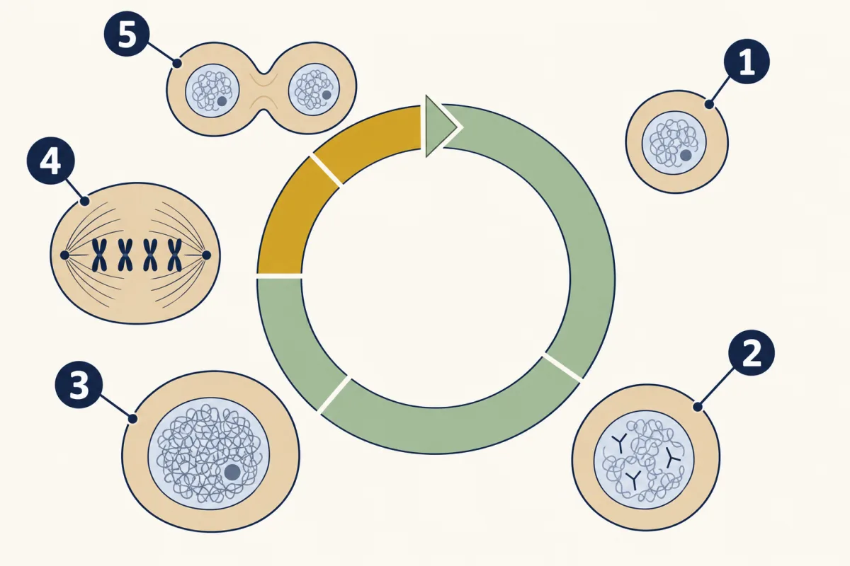
- G1
- S
- G2
- mitosis
- cytokinesis
- G1
- blank
- blank
- blank
- cytokinesis
Word bank: G2, mitosis, S
- blank
- blank
- blank
- blank
- blank
Check your labels
- G1
- S
- G2
- mitosis
- cytokinesis
When the controls on the cycle fail, cells can keep dividing when they should not. That uncontrolled division is a key feature of cancer.
Explain: the stages of mitosis
A stained slide shows only still shapes, so read each shape as evidence. Sister chromatids are held together at the centromere, a narrow region of the chromosome. Spindle fibers are thin protein fibers that attach to chromosomes and move them.
| Stage | Clue in a still cell | What the clue means |
|---|---|---|
| Prophase | Thick, separate chromosomes become visible. | The chromosomes have coiled up. Spindle fibers form, and near the end the nuclear envelope breaks down. |
| Metaphase | Copied chromosomes form one row across the middle. | Spindle fibers have lined them up before separation. |
| Anaphase | Two matching groups sit apart, heading toward opposite ends. | Sister chromatids have separated and are being pulled to opposite ends. Each is now its own chromosome. |
| Telophase | A chromosome set sits at each end as two nuclei appear. | A new nuclear envelope forms around each set, and the chromosomes loosen again. |
The first film showed the animal pinch, the plant plate and when cytokinesis starts. It skipped how each works. In an animal cell, a ring at the middle does the pinching, making a furrow that deepens until the cell splits in two. A plant cell’s stiff wall cannot pinch, so a cell plate grows outward from the middle until it becomes a new wall.
Predict. A stained plant cell has two new nuclei and a plate growing across its middle. Has mitosis finished, has cytokinesis finished, or has only one finished?
Check your prediction
Mitosis has finished because two nuclei have formed. Cytokinesis is still underway because the cell plate has not yet become a complete wall.
Explain: why cells divide and stay small
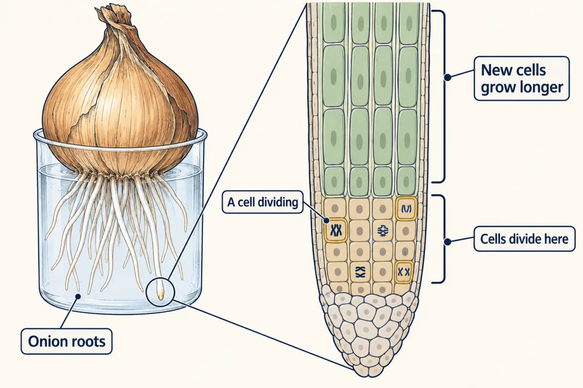
A small cut on your finger usually closes on its own in roughly a week or a little longer. Near an onion root tip, cells divide as the root pushes deeper into the soil. Both depend on adding cells, and cells divide for several reasons.
- Growth. A tree or a teenager grows by adding cells, and many of those new cells then get bigger.
- Repair. Skin cells divide behind a wound to replace those that were lost.
- Replacement. The lining of your gut and the outer layer of your skin are replaced over and over.
- Asexual reproduction. A single-celled amoeba or yeast cell can make a new organism by dividing. Bacteria divide too, by a simpler process covered in How Microbes Multiply.
Why not grow only bigger cells? Oxygen, food molecules and wastes all cross the cell membrane, which you study in Crossing the Membrane. The surface-area-to-volume ratio compares how much outside surface a cell has with how much inside it has.
Predict. A cube’s side length doubles. Does its surface area, its volume, or both grow by the larger factor?
Check your prediction
Volume grows by the larger factor. Surface area becomes four times larger, while volume becomes eight times larger.
Model a cell as a cube:
| Side of cube (units) | Surface area (square units) | Volume (cubic units) | Surface area ÷ volume |
|---|---|---|---|
| 1 | 6 | 1 | 6 |
| 2 | 24 | 8 | 3 |
| 4 | 96 | 64 | 1.5 |
These values come from geometry (area = 6 × side × side, volume = side × side × side), not from measuring cells. Each time the side doubles, volume grows eight times but surface only four times, so a large cell has less membrane for each bit of its inside.
Real cells are not cubes, and long, thin or flat cells keep more surface. The model works best for comparing cells of similar shape. Some cells, such as egg cells and long nerve cells, are very large. Cells also control their own size, so surface area is one limit among several.
The onion root shows both ideas together. Near the tip, cells divide. Just behind that zone, the new cells grow longer, and that lengthening pushes the root through the soil.
Elaborate: count onion root-tip cells
Those dividing cells raise a new question. Between one division and the next, how much time goes to preparing and how much to separating? The second film turns a root-tip snapshot into a clock.
Read the transcript of “A Snapshot Becomes a Clock”
Film transcript: A Snapshot Becomes a Clock
The first film showed how chromosome copies reach matching nuclei. How can a frozen onion root reveal which part of that cycle takes longest? The cells cannot answer, but their patterns can. A stained root tip is a snapshot. Each cell is caught at one moment of its cycle. Some show loose chromosomes inside a yellow nucleus. Others show chromosomes coiling, lining up, separating, or reaching opposite ends. Imagine many runners photographed at random points around a track. A slow section holds more runners because each runner stays there longer.
Cycling cells follow the same counting model. A longer phase should appear in more cells. Two phase lanes now contain different numbers of cells. Suppose every cycling cell moves steadily and the sample is fair. Which lane represents the longer-lasting phase? Hold your answer.
The fuller lane represents the longer phase. More cells were caught there because each one remains there longer. This gives a fraction, not a stopwatch reading. Divide the cells in one phase by all the cells counted, assuming every one is cycling. Now sort one teaching set by visible clues. Intact nuclei go to interphase. Coiled chromosomes go to prophase. A middle row marks metaphase. Separating groups mark anaphase. Two forming nuclei mark telophase. To estimate time, multiply a phase's fraction by the total cycle time. The teaching activity supplies a round total time rather than measuring it. Put that value on screen. The result is a model estimate for this set.
The model can fail if the pictures were chosen to display every stage. It also weakens if cells stopped cycling or changed pace. A random field, a clear sampling rule, and a measured total time would support stronger estimates. One more distinction matters. Mitosis separates chromosomes between nuclei, while cytokinesis separates the cell. Root-tip photographs can show both nearby, but the phase count sorts chromosome patterns. It does not directly time membrane or wall completion. The two processes overlap, so one snapshot can catch both. The count still teaches an important idea.
Cells spend time growing, copying DNA, and preparing before chromosomes visibly separate. That preparation serves the first film's rule. Copy each chromosome first; separate the copies so each nucleus gets one. And the counting rule is just as short. In a fair snapshot, a longer phase holds more cells.
From the film
- In a fair snapshot, a longer phase holds more cells.
- A stained root tip is a snapshot of many cells caught at different moments.
- A phase-time estimate is the fraction of cells in that phase times an assumed or measured total cycle time.
- Hand-picked pictures, cells that stopped cycling, or a changing pace can break the estimate.
Now test the second film’s counting model on a real teaching set.
| Phase | Cells counted | Fraction of all 36 cells |
|---|---|---|
| Interphase | 20 | 20/36 |
| Prophase | 10 | 10/36 |
| Metaphase | 3 | 3/36 |
| Anaphase | 2 | 2/36 |
| Telophase | 1 | 1/36 |
| Total | 36 | 36/36 |
Source: a University of Arizona teaching activity on the onion root-tip cell cycle. The counts are of 36 photographed onion (Allium cepa) root-tip cells on its completed answer page. The page does not say when or at what temperature the roots were grown. The activity assumes one cycle takes 24 hours, a round assumption, not a measurement.
- Which phase holds the most cells? About what fraction of the cells is it?
- One cycle of 24 hours is 1,440 minutes. Estimate the time in metaphase as its fraction times 1,440 minutes. Do the same for the other phases, then check that your five estimates add up to 1,440.
- The activity does not say how these photographs were picked, and they may have been chosen to show every stage. Would a field picked at random probably show more or fewer cells in interphase? How would that change your estimates?
Check item 2 with prophase
Prophase holds 10 of 36 cells, and 10/36 of 1,440 minutes is 400 minutes. This is a model result, not a measured prophase time.
Claim, evidence, reasoning
Make a claim about which part of the cell cycle takes the most time, using the onion counts.
Start with: The counts suggest that blank takes up the most time in the onion root cell cycle.
Start with: Of 36 cells, blank were in interphase and blank were in a stage of mitosis. With a 24-hour cycle, interphase works out to about blank minutes.
Start with: A phase that lasts longer holds more cells at any moment, because blank. This makes sense because in interphase the cell must blank.
Compare with a model answer
Interphase takes up the most time in the cycle. Of the 36 onion root-tip cells, 20 were in interphase and 16 were in a stage of mitosis. With the activity’s 24-hour cycle, 20/36 of 1,440 minutes is 800 minutes, more than half of the cycle. A longer phase holds more cells at any moment, and interphase includes growth and copying all the DNA. The limits are real: these are 36 photographs from a teaching set that does not say how they were picked. They hold far more dividing cells than a random field of a real root tip would, so the times for the stages of mitosis come out much too long. And 24 hours is an assumption, not a measured cycle time.
Saved in this browser only.
Design problem
Does the temperature an onion root grows at change how many of its tip cells are dividing? Plan an investigation. Name your variables, constants, control, sample size and repeated trials, and say how you will stay safe.
Predict. If warmer roots show a larger fraction of cells in mitosis, can that result alone tell you the total cycle length?
Saved in this browser only.
Check your prediction
No. The result would suggest that temperature changes the cycle, but a total cycle length needs a separate measurement.
One way to do it
- Question: Do onion roots grown at a warmer temperature have a larger fraction of tip cells in mitosis?
- Independent variable: the growing temperature, for example a cool room and a warmer incubator, checked with a thermometer.
- Dependent variable: the fraction of tip cells in any stage of mitosis.
- Constants: one onion variety, the same water and days of growth, tips cut to the same length at the same time of day, the same stain and sorting rules.
- Control: roots grown at ordinary room temperature, to compare with.
- Sample size and trials: several roots from each of several onions at each temperature. On each slide, count every cell in a row of fields, not only interesting ones, until you have a few hundred. Have two people count one slide to check agreement.
- Safety: goggles and gloves. The stain and the acid used to soften roots are handled by your teacher, and blades are used with care.
Evaluate: check your thinking
The copy-then-separate rule explains how each new nucleus receives a full chromosome set; cytokinesis then splits the cell. Experiments on mouse skin suggest that new skin covers a wound as cells near the edge move into the gap, while cells farther back divide to replace them (see Going deeper for wound shrinkage and differences among wound types). An onion root grows deeper because cells at the tip divide and the cells behind them stretch. Living things add cells instead of growing giant ones partly because small cells have more membrane for each bit of their inside.
Science practices you used: making predictions and testing them with a model, reading counts and turning them into fractions and estimates, planning a fair test with a large enough sample, and naming the limits of a data set and a model.
Going deeper
How spindle fibers move chromosomes
You can picture spindle fibers as ropes, but they do not reel anything in. They get shorter by losing pieces at their ends.
Where the onion counts come from
The counts come from The Biology Project, University of Arizona, onion root-tip cell cycle activity (page dated 1997, revised August 2004). A 2018 study that gathered published estimates for roots grown from onion bulbs reported values from about 13.5 to 23 hours, so 24 hours is a round assumption, not a measurement.
Wound repair in mouse skin
In mouse skin, the wound also shrinks as its edges are pulled in, and how much each step matters depends on the kind of wound.
Your notes
Select any text in the lesson to highlight it or add a note to it. Your highlights and notes are saved in this browser and collected in your notebook.
This browser is not letting the site save anything (a private window or strict privacy settings), so highlights and notes will be lost when you leave the page.
How we measure it
Counting fixed (preserved) cells under a microscope
Root tips are stained so the chromosomes show up, then each cell is sorted by what its chromosomes look like. A count is a snapshot of many cells at one moment; it does not follow one cell through time.
Estimating phase time from counts
As a simple model, the fraction of counted cells in a phase is multiplied by the total time of one cycle. This assumes every counted cell is dividing at a steady pace. Some cells in a root tip have stopped dividing, so the estimate is only as good as the sample and the total time used.
Labeling cells that are copying DNA
Cells are given a marker (BrdU or EdU) that is built into new DNA, so cells copying DNA in S phase light up. Wound studies in mice used it to map where dividing cells are.
Where this lesson fits
Before thisReview first
- The Cell Theory and Its EvidenceThis course · Lesson 6
You need the idea that all cells come from earlier cells to see why division matters here.
- Organelles as a SystemThis course · Lesson 7
You need to know the nucleus holds the DNA that is copied and split here.
- Crossing the MembraneThis course · Lesson 8
The surface-area-to-volume argument here relies on food and wastes crossing the membrane.
After thisStudy next
- Meiosis and VariationThis course · Lesson 19
It reuses the copy-then-separate rule but separates chromosomes twice, making cells that differ from each other.
- DNA: Structure and CopyingThis course · Lesson 16
It explains how the DNA copying done in S phase actually works.
- One Cell or ManyThis course · Lesson 11
It builds on why cells stay small and how many cells share the jobs of an organism.
Elsewhere on the siteFilms, simulations and method films in other courses
- Heal the scrapeSimulation · Life Science
Heal a scrape to see cell division at work in repairing skin.
- Copy First, Then ShareFilm · Life Science
It shows how a cell copies first and then shares, as in interphase and mitosis.
Suggested by an AI model from the lessons themselves, then checked. How we use AI
Finish line
Test yourself
8 questions
Practice and MCAT-style, with explained answers.
Next · Lesson 11
One Cell or Many
How can one Paramecium do every job of life inside a single cell?
Continue →