Life Science · Unit 1: Cells
How Cells Divide
Rewritten on : new text, flashcards and film. Your notes and highlights stay, but the text around them may have moved.
When you scrape your knee, where does the new skin come from?
Pause when the reading reaches a film, and offer to play it. Saved on this device.
Watch the film now, or keep listening
Builds on:Inside a CellCells Are EverywhereWhy these?
Where this lesson sits
- Virginia SOL LS.2 d
- Practices LS.1 c, LS.1 d, LS.1 e
Essential question: How does my body make new cells to heal a cut without mixing up the instructions?
-
Life Science (grade 7) LS.2 d You are here
This lesson
-
Biology
Preview it at high school level
In Biology you will learn that interphase has three parts. In G1 the cell grows. In S it copies its DNA. In G2 it gets ready to divide. You will learn names for the parts you saw here. The two joined copies of a chromosome are sister chromatids, held together at a spot called the centromere. Spindle fibers are the thin fibers that pull them apart. The splitting of the rest of the cell into two is called cytokinesis. It starts near the end of mitosis and is counted separately from it. You will also learn one reason cells divide instead of growing very large: as a cell grows, its inside grows faster than its outside surface, so materials may not move in and out fast enough. Cells also control their size in other ways.
-
Biology II Biology II: Advanced Survey of Biology Topics, BIOII.2 e; Genetics, GEN.4 a and c
Preview where it goes next
In Virginia's Biology II courses, cell division comes back in two ways. In Advanced Survey of Biology Topics, you study how cells send signals to each other that control when they grow, divide and become specialized. In Genetics, you compare mitosis with meiosis and ask how meiosis makes offspring different from each other. You also study what happens when chromosomes are shared out unevenly, so that a cell ends up with one too many or one too few.
-
College
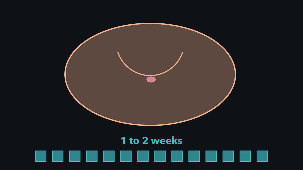 ▶ Watch Copy First, Then Share · 3:21
Read the transcript of “Copy First, Then Share”
Film transcript: Copy First, Then Share
Scrape your knee, and new skin often covers the spot in about 1 to 2 weeks. Where did those new skin cells come from? Zoom in on the edge of the scrape. Skin cells near the gap divide, and each one becomes two. Some cells also crawl into the gap. Every new cell comes from a cell that was already there. Here is the harder question. Most cells carry a full set of instructions. When one cell becomes two, how does each new cell get a complete set? The instructions sit on chromosomes. These are threads inside the nucleus that carry the cell's instructions. Our model cell has four of them: red, blue, green and yellow. A dividing cell goes through the cell cycle.
It has two main parts. Interphase comes first. Then comes mitosis, when the cell divides into two new cells. In interphase the cell is busy, not resting. It grows bigger. And it copies every one of its chromosomes. Each thread gets a matching copy, joined to it. Both stay thin and spread out for now. Look at the counter. Before copying, the cell had four chromosomes. Now each one is a pair of joined copies. Does the cell have four chromosomes, or eight?
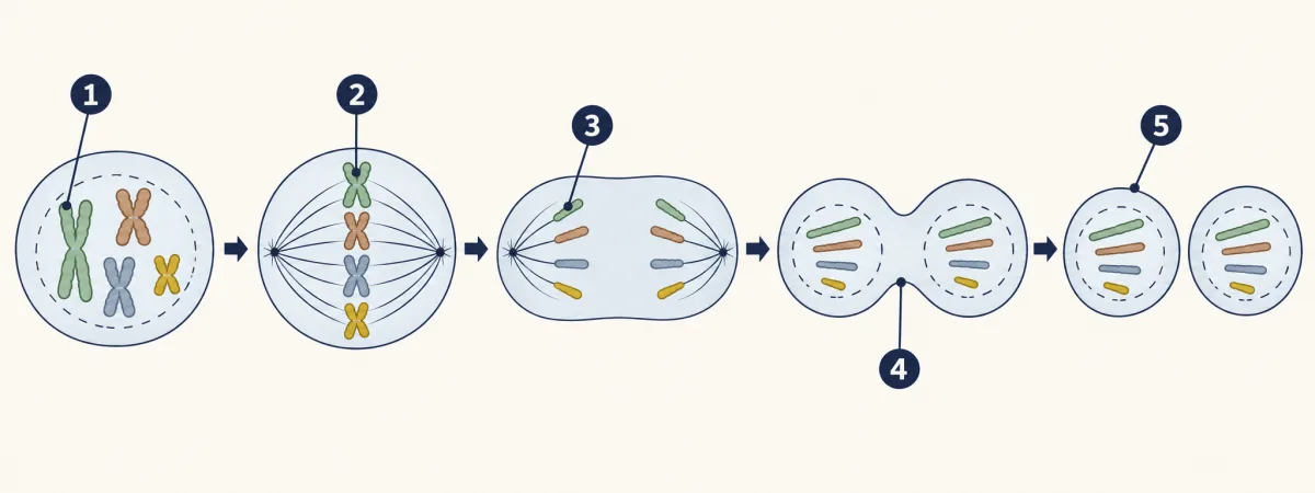
Four. A copied chromosome is still one chromosome. It is made of two identical copies, joined together. So the cell has four chromosomes, and eight copies. Now mitosis begins. Its four stages always come in the same order. In prophase, the chromosomes coil up. Each pair of copies becomes a thick X you can see. In metaphase, the chromosomes line up in a row across the middle of the cell. Red, blue, green and yellow, side by side. In anaphase, the two copies of each chromosome are pulled apart. One copy of each colour moves to each end of the cell. In telophase, two new nuclei form. Then the cell splits into two. Each new cell is called a daughter cell.
Now count each daughter cell. Red, blue, green, yellow. Four chromosomes, one of each. That is the same set as the parent cell. So here is the answer to our question. Copy first, then share. Copying makes two joined copies of every chromosome.
Sharing sends one copy of each to each new cell. Nothing is missing. This is how body cells divide when you grow or when a scrape heals. Mistakes can happen. But normal division gives each new cell a full set.
From the film
- New body cells come from cells that were already there: near a scrape, skin cells divide, and some crawl into the gap.
- In interphase a cell grows and copies every chromosome. A copied chromosome is still one chromosome, made of two joined copies.
- In mitosis the chromosomes coil up (prophase), line up in the middle (metaphase), are pulled apart (anaphase) and end in two new nuclei as the cell splits (telophase).
- Copy first, then share: each daughter cell gets one copy of every chromosome, the same set as the parent cell.
The first film, above, showed where the new skin over a scrape comes from. Its made-up model cell had just 4 chromosomes: threads in the nucleus, the cell’s inner compartment, that carry its instructions (DNA). A typical human body cell that has a nucleus has 46. How does each new cell get a full set, with nothing missing?
What do you think? Decide before you read.
-
Scientists disagree: Before a cell divides, it copies every chromosome. In stained cells under a microscope, the chromosomes look doubled before they separate, and each new cell ends up with the same set as the parent.
What scientists think
Scientists disagree: Before a cell divides, it copies every chromosome. In stained cells under a microscope, the chromosomes look doubled before they separate, and each new cell ends up with the same set as the parent.
-
Scientists disagree: A cell does grow before it divides, often to about twice its starting size. Then it splits into two smaller cells, so most cells stay small. Growing mostly adds more cells, though some cells do get bigger.
What scientists think
Scientists disagree: A cell does grow before it divides, often to about twice its starting size. Then it splits into two smaller cells, so most cells stay small. Growing mostly adds more cells, though some cells do get bigger.
-
Scientists disagree: When scientists photograph a root tip, where cells divide often, most cells are not dividing at that moment. They are in a long stage of growing and copying.
What scientists think
Scientists disagree: When scientists photograph a root tip, where cells divide often, most cells are not dividing at that moment. They are in a long stage of growing and copying.
The one idea: copy first, then share. Before a cell divides, it copies every chromosome. Then mitosis, the dividing step, gives one copy of each chromosome to each new cell. So both get the same full set as the parent: one of every chromosome.
Where it applies: body cells and one-celled living things that divide this way. Mistakes can happen. Sex cells come from a different kind of division (see Going deeper).
Explore: heal a scrape, one cell at a time
In the simulation, the left panel fills a skin gap with dividing cells; you set the cell-cycle time and watch a cell counter. The right panel steps one cell with 4 chromosomes and shows whether each is copied. A switch skips the copying.
1. Fill the gap
- Cells now
- 4
- Gap spaces filled
- 0 of 12
- Time so far
- 0 time units
A strip of skin cells with a gap. Press Start.
2. Follow one cell
In this sketch, the strip starts with 4 cells, every cell divides at the same time, the gap has 12 spaces, and each cell divides twice. A model cell has 4 chromosomes instead of the 46 in most human body cells. Cells are flat shapes, and time units are not to scale. Interphase is drawn as the longer part of the cycle, because the lesson says it is usually the longest; the exact split is not to scale.
Rule to keep: a cell copies its chromosomes first, then each daughter cell gets one copy of each, so it has the same number as the parent cell. A copied chromosome is still one chromosome. In a real scrape some cells also crawl into the gap.
Try this.
- Predict how a shorter cell cycle changes the time to fill the gap. Then test it.
- Before you step the single cell, predict how many chromosomes each new cell will have at the end. Step through and count.
- Turn on “skip the copying.” Predict how many chromosomes each new cell will get now. Then run it and compare.
Explain: the cell cycle
- cell cycle
- interphase
- mitosis
- chromosome
- daughter cell
- prophase
- metaphase
- anaphase
- telophase
All living things grow and reproduce. When you grow, the number of cells in your body goes up; when you heal, new cells replace the ones you lost. Either way, cells that already exist divide. Key words:
- The cell cycle is a cell’s steps from when it forms until it divides.
- Interphase is when the cell grows and copies its chromosomes.
- Mitosis is when the cell divides into two new cells with the same chromosomes.
- Each new cell is a daughter cell; the cell that divided is the parent cell.
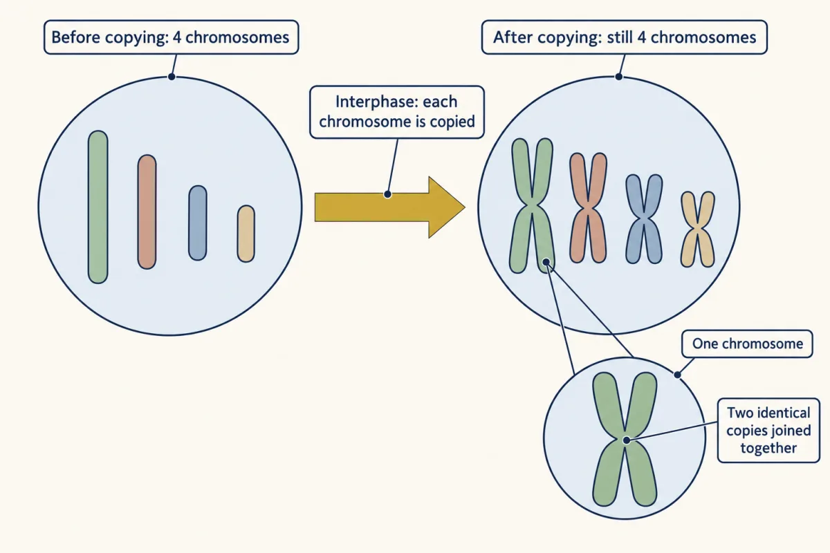
Think of a recipe book. Tear it in half, and each person gets half the recipes. Copy it first, and each person gets the whole book. In a cell the “book” is many separate chromosomes, and each one is copied.
Explain: spotting the four stages of mitosis
To sort cells in a photo, you need what each stage looks like frozen:
| Stage | What you see in a stained cell |
|---|---|
| Prophase | Thick, coiled chromosomes, scattered, not in a row. |
| Metaphase | Chromosomes lined up in a row across the middle. |
| Anaphase | Two matching groups of chromosomes pulled to opposite ends. |
| Telophase | Two new nuclei forming at the ends as the cell splits in two. |
Two things the first film left out: thin fibers in the cell do the pulling in anaphase, and a plant cell, with its stiff wall, cannot pinch in two in telophase; a new wall forms across its middle instead.
Name the numbered stages and the cells at the end.
- prophase
- metaphase
- anaphase
- telophase
- daughter cell
- blank
- metaphase
- blank
- blank
- daughter cell
Word bank: anaphase, prophase, telophase
- blank
- blank
- blank
- blank
- blank
Check your labels
- prophase
- metaphase
- anaphase
- telophase
- daughter cell
Why copy first? Check your answer to Try this 3. Skip the copying, and the red, blue, green and yellow chromosomes must be split: say red and blue to one cell, green and yellow to the other. Each cell misses two whole chromosomes and their instructions; no split gives both cells all four colours. (This is a made-up test, not normal mitosis.) What matters is not the count alone: each new cell needs the right number, one of every kind.
Elaborate: count onion root-tip cells
Reminder: copy first, then share. But how long does copying take, compared with dividing? Cells near an onion root tip divide often, so a root tip can tell you.
Predict. Someone photographed 36 onion root-tip cells and sorted each into a stage. How many of the 36 do you think were in interphase: fewer than 10, 10 to 18, or more than 18? Write down your guess and one reason.
The second film shows how a still photo can tell time.
Read the transcript of “A Photo That Tells Time”
Film transcript: A Photo That Tells Time
Can a still photo tell you how long something takes? A photo freezes one moment, and nothing in it moves. So where would the time be hiding? The first film followed one model cell through the cycle, step by step, in order. A stained root tip cannot show you that. It gives you only a snapshot. Picture a huge school with no shared bell. Each student keeps a different timetable.
Snap a photo at a random moment. Most students are in class. Only a few are in the hallway. Why? Class lasts much longer than the walk between classes. So at any random moment, a student is more likely to be in class. Try it yourself. Stage A lasts twice as long as Stage B. In a random snapshot, which stage will you see more of?
Stage A. You should see about twice as many things in Stage A. A stage that lasts longer tends to hold more of them in a snapshot. Now swap the students for cells. Set an onion on a glass of water, and roots grow down. Near each root tip, cells divide often. A thin root tip is dyed, or stained, so the chromosomes show up. Under a microscope, each cell is caught at one moment of its own cycle. Each cell is sorted into a stage by what its chromosomes look like. Thick, coiled chromosomes, not yet in a row, mean prophase. A row across the middle means metaphase. Let's work one stage together, using real counts. In one set of 36 photographed onion cells, 10 were in prophase, the coiled stage.
Turn that count into a share of all the cells. Follow the steps on the screen. Prophase holds about 28 percent of the cells. The activity says to assume one cycle takes 24 hours. Take each stage's share of the cells as its share of the time. Then prophase lasts about 7 hours. Treat this as a rough guide. There are only 36 photos, and nobody says how they were picked. And the 24 hours is assumed, not measured. Still, it works best with many fairly picked cells, dividing out of step in a steadily growing root. Then a longer stage tends to hold more cells in a snapshot. So a stage's share of the cells is only a rough guide to its time. That was one stage.
Four more are waiting, along with the biggest question in the lesson. Now count the rest yourself.
From the film
- A stained root tip is a snapshot: each cell is caught at one moment of its own cycle.
- When cells divide out of step, each at its own point, a longer stage tends to hold more cells in a snapshot.
- A stage's share of the cells is a rough guide to its share of the time: 10 of 36 cells in prophase suggests about 7 of 24 hours.
- The guide is rough: there are only 36 photos, nobody says how they were picked, and the 24-hour cycle is assumed.
The second film worked out prophase only. Here are all 36 cells.
Each bar counts cells, one per photo. Was your prediction right?
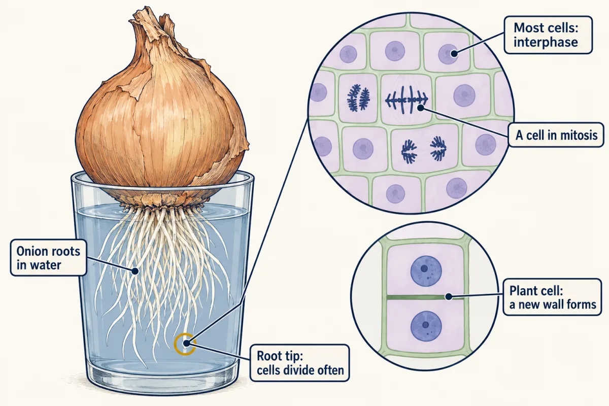
Use the second film’s steps: cells ÷ 36, then × 24 hours.
- Which stage has the most cells? What percent of the 36 cells is that?
- Find the percent for each stage. Do your five percents add up to about 100? (Rounding can make it 99 or 101.)
- If one cycle takes 24 hours, about how many hours does interphase take?
The second film’s rule needs cells that divide out of step. If they all divided together, like a school moving at one bell, one photo could catch nearly all in one stage and say nothing about time.
- The stage with the most cells is blank.
- This stage takes the most time because blank.
- One limit of these counts is blank.
Claim, evidence, reasoning
Make a claim about which part of the cell cycle takes the longest, and what that tells you about how a cell gets ready to divide.
Start with: The counts show that blank takes the longest part of the cell cycle.
Start with: Out of 36 cells, blank were in interphase. That is about blank percent.
Start with: A longer stage tends to hold more cells at one moment, because blank. During this stage the cell blank.
Compare with a model answer
Interphase takes the longest part of the cell cycle. Out of 36 onion root-tip cells, 20 were in interphase, about 56 percent. That is more than all four stages of mitosis together (20 cells against 16). When cells divide out of step, a longer stage tends to hold more cells in a snapshot. In interphase the cell grows and copies every chromosome, so each daughter cell can get a full set. One limit: there are only 36 photos, and nobody says how they were picked. The 24-hour cycle is also an assumption.
Saved in this browser only.
Design problem
Does temperature change how fast onion roots grow longer? Plan an investigation.
Saved in this browser only.
One way to do it
- Question: Do onion roots grow longer each day in a warm place than in a cool place?
- Independent variable: temperature, such as a cool spot near 15 °C and a warm spot near 25 °C, checked with a thermometer.
- Dependent variable: root length in millimetres, measured with a ruler each day for 5 days.
- Constants: the same kind and size of onion, the same cup and amount of water, the same light.
- Control: onions kept at normal room temperature, to compare with.
- Repeated trials: at least 5 onions at each temperature. Find the mean length for each group.
- Safety: wash your hands after touching onions. Do not taste anything. Your teacher handles any blades.
Roots grow when cells near the tip divide and the new cells stretch. Faster growth in the warm group would suggest that temperature speeds this up. Seeing the dividing cells themselves would need stained slides, which your teacher prepares.
Evaluate: check your thinking
Interphase, when a cell grows and copies every chromosome, usually outlasts all of mitosis, so a snapshot of cells dividing out of step tends to catch more in interphase: here, 20 against 16.
Science practices you used: making predictions and testing them with a model, reading counts in a bar graph, finding percents to compare stages, and planning a fair test with repeated trials.
Going deeper
Where the onion numbers come from
The counts come from The Biology Project, University of Arizona: an onion (Allium cepa) root-tip cell cycle activity, completed-photo page dated 1997 and revised August 2004, which sorts 36 photographed cells. Its 24-hour cycle is a round number for the activity, not a measured time, and the page does not say how the photos were picked. The photos also seem to hold more dividing cells than usual, because in a real root tip most cells are in interphase.
When a snapshot can tell time
The second film named the share-of-cells rule’s limits. A stained root tip is frozen, but scientists can film living cells and follow one through every stage.
Two fine points
Strictly, mitosis shares out the chromosomes into two new nuclei, and the rest of the cell splits at the end. Where the recipe book breaks: a cell does not decide to copy, as a person would; its molecules do it, step by step.
One-celled living things and sex cells
A one-celled living thing, such as an amoeba, reproduces by dividing. Its two daughter cells are two new amoebas. A different kind of division, meiosis, makes sex cells with half the parent’s genetic material. You study it in Reproduction and Meiosis.
Your notes
Select any text in the lesson to highlight it or add a note to it. Your highlights and notes are saved in this browser and collected in your notebook.
This browser is not letting the site save anything (a private window or strict privacy settings), so highlights and notes will be lost when you leave the page.
How we measure it
Light microscope and stained root tips
A thin onion root tip is stained so the chromosomes show up, and each cell is sorted into a stage by what its chromosomes look like. The photo is a snapshot of many cells at one moment, not a movie of one cell.
Counting cells in each stage
The number of cells in each stage is divided by the total to give a percent. When cells divide out of step, each at its own point, a longer stage tends to hold more cells in a snapshot. The percent of cells is only a rough guide to the percent of time.
Where this lesson fits
Before thisReview first
- Inside a CellThis course · Lesson 2
You need to know the nucleus holds the chromosomes before following how they are copied and shared.
- Cells Are EverywhereThis course · Lesson 1
You need the idea that living things are made of cells, and that cells make more cells.
After thisStudy next
- Reproduction and MeiosisThis course · Lesson 28
It takes copy-then-share further to a different division that gives sex cells half the chromosomes.
- DNA: The Code of LifeThis course · Lesson 27
It explains what the copied chromosomes carry and why both daughter cells need the same instructions.
- Mutations and VariationThis course · Lesson 31
It shows what happens when a mistake is made while DNA is copied before division.
Elsewhere on the siteFilms, simulations and method films in other courses
- How One Set Becomes TwoFilm · Biology
It shows how copying each chromosome first gives both new nuclei one full set.
- Mitosis stepper and phase counterSimulation · Biology
You can step a cell through mitosis and count the cells in each phase.
- One Cell, Many JobsFilm · Biology
It shows how one cell does every job, while many cells divide up the work.
Suggested by an AI model from the lessons themselves, then checked. How we use AI
Finish line
Test yourself
8 questions
Practice and MCAT-style, with explained answers.
Next · Lesson 5
Diffusion and Osmosis
Why does salted cucumber get wet, while limp lettuce in plain cold water turns crisp?
Continue →