Developmental Neuroscience · Unit 2: Becoming neural
Neurulation and Neural Tube Defects
Rewritten on : new text, flashcards and film. Your notes and highlights stay, but the text around them may have moved.
In a human embryo the neural tube has closed by the end of about the fourth week after fertilization. What happens if it doesn't? Part of the brain or spinal cord stays open, and folic acid taken before conception lowers the risk.
Pause when the reading reaches a film, and offer to play it. Saved on this device.
Watch the film now, or keep listening
Builds on:Neural Induction: The Default ModelThe Organizer: Spemann and MangoldWhy these?
 ▶ Watch Narrow First, Then Fold · 2:15
Read the transcript of “Narrow First, Then Fold”
Film transcript: Narrow First, Then Fold
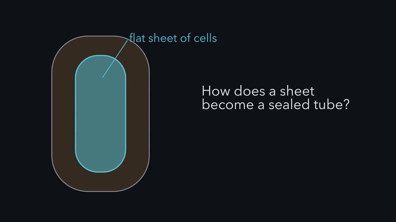
The brain and spinal cord begin as a flat sheet of cells. By about the fourth week, how do they become a sealed tube? This sheet is the neural plate, along the midline of the embryo's back. On either side, surface ectoderm will form the skin. Step one, shaping. The cells grow taller, and the plate narrows from side to side and lengthens from head to tail. Hold on to this step. If the plate's middle stays wide, the folds may have too far to go. Step two, bending. The plate creases at hinge points, and its edges rise as the neural folds. Step three. The folds move toward the midline, meet, and fuse. Step four. The tube separates from the skin, which seals over it.
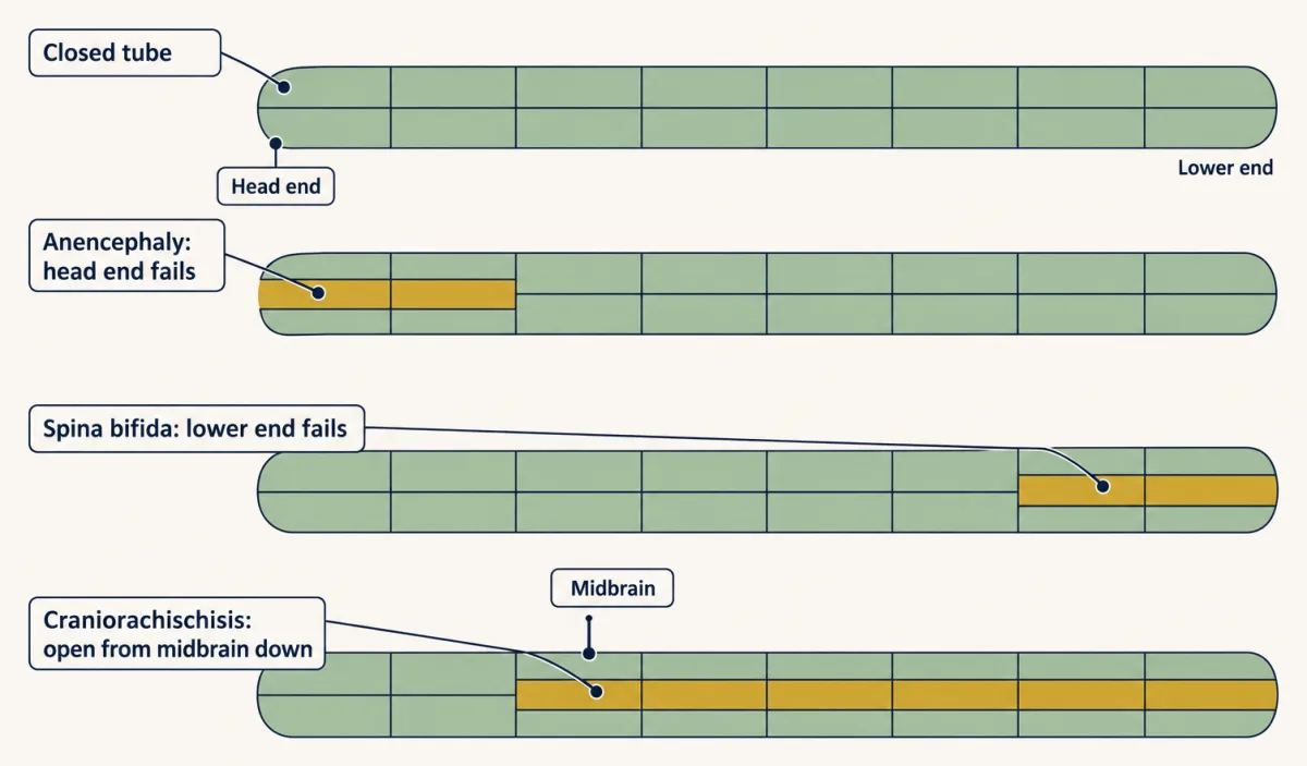
Now look from above. In the mouse, closure begins at several sites and spreads from each like a zipper. The first site is called Closure 1. It sits at the boundary between the hindbrain and the spinal cord. A stretch that does not close stays open for the rest of pregnancy. Where it stays open names the defect. If the head end fails, the result is anencephaly. The exposed brain tissue degenerates. If the lower end fails, the cord itself lies in an open lesion. This is myelomeningocele, the open form of spina bifida. In the mouse, if Closure 1 never happens, the tube stays open from the midbrain down the spine. This is craniorachischisis. So why would a tube fail?
One reason: the folds can close only a gap their bending can cover. And the midline between them sets that gap. Narrowing it comes early and overlaps folding. Failures of bending, fusion or sealing are other routes. Takeaway: narrowing starts first and keeps going as the folds rise. Where closure fails names the defect.
From the film
- The plate narrows and lengthens first, and narrowing keeps going as the folds rise.
- A stretch of the tube that does not close stays open, and where it stays open names the defect.
- The folds can close only a gap their bending can cover, and the midline between them sets that gap.
In a human embryo, the tissue that becomes the brain and spinal cord starts as a flat sheet of cells, and by the end of about the fourth week after fertilization all but its lowest part has rolled into a closed tube. That is often before a woman knows she is pregnant.
This lesson asks one question: what happens if the tube does not close? To answer it you need to know how closing works, where it can fail, and when it happens.
From a flat plate to a tube
We will look at the plate two ways: from above, to see its length and width, and in cross-section, as if sliced across the back, to see it fold.
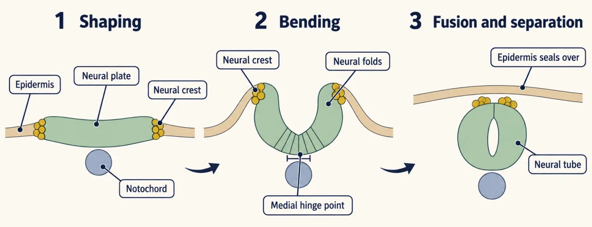
Three tissues come first. The ectoderm is the outermost of the embryo’s three germ layers. After neural induction (the signals that make ectoderm neural), a band of it down the centre of the back becomes the neural plate, the sheet that will form the brain and spinal cord. The ectoderm on either side becomes surface ectoderm, which forms the epidermis, the future skin, and in the head the sensory placodes. Beneath the midline runs the notochord, a rod of mesoderm.
Primary neurulation turns the plate into a tube in overlapping steps, worked out mostly in chick and mouse embryos:
- Shaping. The plate cells grow taller, so the plate thickens. At the same time the plate narrows from side to side and lengthens from head to tail. Hold on to this step.
- Bending. The plate creases along hinge points. In chick and mouse a medial hinge point lies over the notochord, and paired dorsolateral hinge points form at some levels of the body. Cells at a hinge become wedge-shaped. The edges rise as the neural folds.
- Meeting and fusion. The folds move toward the midline, meet and fuse.
- Separation. The tube detaches from the epidermis, which seals over it.
Neural crest cells arise at the border where the neural plate meets the epidermis. In the mammalian head they leave the folds before the folds fuse; in the trunk, and in chick, they leave around or after fusion. They form much of the peripheral nervous system, and how they leave is the subject of the lesson on neural crest.
Now look from above. Closure does not happen all at once. In the mouse, fusion begins at several separate sites and spreads from each like a zipper. The first, called Closure 1, is at the boundary between the hindbrain and the spinal cord. The open stretches left between and beyond the zipped regions, called neuropores, close in turn: the head-end neuropore, then the tail-end one last. Whether human closure starts from the same set of sites is debated, so the mouse map should not be read straight onto the human embryo.
The lowest part of the cord is built another way. There, cells of the tail bud gather into a solid rod that then hollows out and joins the tube made above it. This is secondary neurulation, and in humans it is thought to form the lowest sacral and tail-end segments of the cord.
Where closure fails
If one stretch of the tube does not close, it stays open for the rest of pregnancy. Where it stays open gives the defect its name.
Predict. Use the zipper picture. What would you expect at birth in each case? (a) The head end never closes. (b) The lower end never closes. (c) Closure 1 never forms, so the zipper that should spread from it never starts. (d) The tube closes everywhere, but the bony arch of one vertebra fails to close over it, under normal skin.
Check your prediction
(a) is anencephaly, (b) is myelomeningocele and (c) is craniorachischisis, all open defects, described below; in (c) the forebrain can still close from its own sites.
(d) is the trap: spina bifida occulta, which is not a failure of the tube at all. The tube closed; the bone over it did not. Open and closed defects are easy to blur because both can carry the name spina bifida.
Open defects: the tube itself never closed.
- Anencephaly. The head end fails to close. The exposed brain tissue degenerates, and most of the forebrain and the top of the skull are missing. Affected babies are stillborn or die soon after birth.
- Myelomeningocele (open spina bifida). The lower end fails to close, and the cord itself is in the open lesion. It commonly brings weakness or paralysis below the lesion, loss of bladder and bowel control, and often hydrocephalus.
- Craniorachischisis. The tube is open from the midbrain down the spine. In mouse terms, Closure 1 never happens, as in the loop-tail mouse you will meet below.
Closed defects: the cord is not exposed. Two forms of spina bifida are usually covered by skin and are generally less severe than myelomeningocele, though not always harmless.
- Spina bifida occulta. One or more vertebral arches are unclosed, but the cord lies in place under normal skin. It is often found by chance and is, strictly, a defect of bone over a closed tube.
- Meningocele. The meninges, the membranes around the cord, bulge through the gap in the bone in a sac, while the cord stays in place.
The next question is why closure fails at all.
Narrow first, then fold
Read the transcript of “Where in the Plate?”
Film transcript: Where in the Plate?
How does the width of the midline between the folds affect whether they can meet? A simple cartoon shows the idea. Put your hands flat, thumbs touching, and roll each up onto its thumb edge. The top edges meet, however broad your hands.
Now put a book between your thumbs. Upright, the hands stop apart by the book's width. Broader hands do not help unless you tip them past upright. The book is the midline. How does the midline narrow? Cells crawl in between their side neighbours. This is intercalation. Same cells, rearranged: narrower across, longer along. This is convergent extension.
But which way is across? On a fly's wing, every hair points toward the tip. In mutant flies, the hairs swirl. Frizzled and Dishevelled gather on one side of each cell. Van Gogh and Prickle gather on the other. This is planar cell polarity, a sense of direction within a sheet.
In 2002, John Wallingford and Richard Harland asked where in the frog plate it matters. Frog cell fates are mapped, so an injection can be aimed at one part of the plate. They injected a Dishevelled built to block planar polarity. In some embryos it reached the midline; in others, the sides.
Predict. Which embryos keep an open tube: those blocked at the midline, or those blocked at the sides? Blocked at the midline, the plate stayed wide. The folds rose and moved inward. But the tube stayed open. Blocked at the sides, where the folds form and fuse, closure was normal.
The hands rose with the book still between them. Dishevelled is needed at the midline. A mouse points the same way. The loop-tail mouse carries a mutation in Vangl2, a relative of Van Gogh. With two copies, the tube is open from the midbrain down the spine.
In 2007, Andrew Copp's laboratory labelled that midline. Before neurulation began, it had failed to extend. And Closure 1 never happened. Narrowing starts early and overlaps folding. The plate must still bend, and the fold tips must stick together.
Takeaway: the midline must be narrow enough for the folds to close the gap. Frog and mouse both tie closure to midline narrowing.
From the film
- The midline must become narrow enough, in time, for the folds to meet.
- Convergent extension narrows the midline, and planar cell polarity tells each cell which way is across.
- In frogs, blocking Dishevelled at the midline left the tube open, but blocking it at the sides did not.
- In mice with two copies of the loop-tail Vangl2 mutation, the tube is open from the midbrain down the spine.
The one idea to carry through this lesson. Narrow first, then fold. The two neural folds can close only a gap their bending can cover, and the midline between them sets that gap. Convergent extension narrows the midline; bending, rising and fusion close what is left. The steps overlap: narrowing starts before the folds rise and keeps going while they rise.
What the rule covers.
- Which tube. Primary neurulation: the brain and all of the spinal cord except its lowest part.
- Which failures. Defects in which the folds rise but start too far apart, as in the loop-tail mouse below. Failures of bending, fusion or sealing are other routes. The rule says only that the midline must become narrow enough, in time, for the folds to meet.
- Which width. The midline is the middle strip of the plate, over the notochord, between the two edges that rise as folds.
- Not folate. It says nothing about how folic acid lowers risk.
Why the midline? Put your hands flat on a table, palms down, thumbs touching. Roll each hand up onto its thumb edge until it stands upright, palms facing. The little-finger edges, now on top, meet, however broad your hands are. Now lay a book between your thumbs and do it again. The hands stop apart by the width of the book, and broader hands help only if you lean them inward past upright. Your hands are the folds, your thumbs are where the folds rise, and the book is the midline: whatever width it still has when the folds rise is a gap they must cover. The cartoon is rigid. In the embryo the midline creases at the medial hinge, and fold size, hinge bending and forces from surrounding tissue also matter, but a broader midline still leaves the folds farther apart.
How the midline narrows. Cells in the plate crawl in between their neighbours on either side, a move called intercalation. It rearranges cells without adding or removing them. In an idealized patch with no cell division, 40 cells across and 50 long is 2,000 cells; the same 2,000 cells at 20 across are 100 long. This is convergent extension: converge across, extend along. In the real plate cells also divide and change shape, and cells growing taller, which makes each take up less of the sheet’s surface, also narrow the plate; the share of each differs by species and level of the body. Convergent extension is the narrowing the experiments below test.
How the cells know which way is across. Look at a fly’s wing. Every cell grows one hair, and all the hairs point toward the wingtip. In flies mutant for genes such as frizzled or Van Gogh, the hairs swirl. The proteins these genes make sort to opposite sides of each cell: Frizzled and Dishevelled gather on one side, and Van Gogh (called Vangl in vertebrates) and Prickle on the other. This planar cell polarity (PCP) is a sense of direction within the plane of a sheet. In the neural plate it tells each cell which way is side to side, and so which neighbours to crawl between. Two of these proteins matter here: Dishevelled in the frog and Vangl2 in the mouse.
Predict. In frog embryos (Xenopus laevis) the fate of each early cell is mapped, so messenger RNA injected into one chosen cell ends up in a known part of the neural plate. In 2002 John Wallingford and Richard Harland injected a form of Dishevelled built to block its planar-polarity signaling into cells that would form either the midline of the plate or its sides, where the folds rise and fuse. Use the rule to fill in the table before you open the answer. One cell was not reported, so it cannot be predicted.
| Where PCP is blocked | Width of the plate | Do the folds rise and move inward? | Does the tube close? |
|---|---|---|---|
| Midline | ? | ? | ? |
| Sides | not reported | ? | ? |
| Not blocked | normal | yes | yes |
Check your prediction
| Where PCP is blocked | Width of the plate | Do the folds rise and move inward? | Does the tube close? |
|---|---|---|---|
| Midline | stays wide | yes | no |
| Sides | not reported | yes (closure was normal, so the folds must have met) | yes |
| Not blocked | normal | yes | yes |
Blocking at the midline blocked convergent extension. The plate stayed wide, and the tube stayed open even though the folds rose and moved inward: the hands turned up with the book still between them. Blocking at the sides left closure intact. Under these conditions, the requirement for Dishevelled is at the midline.
The year before, the same pair had shown that a mutant Dishevelled targeted to neural tissue stopped it from lengthening, both in grafts and in explants, pieces of tissue cut out and grown in a dish. Marker genes showed the tissue was still neural: the defect is one of shape, not of fate.
Evidence from a mammal. The loop-tail mouse was named for the kinked tail of animals carrying one copy of its mutation. In 2001 the mutation was traced to Vangl2, the mouse relative of fly Van Gogh.
Predict. Loop-tail mice with two copies of the mutation carry a faulty PCP protein in every cell. Would you expect the tube to close? If not, guess where along the tube the open defect starts, then check.
Check your prediction
They have craniorachischisis: a tube open from the midbrain down the whole spine. In 2007 Andrew Copp’s laboratory labelled the midline of these embryos. It failed to extend before neurulation began, and Closure 1 never happened.
Two species and two PCP proteins both tie closure to convergent extension of the midline.
Narrowing is not the whole job. The plate still has to bend at its hinges. At some hinges, cells constrict their apical face, the face on the top surface of the plate that will line the inside of the tube; other hinges bend without it (see Going deeper). Signals from the surface ectoderm beside the plate contribute, and the fold tips must stick together where they meet. The share of each differs by species and by level of the body. Mice lacking the actin-binding protein Shroom3, which takes part in apical constriction, fail to close the head folds, and some also develop spina bifida.
When the tube closes
Read the transcript of “Four Weeks, and a Vitamin”
Film transcript: Four Weeks, and a Vitamin
The tube closes very early. So when does prevention have to start? Embryologists date human embryos by Carnegie stages, defined by structure, not by age. Counted from fertilization, primary closure is complete by the end of about the fourth week. Doctors count pregnancy from the last menstrual period. That clock starts about two weeks earlier. Predict. On the doctor's clock, how far along is the pregnancy when the tube has closed?
About six weeks of pregnancy. Many pregnancies are not yet recognized at that point. So the vitamin has to be there already. The first trial enrolled women who had already had an affected pregnancy, so their risk was high. Folic acid, started before conception, cut recurrence by about 72 Were 72 of every 100 women spared? No. That is a relative change. Without folic acid, about three and a half and a hundred pregnancies were affected. With it, about 1. The absolute fall was about 2.5 percentage points. The relative figure alone does not say how many women benefit.
But most affected babies have mothers with no earlier affected pregnancy. Can a first case be prevented? In Hungary, women planning a pregnancy took a multivitamin with folic acid, or trace elements. There were six affected pregnancies in the trace-element group, and none with the multivitamin. But the numbers were small, and a multivitamin cannot credit folic acid alone. Folic acid lowers the risk; it does not remove it. Most mothers of affected babies are not folate deficient by ordinary tests. In mice, some mutants respond to folate and others do not. How folate prevents defects is still not known.
Takeaway: the tube often closes before a woman knows she is pregnant. So folic acid must come first.
From the film
- Primary closure is complete by about the end of the fourth week after fertilization, about six weeks of pregnancy by the doctor's count.
- Many pregnancies are not yet recognized by then, so folic acid has to be in place before conception.
- In the first trial, folic acid cut recurrence by about 72 percent, an absolute fall of about 2.5 percentage points.
- Folic acid lowers the risk but does not remove it, and how it works is not settled.
Embryologists date human embryos by Carnegie stages, a series of 23 stages defined by structure, not by age, covering the first eight weeks after fertilization. Closure runs over a few of them:
| Event | Carnegie stage |
|---|---|
| Neural folds first meet | about 10 |
| Head-end neuropore closes | about 11 |
| Tail-end neuropore closes, completing primary neurulation | about 12 to 13 |
Exact days differ between sources. Now put the end of primary closure on two clocks. Embryologists count from fertilization. Doctors usually count pregnancy from the first day of the last menstrual period, which adds about two weeks.
| Clock | Starts counting at | Primary closure is complete by |
|---|---|---|
| Embryologist’s | fertilization | the end of about the fourth week (about day 28) |
| Doctor’s (obstetric) | the last menstrual period, about two weeks earlier | ? |
Predict. Fill in the last cell. Then say what it means for a woman who has not yet realized she is pregnant.
Check your prediction
About six weeks of pregnancy by the obstetric count: four weeks after fertilization, plus the two weeks the doctor’s clock adds. So by the time many pregnancies are confirmed, closure is already under way or finished. Any prevention has to be in place before then.
The folic-acid trials: strong evidence
Folate is a B vitamin. Folic acid is the synthetic form used in tablets and fortified food. Two randomized trials, in which women were assigned to a supplement or a comparison by chance, carry most of the evidence that it prevents neural tube defects.
Recurrence. The first trial enrolled women who had already had an affected pregnancy, whose risk of another was much higher than average. In the Medical Research Council Vitamin Study (1991), 4 milligrams of folic acid a day, started before conception, cut recurrence by about 72 percent.
Predict. Does “about 72 percent fewer” mean that 72 of every 100 women were spared an affected pregnancy? What else would you need to know?
Check your prediction
No. Seventy-two percent is a relative reduction; to know how many women benefit you need the starting risk. In the trial, about 3.5 in 100 pregnancies were affected in the groups without folic acid, against about 1 in 100 with it. That is a relative fall of about 72 percent but an absolute fall of about 2.5 percentage points: roughly 40 high-risk women (100 divided by 2.5) took folic acid for each affected pregnancy prevented. Where the starting risk is far lower, as in women with no earlier affected pregnancy, the same relative cut spares far fewer per 1,000.
First occurrence. Most babies with a neural tube defect are born to women who have never had an affected pregnancy, so a second question remained. In Hungary, through a family-planning service, women planning a pregnancy took a multivitamin with 0.8 milligrams of folic acid or a trace-element supplement, starting at least a month before conception. There were no affected pregnancies in the multivitamin group and six in the trace-element group (Czeizel and Dudás, 1992). The numbers are small, and the supplement held several vitamins, so this trial alone could not credit folic acid by itself. Read beside the British trial, it is evidence that a first occurrence can also be prevented.
Since many pregnancies are unplanned, the United States and Canada began requiring folic acid in enriched grain products in 1998, and many countries have since done the same. Recorded rates fell after fortification, though the size of the fall depends on how and where defects are counted.
What is still open
Folic acid lowers the risk; it does not remove it. Much remains unexplained.
- How folate works. Its reduced forms carry one-carbon units for making thymidylate and purines and for methylation reactions, as that lesson explains, and either could limit a fast-growing plate. How folate prevents defects is not settled. Most mothers of affected babies are not folate deficient by ordinary tests, and some defects persist despite fortification.
- Defects folate does not fix. In mice, some mutants respond to folate and others do not. The curly tail mouse develops spina bifida that folic acid does not prevent, and another nutrient, inositol, lowered its rate (see the anecdote below).
- Other routes. Valproic acid, an anticonvulsant taken in early pregnancy, raises the risk of spina bifida, and so does diabetes in the mother before pregnancy.
- Human PCP genes. Rare variants in human genes for planar cell polarity have been reported in some affected people.
Design problem
Two mouse mutants both have open neural tubes. In the first, the neural plate is short and wide from the start. In the second, the plate narrows normally, but the folds never meet. Say what failed in each, and propose one measurement and one experiment that would tell them apart.
Saved in this browser only.
One way to do it
The first mutant’s short, wide plate suggests that convergent extension failed, the PCP-dependent narrowing. If its midline is wide too, its folds start too far apart to meet; whole-plate shape alone does not show that, so the measurement below checks it. The second points to a failure after shaping: the folds do not bend at the hinges, rise or move inward far enough to meet.
The measurement: in staged embryos fixed just before the folds rise, measure the width of the midline and the width and length of the neural plate against wild-type littermates. A convergent extension mutant has a wider midline and a wider, shorter plate before folding begins; the second mutant matches wild type at that stage.
The experiment: culture embryos and film the folds, or label midline cells with a dye and follow them. In the first mutant, labelled cells fail to intercalate and the midline stays wide while the folds, once raised, still move inward. In the second, the plate narrows on schedule but the folds stall, and you can then ask whether hinge cells constrict their apical faces or whether the folds rise but fall short of the midline.
Going deeper
Hinge cells: what squeezes and what does not
A hinge cell’s wedge is narrow at the apical face and wide at the base. At the midline hinge of chick and mouse, much of that wedging comes from nuclei sitting near the base of the cells during part of the cell cycle. Actin and myosin help squeeze the apical face at some hinges and levels, with Rho kinase helping in chick, as that lesson describes for other cells, and Shroom- and Rho kinase-dependent apical constriction matters in the head and in frogs. Yet chick midline hinge cells wedge even without actin filaments, and mouse spinal hinges still bend without them.
Planar polarity proteins in a living plate
In vertebrates, planar polarity is the branch of Wnt signaling that runs through Dishevelled without beta-catenin and reaches the cytoskeleton, partly through Rho; that lesson covers the branches. In his own laboratory, John Wallingford and Mitchell Butler later filmed fluorescent planar-polarity proteins in living frog neural plates and found them enriched at shrinking cell junctions (Butler and Wallingford, 2018). The imaging links planar cell polarity (PCP) protein enrichment to junction shrinkage and actomyosin, without settling which drives which.
Trial details and policy dates
Before the British trial, a non-randomized study led by Richard Smithells had suggested that women who took a multivitamin before conceiving had fewer repeat defects; the women who chose vitamins might have differed in other ways. An earlier trial in Cardiff was too small to give an answer. The MRC trial was led by Nicholas Wald and used a factorial design, folic acid alone, other vitamins alone, both, or neither; the other vitamins showed no clear effect. In 1992 the US Public Health Service advised 0.4 milligrams of folic acid a day for women who could become pregnant.
Your notes
Select any text in the lesson to highlight it or add a note to it. Your highlights and notes are saved in this browser and collected in your notebook.
This browser is not letting the site save anything (a private window or strict privacy settings), so highlights and notes will be lost when you leave the page.
How we measure it
Targeted microinjection in frog embryos
Messenger RNA for a normal or mutant protein is injected into one chosen cell of an early Xenopus embryo. The fate map says which tissue that cell's descendants will form, so the protein ends up in a known region, such as the midline or the sides of the neural plate.
Explants (Keller sandwiches)
Two pieces of the dorsal region of an early frog embryo are cut out and pressed together, inner faces touching, and grown in a dish. The tissue elongates on its own, so its convergent extension can be measured apart from the rest of the embryo.
Positional cloning of a mouse mutation
A gene known only from a mutant animal is located by tracking the mutation against marked stretches of chromosome over many crosses, then narrowing to one gene. This is how the loop-tail mutation was traced to Vangl2.
Randomized controlled trial
Participants are assigned to a treatment or a comparison by chance, so differences that people bring with them tend to be spread evenly between groups. A difference in outcome can then be credited to the treatment, after allowing for chance, within the population the trial enrolled.
Practice questions on this lesson (3) → MCAT-style, original, each with an explanation
Where this lesson fits
Before thisReview first
- Neural Induction: The Default ModelThis course · Lesson 6
Neural induction makes the ectoderm neural; this lesson starts from the neural plate that induction produces and rolls it into a tube.
- The Organizer: Spemann and MangoldThis course · Lesson 5
The organizer and notochord set up the midline plate and the tissues beneath it that the folds close over.
After thisStudy next
- The Neural Crest: Cells That Leave HomeThis course · Lesson 20
Neural crest cells arise at the plate border and leave the folds around closure, which this lesson only mentions.
- Head to Tail: Hox Genes, Retinoic Acid and RhombomeresThis course · Lesson 8
After the tube closes, its head-to-tail axis is patterned into hindbrain segments and the spinal cord.
- Ventral to Dorsal: Sonic Hedgehog and BMP in the Neural TubeThis course · Lesson 10
It uses the closed tube and the notochord below it to explain how the tube is patterned from floor to roof.
Suggested by an AI model from the lessons themselves, then checked. How we use AI
Finish line
Test yourself
8 questions
Practice and MCAT-style, with explained answers.
Next · Lesson 8
Head to Tail: Hox Genes, Retinoic Acid and Rhombomeres
Rhombomeres r2 and r4 start out looking alike. What tells one to build jaw motor neurons and the other facial ones?
Continue →