Cell Biology · Unit 4: Division and cell loss
New to this? Review it at high school level: The Cell Cycle and Mitosis
Mitosis and Chromosome Segregation
Rewritten on : new text, figures, flashcards and film. Your notes and highlights stay, but the text around them may have moved.
How does a dividing cell give each daughter exactly one sister of every chromosome, and what happens when one attachment goes wrong?
Pause when the reading reaches a film, and offer to play it. Saved on this device.
Watch the film now, or keep listening
Builds on:Cyclin-CDK Control of the Cell CycleCytoskeletal Polymers and DynamicsCheckpoints and Irreversible TransitionsWhy these?
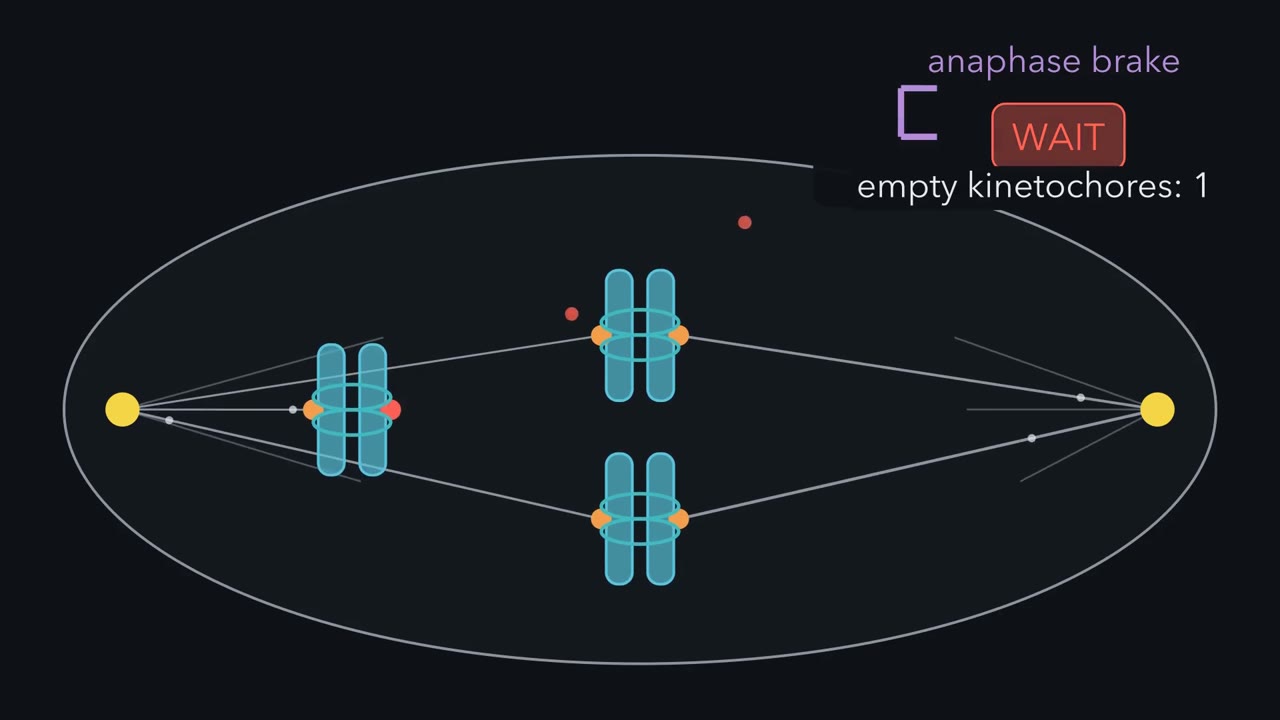 ▶ Watch What the Cell Waits For · 5:06
Read the transcript of “What the Cell Waits For”
Film transcript: What the Cell Waits For
This cell has copied every chromosome. Each daughter must get exactly one copy of each. But one chromosome is attached to only one pole. And the whole cell waits for it. What is it waiting for? Look closer at one copied chromosome. Two sister chromatids lie side by side, held by cohesin rings. Each sister carries a kinetochore, the patch where spindle fibres grip. The two face opposite ways. Fibres reach the kinetochores at random. The goal is biorientation, each sister gripped by the opposite pole. But both sisters can be gripped by the same pole. That is syntelic, an error. How can a cell tell right from wrong? It can sense force. Think of the pair as a short rope, with cohesin resisting.
Pulled from opposite ends, it goes taut. That stretch is tension. Now go back to the syntelic pair. Both sisters are pulled toward the left pole. Is it under tension? In the rope picture, no. Same-way pulls drag the pair along without stretching it. Real pairs can still bear some load from other forces. So the cell should let go and try again. A kinase called Aurora B sits mostly between the sister kinetochores. It phosphorylates the proteins that grip the fibres, weakening their grip.
Slack grips tend to let go and be remade. Under tension, Aurora B weakens the grip less. Force also steadies a grip directly. So taut grips tend to stay. Here is the whole rule. Keep what is pulled from both sides. Remake what is not. And cut only when no kinetochore is left empty. Turning over a wrong grip can leave its kinetochore empty. Empty means without enough end-on attachment. An empty kinetochore sends a wait signal. It holds back the APC, or anaphase-promoting complex. While even one is empty, the cell waits. In a stuck rat kangaroo cell, Conly Rieder's team destroyed that one empty kinetochore with a laser. About twenty minutes later, anaphase began.
The damaged chromosome never reached the middle. So the cell was not waiting for alignment. The wait signal came from in or near that empty kinetochore. A human cell has 92 kinetochores before anaphase. 91 are attached. One is empty. Does the cell go?
No. One in 92 is about 1.1 percent. A majority vote would miss it. The checkpoint works the other way: any empty kinetochore means wait. When none is empty, the brake on the APC comes off. It tags securin for destruction. Securin had kept an enzyme called separase inactive. Now separase is free. Separase is a protease. It cuts one subunit of the cohesin ring, and the ring opens. In vertebrates, the cut acts mainly at the centromere. And a cut ring cannot be rejoined. In a normal cycle, sisters are paired only as DNA is copied. The sisters part. In anaphase A, each moves toward its pole. In anaphase B, the poles themselves move apart. One error slips through.
Sometimes one kinetochore is gripped from both poles, called merotelic. It is attached, so it sends no wait signal. The pair is partly under tension, so Aurora B can miss it. Anaphase can start on time, error and all. That kinetochore is pulled toward both poles, and its chromatid can lag. In two human cell lines, most lagging chromosomes rejoined the main nucleus. A minority ended up in a small separate nucleus. The checkpoint never reads right or wrong. Tension sorts the grips: taut ones tend to stay, slack ones are remade. It just waits until no kinetochore is empty. Then one cut, and the sisters part for good.
From the film
- A sister pair gripped from opposite poles is under tension, seen as stretch between the kinetochores.
- Slack grips, like a syntelic pair, tend to be loosened by Aurora B and remade; taut grips tend to stay.
- The checkpoint keeps the cell waiting while even one kinetochore is empty; it does not read alignment or correctness.
- When none is empty, the brake on the APC/C comes off, separase is freed, and its cut of cohesin cannot be rejoined.
- A merotelic kinetochore is attached, so it sends no wait signal and its chromatid can lag in anaphase.
In 1995 Conly Rieder and colleagues watched a dividing rat kangaroo (PtK1) cell that was stuck. A cell normally delays anaphase while even one kinetochore lacks firm end-on attachment to the spindle. Here one chromosome was attached to only one pole, and the whole cell waited for it.
The first film above follows that cell and what the laser showed, from grip to cut. It explains how a cell gives each daughter exactly one copy of every chromosome, and why division sometimes fails. This page adds the evidence, the numbers and the cases the film skips, such as cyclin B and the arm-versus-centromere split in cohesin.
The picture: one duplicated chromosome, five phases
You arrive at mitosis with every chromosome already copied. The film zooms in on one; its parts are:
- Each rod is a sister chromatid, one complete DNA copy.
- The sisters are held together by cohesin, a protein ring that encircles DNA.
- At one constriction, the centromere, each sister carries a kinetochore, a protein machine that will grip spindle fibres. A pair of sisters therefore has two kinetochores, facing opposite ways.
CDK1-cyclin B has driven the cell into mitosis (see cell-cycle control). In a typical animal cell, mitosis has five named phases. (Budding yeast keeps its nuclear envelope through mitosis, so the envelope events below do not apply there.)
| Phase | What you would see | What the cell is solving |
|---|---|---|
| Prophase | Chromosomes condense into visible rods; the spindle starts to form | Package the sisters; start building the machine |
| Prometaphase | The nuclear envelope breaks down; microtubules reach kinetochores | Grip every pair, correct wrong grips, and wait while any kinetochore is empty |
| Metaphase | Chromosomes lined up in the middle, at the metaphase plate | Keep every pair bioriented while the last wait signal clears |
| Anaphase | Sisters part and move to opposite poles; the poles move apart | Cut cohesin, then move |
| Telophase | Chromosomes reach the poles; a nuclear envelope forms around each set | Rebuild two nuclei; the cell itself splits in cytokinesis |
Predict. A normal diploid human cell in metaphase: how many chromosomes, and how many chromatids? How many chromosomes does each daughter get?
Check your prediction
46 chromosomes and 92 chromatids; each daughter gets 46 chromosomes. While two sisters are joined, they count as one duplicated chromosome, even though each sister already has its own centromere and kinetochore. Once anaphase separates them, each sister counts as a chromosome in its own right. The DNA did not double at anaphase; only the counting rule changed.
Meiosis is in chromosomes and meiosis; what a wrong count does to an organism is in chromosome changes.
The one idea to carry through this lesson. Keep what is pulled from both sides, remake what is not, and cut only when no kinetochore is left empty. The film above shows the rule in motion; the sections below supply its evidence and limits.
Three limits on that rule:
- The checkpoint reads emptiness, not correctness, and tension is a strong sign, not proof. That gap lets some errors through and produces lagging chromosomes (last section).
- “Cannot be undone” refers to sister cohesion, not to cohesin itself (“Cut”).
- In vertebrates most arm cohesin leaves earlier, so the final cut acts mainly at the centromere (“Cut”).
Grip: the spindle and the kinetochore
The spindle is a two-poled array of microtubules, the dynamic polymers described in cytoskeletal polymers and dynamics. Their minus ends gather at the two poles and many plus ends point toward the chromosomes. The kinetochore couples the chromosome to growing and shrinking microtubule tips. A k-fibre is the bundle of microtubules that connects one kinetochore to a pole.
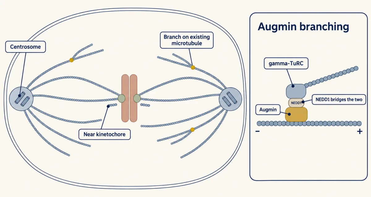
In the classic model, search-and-capture, centrosomes at the poles fire microtubules in random directions until a plus end happens to hit a kinetochore and is stabilized. Like casting fishing lines at random, it works, but alone it would be slow. Cells speed it up by also starting microtubules near chromatin and kinetochores and as branches off existing microtubules.
Test and correct: how tension helps stabilize biorientation
In prometaphase microtubules reach kinetochores at random. Four common ways a pair of sisters can end up gripped are worth knowing (there are in-between states too, such as side-on contacts).
- Biorientation, the goal: each sister’s kinetochore is attached to the opposite pole.
- Monotelic: one sister is attached and the other is still free. This is often a normal step on the way to biorientation.
- Syntelic: both sisters are attached to the same pole. This is an error.
- Merotelic: one kinetochore is bound by microtubules from both poles. This is also an error, and the last section returns to it.
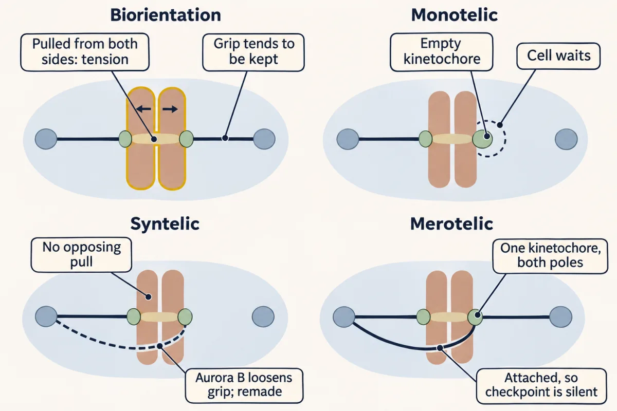
How can a cell tell which is right? It does not need to know which pole a microtubule comes from: it can sense force.
The film’s rope picture answers that: a bioriented pair is pulled by one pole on each sister while cohesin resists, so it is under tension, seen as stretch between the two kinetochores.
Predict. A pair is syntelic: both kinetochores face the left pole. Is it under tension? What should the cell do with it?
Check your prediction
In the rope picture, no: same-direction pulls do not stretch the pair between its two ends, so the cell should let go and try again. A monotelic pair also lacks the opposing pull. Real syntelic and monotelic attachments can still bear load, from pushes on the chromosome arms and other forces, so biorientation is the usual source of opposed sister tension, not the only way to load a grip.
Cells act on that difference in two ways.
Without tension, Aurora B loosens the grip. Aurora B, the kinase the film shows between the two sister kinetochores, sits mostly at the inner centromere. Among the outer-kinetochore proteins it phosphorylates is the Ndc80 complex. Lampson and colleagues blocked Aurora kinase in living vertebrate cells with a reversible inhibitor, let wrong attachments pile up, then washed the inhibitor out. The reactivated kinase selectively took apart the wrong k-fibres, and the chromosomes went on to biorient. Under tension Aurora B weakens the grip less; exactly how is still being worked out (Going deeper).
Under tension, the grip holds. Force also stabilizes attachment directly. In a laser trap, with no detectable Aurora B and no ATP, purified budding-yeast kinetochore particles stayed attached to microtubules longer when Akiyoshi and colleagues pulled harder. In cells both mechanisms are thought to act together.
So wrong attachments tend to be remade and right ones tend to stay. The sorting is probabilistic: right grips still turn over, and some errors last into anaphase. It matters because first tries are unreliable. If each of 46 duplicated chromosomes independently had a one-in-two chance of biorienting on the first try (a crude toy number), all 46 would be right at once about 1 time in 70 trillion. Error correction gives wrong grips another try.
The link to the checkpoint. Wrong grip → little tension → Aurora B turns it over → kinetochore empty → wait. The wait-anaphase signal is that of the spindle-assembly checkpoint (the APC/C-Cdc20 is under “Cut”). A kinetochore with enough end-on attachment stops sending it, whether its attachment is right or wrong; side-on contacts are not enough. Correction can also strip only the wrong microtubules from a kinetochore without emptying it, as in merotely (last section); then the checkpoint never hears about the error.
Wait: the checkpoint reads emptiness
Return to Rieder’s cell, where the laser removed the wait signal with the one empty kinetochore. Destroying an attached kinetochore instead, while a different chromosome stayed unattached, did not release the delay. So the signal comes from in or near an unattached kinetochore. Whether tension also feeds the checkpoint directly is the question of the design problem at the end.
Predict. A diploid human cell before anaphase has 92 kinetochores. Suppose 91 are attached and one is empty. Does the cell go?
Check your prediction
No. One empty kinetochore is 1 in 92, about 1.1% of the total, so a majority vote would miss it. The checkpoint’s logic is the opposite: any empty kinetochore means wait, as one did in Rieder’s cell.
Why a sensor, and not a timer? Take a toy model: each of the 46 duplicated chromosomes finishes attaching both sisters after its own random, exponentially distributed wait with average τ, independently of the others, and stays attached. The last one finishes at about 4.4 τ on average. A timer set to that average would start anaphase with an empty kinetochore in about 43% of cells; to be safe in 99% of cells it would have to wait about 8.4 τ. In this model, waiting for the last kinetochore is both faster and safer.
Note what the checkpoint does not read: alignment. In a normal cell bioriented pairs gather at the metaphase plate, so lining up comes with biorientation, but it is a symptom, not the signal, as the film’s laser cell showed. How the signal is made and relayed to the APC/C belongs to checkpoints.
Cut: securin, separase and cohesin
Once no kinetochore is empty and the wait signal already made has cleared, the brake comes off the APC/C, or anaphase-promoting complex/cyclosome, an E3 ubiquitin ligase: it tags target proteins with chains of ubiquitin that send them to the proteasome (the chemistry is in ubiquitin and proteolysis). APC/C bound to its coactivator Cdc20 tags two key targets.
- Securin. Securin binds separase and keeps it inactive. Once securin is destroyed, separase is released from it.
- Cyclin B. Its destruction inactivates CDK1, which lets the cell leave mitosis. In vertebrates it also removes a second brake on separase, because CDK1-cyclin B inhibits separase there.
Separase is a cysteine protease. It cuts the kleisin subunit of cohesin, Scc1 (called Rad21 in vertebrates), and the ring opens. Keep the division of labour straight: the APC/C and the proteasome destroy securin; separase cuts cohesin.
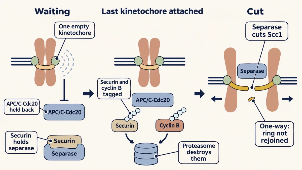
Is the cut enough by itself? Uhlmann and colleagues caused Scc1 to be cleaved in budding yeast arrested at metaphase, where separase is held inactive, and the sisters separated and segregated. That sufficiency test is stronger than watching a protein vanish.
Why is the step one-way? A cut cohesin ring cannot be rejoined. Cohesin can load onto DNA again in the next G1, but in a normal cycle sister cohesion is set up only during replication, so the next S phase pairs new sisters, not these. (Cells can build some cohesion after DNA damage, but that does not reverse anaphase.) The cyclin B destroyed at the same step commits the cell to leaving mitosis.
Predict. A human cell’s securin is replaced with a mutant that the APC/C cannot ubiquitylate. Its chromosomes align and the checkpoint is satisfied. What happens to the sisters?
Check your prediction
They stay joined at their centromeres. Securin is never destroyed, so separase stays off and the protected centromeric cohesin is never cut. A silent checkpoint is not enough: the go signal acts through the destruction of securin.
Why “centromeric”? Vertebrates do not wait for separase to remove all their cohesin. During prophase and prometaphase most arm cohesin leaves without a cut, by a phosphorylation route called the prophase pathway. A protector called shugoshin keeps centromeric cohesin in place until separase cuts it, along with the small amount left on the arms. In budding yeast, by contrast, most cohesin is cleaved at anaphase.
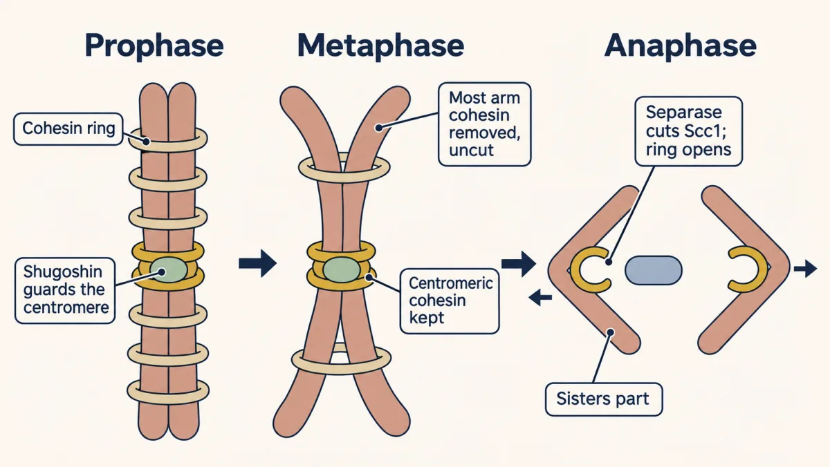
With the remaining cohesin cut, cohesin no longer holds the sisters together.
The second film picks up here: once the sisters are free, what actually moves them apart?
Read the transcript of “Two Motions Pull the Sisters Apart”
Film transcript: Two Motions Pull the Sisters Apart
In chapter one, separase cut the cohesin links, and the sisters began to part. But what carries them apart? Two things move at once. Watch two distances. In anaphase A, each sister moves toward its pole. In anaphase B, the poles themselves move apart. The first changes chromosome-to-pole distance. The second changes pole-to-pole distance. In anaphase A, each k-fibre, the bundle from a kinetochore to its pole, gets shorter. A k-fibre contains many microtubules, not one rope. But where does it lose its subunits? In 1988, Gorbsky, Sammak and Borisy made the fibres glow in cultured mammalian cells. Then they bleached a dark stripe across the anaphase spindle.
They tracked poles, chromosomes, and the stripe in living cells. If the pole reeled the fibres in, the stripe would ride toward the pole with the chromosome. So which way does the stripe go? The stripe did not move detectably relative to the near pole. The chromosome moved toward it, shortening the fibre between kinetochore and stripe. So these fibres lost subunits mainly at the kinetochore. A small loss elsewhere was not ruled out. Poleward flux means the microtubule lattice itself moves toward the pole. In fruit-fly embryos and frog egg-extract spindles, flux can contribute much more. Anaphase B has its own engines. In many animal cells, motors slide overlapping midzone fibres apart.
Dynein at the cell cortex, its edge, pulls on astral fibres. Their balance differs among cell types. Now read a movie, with example numbers. Each kinetochore closes on its pole at 1 µm a minute. The poles separate at 1 µm a minute. These are illustrative, not a universal speed. So how fast do the sisters separate? Each closes on its own pole while the poles move apart. 2 × 1, plus 1: 3 µm a minute. In this symmetric movie, sister separation is twice A plus B. Hide the poles. A movie tracking only chromosomes sees that three, but cannot tell how much came from the poles. Without that landmark, A and B are indistinguishable. The two motions add: chromosomes travel along shortening fibres while the poles spread apart. Measure each against its own landmark.
Those landmarks keep the rates from being confused. Sisters part by two motions: chromosomes to poles, poles apart.
From the film
- In anaphase A each sister moves toward its pole; in anaphase B the poles themselves move apart.
- In the mammalian cells Gorbsky and colleagues studied, k-fibres shortened mainly by losing subunits at the kinetochore.
- In fruit-fly embryos and frog egg-extract spindles, poleward lattice flux can contribute much more.
- Depending on the animal cell, midzone motors, cortical dynein or both can help drive anaphase B.
- In the film's symmetric example, sister separation is twice the anaphase A rate plus the anaphase B rate.
Move: anaphase A and B, read from a movie
The second film separated anaphase A (each kinetochore-to-pole distance shrinks) from anaphase B (the poles move apart) and plotted illustrative positions and rates. Ask what moves relative to what, and the two stay distinct.
It also showed the mechanisms. In anaphase A the k-fibres shorten. In the mammalian cells Gorbsky and colleagues studied, the fibres lost subunits mainly at the kinetochore; in some other cells poleward lattice flux, with subunits lost near the poles, contributes much more. In anaphase B, in many animal cells, motors slide apart overlapping microtubules in the spindle midzone, and dynein at the cell cortex pulls on astral microtubules (see molecular motors). Work through the film’s numbers below.
Worked problem: two rates from one movie
The numbers here are illustrative. You film a cultured mammalian cell with labelled poles and kinetochores. At anaphase onset (t = 0) the poles are 12.0 um apart and each sister kinetochore is 5.5 um from its pole. At t = 4.0 min each kinetochore is 1.5 um from its pole and the poles are 16.0 um apart. Assume the spindle is straight and movement is steady.
Step 1, anaphase A. Kinetochore-to-pole distance fell from 5.5 to 1.5 um in 4.0 min.
anaphase A rate = (5.5 - 1.5) / 4.0 = 1.0 um/min
Step 2, anaphase B. Pole-to-pole distance rose from 12.0 to 16.0 um.
anaphase B rate = (16.0 - 12.0) / 4.0 = 1.0 um/min of pole separation
Each pole moved 0.5 um/min relative to the spindle centre.
Step 3, sister separation. The distance between sisters is the pole distance minus both kinetochore-to-pole distances. At t = 0 it is 12.0 - 2(5.5) = 1.0 um. At t = 4.0 min it is 16.0 - 2(1.5) = 13.0 um. The rate is (13.0 - 1.0) / 4.0 = 3.0 um/min.
Check. Each sister moves toward its pole and the poles move apart, so sister separation should equal twice anaphase A plus anaphase B: 2(1.0) + 1.0 = 3.0 um/min. It does.
A movie that tracks only chromosomes cannot tell you how much of their separation came from the poles.
- Anaphase A (kinetochore to pole)
- Anaphase B (pole to pole)
- Sister separation
- Check: 2 × A + B
Sister separation is the pole distance minus both kinetochore-to-pole distances, so its rate is twice anaphase A plus anaphase B. A movie that tracks only chromosomes cannot say how much of that came from the poles. The picture assumes a straight spindle and steady movement, as the lesson does. All numbers here, the lesson’s included, are illustrative, not measurements, and frames between 0 and 4 min are interpolated by the steady-movement assumption.
The rule’s blind spot: merotely, laggards and micronuclei
A checkpoint that reads attachment, and a correction system that trusts tension, leave one gap. Here is the price.
Predict. One kinetochore is merotelic: it is bound by microtubules from both poles, while its sister is bound to one pole. Does the checkpoint see it? Does Aurora B?
Check your prediction
The checkpoint does not: the kinetochore is attached, so it sends no wait signal. Aurora B can miss it too, because the pair is partly under tension. The cell can enter anaphase on time with the error in place, and that kinetochore is then pulled toward both poles at once.
Cimini and colleagues showed this in PtK1 cells. Lagging single chromatids appeared in 1.16% of untreated anaphases and 17.55% after recovery from a nocodazole block, and electron microscopy confirmed microtubules from both poles on the lagging kinetochores.
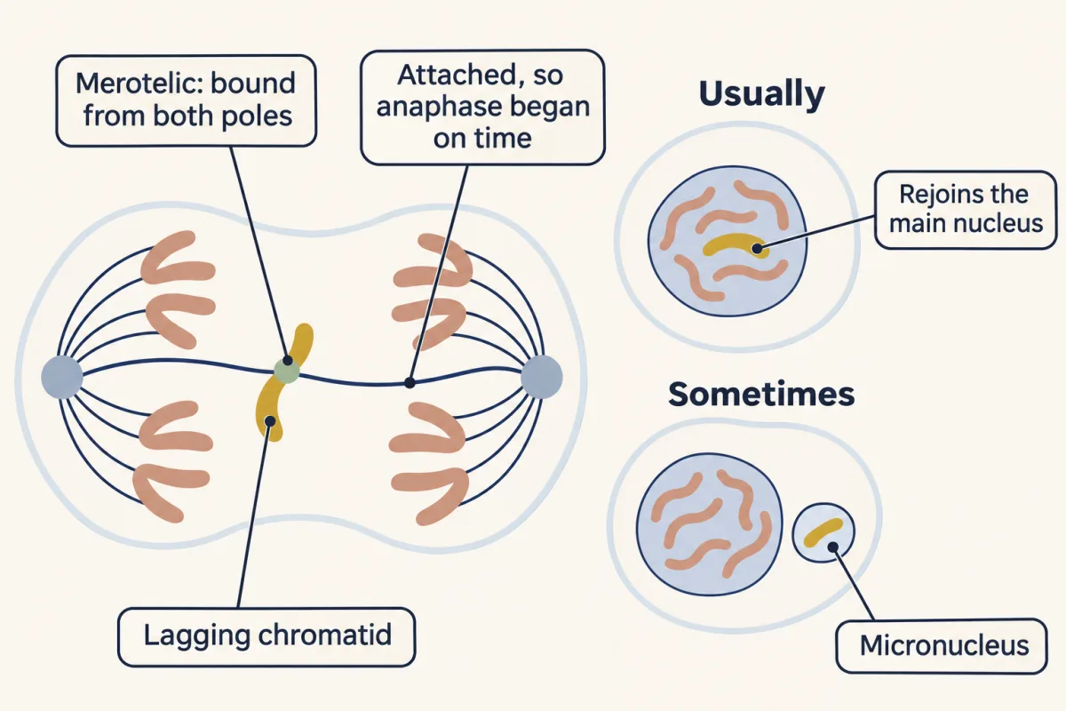
A lagging chromosome seen in one frame is late, but its fate is open. It can reach the main nucleus late or end up in a micronucleus, a small separate nucleus. In human RPE1 and U2OS cells, Orr and colleagues found that most transient laggards rejoined the main nucleus; a midzone Aurora B mechanism helped correct errors in anaphase and delayed the completion of nuclear-envelope assembly on laggards. Where this leads: persistent errors and chromosomal instability in tumours (cancer genetics), spindle orientation and cell fate (asymmetric division), and mitotic defects in neural progenitors that shrink the brain (microcephaly and malformations).
Worked problem, continued: watch the denominators. Orr and colleagues saw at least one transient laggard in 9% of RPE1 cells. That number is per cell. They found that 6% of lagging chromosomes formed micronuclei. That number is per chromosome. If every affected cell had exactly one laggard, the fraction of all cells with a laggard-derived micronucleus would be 0.09 x 0.06 = 0.0054, about 0.5%. If some cells had two or more laggards, the true figure could be higher (if the extra laggards are independent) or lower (if micronuclei cluster in a few cells with many laggards). Say the assumption out loud whenever you multiply rates with different denominators.
Design problem
Is anaphase held back by a signal from unattached kinetochores, or by lack of tension? Rieder and colleagues destroyed the last unattached kinetochore in PtK1 cells, and anaphase followed in a reported 20 plus or minus 2.5 minutes. Li and Nicklas pulled on a mono-oriented chromosome with a microneedle in praying-mantis spermatocytes, and the delay ended. Do these results contradict each other, and what experiment would separate the two ideas?
Saved in this browser only.
One way to do it
They need not contradict each other. Tension stabilizes attachment, so in a cell, pulling on a kinetochore can change how many microtubules stay bound as well as the force. Firm end-on attachment normally silences a kinetochore, whereas a side-on attachment can keep signalling even under tension. Attachment and tension are coupled.
To separate them, change one while holding the other fixed. In vitro, reconstituted yeast kinetochores in a laser trap (Akiyoshi 2010) let you set force while the number of attached microtubules is known. In cells, you could ablate the last unattached kinetochore and compare it with a cell in which that kinetochore is held attached but under low tension, for example after a drug that dampens spindle forces.
Readout: time from last attachment, or from the lesion, to anaphase onset in single cells. Control: irradiate a spot of the same size near the kinetochore but off it, which should not release the delay. Limit: mantis spermatocytes in meiosis and PtK1 cells in mitosis are different systems, so a difference between the two papers may reflect the cell, not the mechanism.
Going deeper
Where spindle microtubules come from
- Near chromatin and kinetochores. In human RPE1 cells, Sikirzhytski and colleagues used correlative light and electron microscopy and found short non-centrosomal microtubules near kinetochores from the start of spindle assembly. End-on attachments formed within the first three minutes of prometaphase. Inhibiting CENP-E, a kinetochore kinesin (see molecular motors), impeded the conversion of side-on contacts into end-on attachments.
- Branching on existing microtubules. Augmin is a protein complex that recruits the gamma-tubulin ring complex (gamma-TuRC), the template that starts new microtubules, onto the side of an existing one, so a new microtubule branches off in the same direction. In HeLa and RPE1 cells, David and colleagues tracked growing ends and found that most spindle microtubules nucleated away from centrosomes; augmin-dependent amplification almost entirely explained where the growing ends were. In vitro, purified human augmin and gamma-TuRC were sufficient for branching. In that reconstitution NEDD1 bridged augmin and gamma-TuRC, and phosphorylation by CDK1 and PLK1 was needed for NEDD1 to bind augmin.
Keep the scope: these results come from two human cell lines and a test tube. Some cells, such as many oocytes, build spindles without centrosomes at all, and the share from each route differs between cell types.
How tension switches off Aurora B
Aurora B is a kinase of the chromosomal passenger complex. Much of it sits at the inner centromere, and smaller pools sit closer to the kinetochores. How tension turns its effect off is still argued. One popular model says stretching pulls the outer kinetochore out of reach of the kinase at the inner centromere; other evidence points to changes in kinetochore structure under force.
In the laser-trap experiment, purified budding-yeast kinetochore particles stayed attached for a mean of 21 plus or minus 5 minutes at 1 piconewton and 50 plus or minus 17 minutes at 5 piconewtons, means from only 15 and 9 detachments. The result does not show that Aurora B is unnecessary in cells.
Cohesin removal in vertebrate prophase
Hauf and colleagues showed in human cultured cells, including HeLa, that phosphorylation of the cohesin subunit SA2 was essential for the prophase pathway. It also needs a cohesin-bound protein called Wapl: when Kueng and colleagues depleted Wapl from human cells, cohesin stayed on the arms until anaphase. When McGuinness and colleagues depleted shugoshin from HeLa cells, centromeric cohesin was lost early, and their results suggested that shugoshin works by preventing SA2 phosphorylation at centromeres.
Where k-fibres lose subunits
Gorbsky, Sammak and Borisy bleached a mark on kinetochore microtubules in cultured mammalian cells and found that the chromosomes moved toward the mark, though a small loss of subunits away from the kinetochore was not excluded. In the cells where poleward flux contributes much more, for example Drosophila embryos and Xenopus egg-extract spindles, the microtubule lattice moves toward the poles while subunits are lost from the minus ends there.
Your notes
Select any text in the lesson to highlight it or add a note to it. Your highlights and notes are saved in this browser and collected in your notebook.
This browser is not letting the site save anything (a private window or strict privacy settings), so highlights and notes will be lost when you leave the page.
How we measure it
Live-cell time-lapse microscopy
Label chromosomes, kinetochores or microtubules with fluorescent proteins and image the same cell every few seconds or minutes through division. It gives positions over time, so you can measure rates and follow the fate of a single chromosome; Orr and colleagues sampled human cells every 30 seconds.
Laser microsurgery
Focus a laser to destroy a chosen structure, such as one kinetochore, in a living cell and watch what happens next. It tests whether a structure is needed for an event, provided a lesion placed nearby but off the target is used as a control.
Laser-trap force clamp
Hold a bead attached to a purified kinetochore particle in a focused laser beam and keep the force on it constant while it tracks a dynamic microtubule end. It measures how long an attachment survives at each force; with no ATP present, no new phosphorylation can happen during the measurement.
Correlative light and electron microscopy
Film a cell live, fix it at a chosen moment, then section the same cell for electron microscopy. The light movie gives the stage and the history; the electron micrographs show which microtubules end at which kinetochore.
Practice questions on this lesson (3) → MCAT-style, original, each with an explanation
Where this lesson fits
Before thisReview first
- Cyclin-CDK Control of the Cell CycleThis course · Lesson 17
CDK1-cyclin B drives the cell into mitosis, and its destruction at anaphase is used here.
- Cytoskeletal Polymers and DynamicsThis course · Lesson 14
Spindle microtubules grow and shrink by dynamic instability, which the kinetochore grip depends on.
- Checkpoints and Irreversible TransitionsThis course · Lesson 18
This lesson uses the idea of a wait signal that holds the cell until the last kinetochore is attached.
After thisStudy next
- Cytokinesis and Organelle InheritanceThis course · Lesson 20
The cell splits after sisters separate, and the spindle position from this lesson sets where the furrow forms.
- Cell Identity, Cell State and DiseaseThis course · Lesson 25
Merotelic errors and lagging chromosomes feed into chromosomal instability and disease states.
- Cell Death and ClearanceThis course · Lesson 21
A cell with persistent mitotic errors can be removed by death, which this lesson's error rates lead into.
Elsewhere on the siteFilms, simulations and method films in other courses
- Mitosis stepper and phase counterSimulation · Biology
Its mitosis stepper and phase counter practise counting chromosomes and chromatids at each phase.
- How One Set Becomes TwoFilm · Biology
It shows how a copied chromosome is separated so each nucleus gets one set.
- One of Each Pair, by Coin TossFilm · Genetics and Molecular Biology
It separates sister chromatids from homologs by counting centromeres, which you need when naming what segregates.
- When Chromosomes Miss Their TurnFilm · Genetics and Molecular Biology
It shows how a missed separation at anaphase leaves a cell with an extra or missing chromosome.
Suggested by an AI model from the lessons themselves, then checked. How we use AI
Finish line
Test yourself
8 questions
Practice and MCAT-style, with explained answers.
Next · Lesson 20
Cytokinesis and Organelle Inheritance
Push the spindle off-centre in a sea urchin embryo cell, and the signal for the furrow follows it before the furrow does.
Continue →