Cell Signaling · Unit 5: Signals that build and defend an animal
Wnt: β-Catenin and Planar Polarity
Why would a cell make a protein all day only to destroy it?
Pause when the reading reaches a film, and offer to play it. Saved on this device.
Watch the film now, or keep listening
Builds on:Ubiquitin and Regulated Proteolysis as SignalsAdaptors, Docking Proteins and ScaffoldsPI 3-Kinase, Akt and InsulinWhy these?
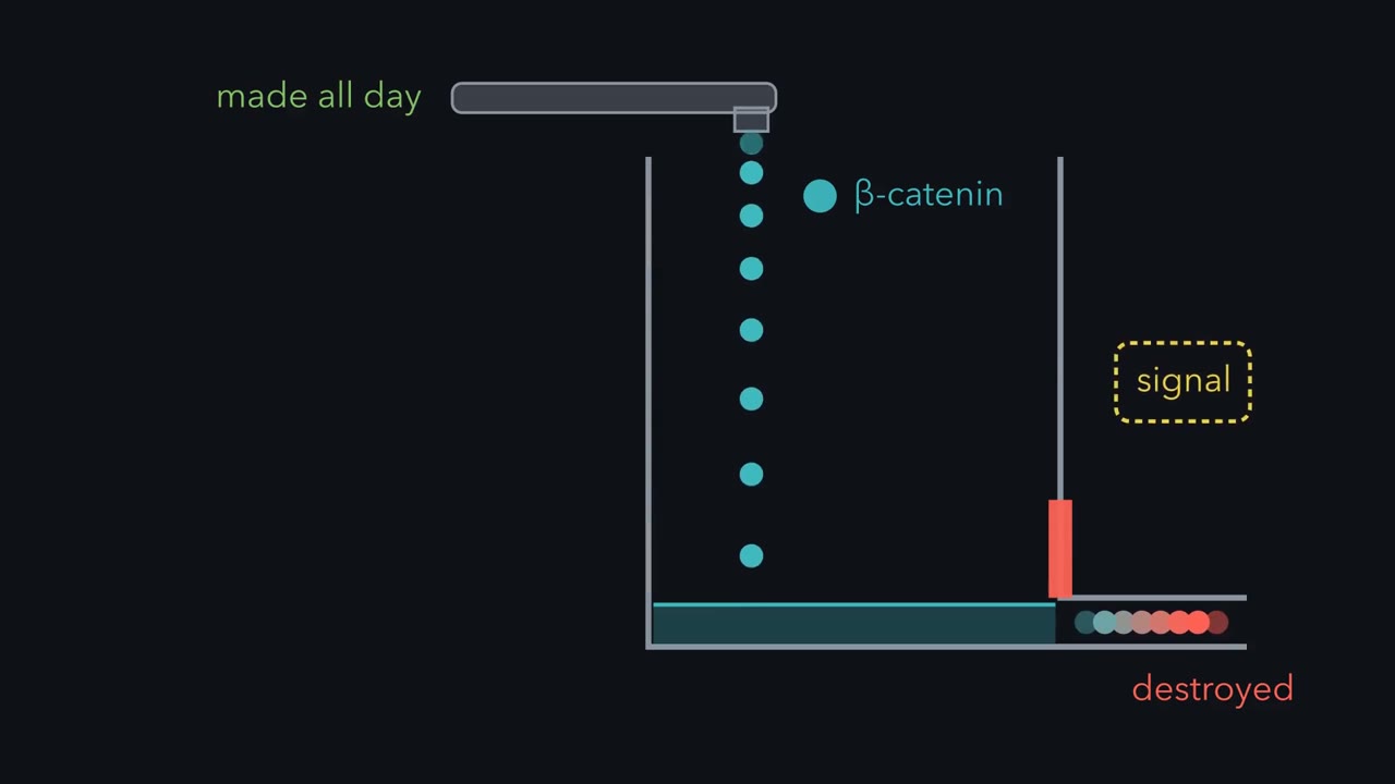 ▶ Watch Wnt Plugs the Drain · 2:58
Read the transcript of “Wnt Plugs the Drain”
Film transcript: Wnt Plugs the Drain
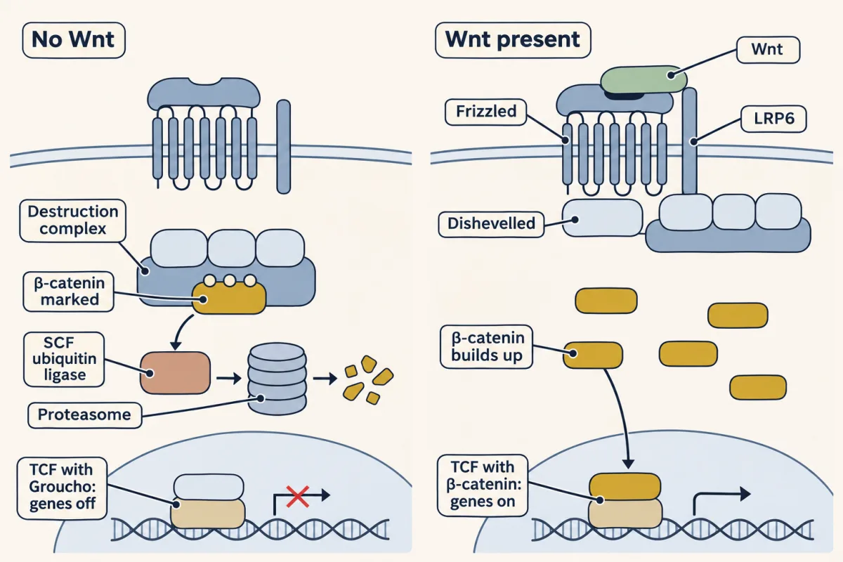
Why would a cell make a protein all day, only to destroy it? Most of your cells do exactly that with a protein called beta β-catenin. Unless a signal tells them to stop, they destroy the free copies soon. That looks like waste. It is a design. Picture a bathtub with the tap running and the drain open. The water level depends on both. The steady level of beta β-catenin is its rate of synthesis divided by its rate constant of destruction. Slow the destruction ten-fold, and the level rises ten-fold, with no change in how fast the protein is made. Take a cell that receives no signal. Every free copy of beta β-catenin meets the destruction complex, where the scaffold Axin holds two kinases beside it.
CK1 puts a phosphate on serine 45, which primes GSK3 to add three more. A protein called beta β-TrCP reads the phosphates on serines 33 and 37. It belongs to SCF, a ligase that adds a chain of ubiquitin, and the proteasome destroys the protein. That signal is a Wnt, a protein that one cell secretes and a nearby cell reads. Its receptor is Frizzled, and Wnt brings it together with a partner, LRP5 or 6. Frizzled recruits a protein called Dishevelled, and the tail of LRP6 is phosphorylated and then binds Axin. The destruction complex is pulled to the receptor, and there it stops delivering beta β-catenin to the ligase. How it is stopped is still argued, but newly made beta β-catenin survives and builds up.
In the nucleus, a protein called TCF binds the DNA of many Wnt target genes, often before a signal comes. Without beta β-catenin, TCF holds a co -repressor called Groucho and keeps those genes off. In the simple model, beta β-catenin enters the nucleus, pushes Groucho off and brings in the proteins that start transcription. So TCF turns from a repressor into an activator. Without a signal, the drain stays open and the level stays low. With Wnt, the drain is plugged and the level rises. The control is on destruction, not on synthesis. A protein that is always made and always destroyed can be raised by one move. Stop the destruction.
Most of your cells make a protein called β-catenin all day long and, unless a signal tells them to stop, destroy the free copies soon after they are made. That looks like waste. It is a design: a protein that is always being made and always being destroyed can be raised quickly by one move, which is to stop the destruction. A secreted signal called Wnt works this way, and embryos use it to lay out pattern and tissues such as the lining of the gut use it to keep renewing themselves. This lesson follows Wnt from the destruction complex to the nucleus and into colon cancer, where losing APC leaves the move stuck on. It ends with a second use of the same receptor, in which the cells of a sheet work out which way to point.
The name: a mouse tumour gene and a fly gene were one gene
Wnt began as two genes that turned out to be one. The mouse gene, int-1, was named for the site where a tumour virus kept inserting itself in mammary tumours. What it does in a healthy animal stayed unknown for five years. Then Frank Rijsewijk, in Roel Nusse’s group, cloned the fly version, and it proved to be a gene fly geneticists already knew as wingless: weak mutations give adult flies that often lack wings, and stronger ones kill the embryo and scramble the pattern of its segments.
So one gene, kept in both lineages for more than 500 million years, causes tumours in a mouse when it is switched on in the wrong tissue and lays out pattern in a fly embryo. In 1991 eight researchers signed a one-page note asking the field to use one name, spliced from wingless and int: Wnt. The fly match was also practical. Other fly mutants with the same segment defect became candidates for the rest of the pathway, and one of them, armadillo, is the fly’s β-catenin.
The idea: β-catenin is made all the time and destroyed all the time
A Wnt is a protein that one cell secretes and a nearby cell reads. To see what it changes, start with a cell that receives none.
β-Catenin has a second job, linking the adhesion proteins called cadherins to the cytoskeleton at cell junctions, and the copies doing that job are stable. Every other copy meets the destruction complex. Its scaffold is Axin, which holds an enzyme next to its substrate as the scaffolds in the lesson adaptors-and-scaffolds do. A second large protein, APC, also binds β-catenin and is needed for the complex to work. The enzymes are two kinases, casein kinase 1 (CK1) and glycogen synthase kinase 3 (GSK3).
The order matters. CK1 puts a phosphate on serine 45 of β-catenin. That phosphate primes GSK3, which adds three more, on threonine 41, serine 37 and serine 33. Then comes the step to remember: SCF targets β-catenin. The phosphates on serines 33 and 37 are read by β-TrCP, the part of an SCF ubiquitin ligase that chooses the substrate. SCF adds a chain of ubiquitin, and the proteasome destroys the protein. The lesson ubiquitin-and-proteolysis gave the general rule, and these are the particulars.
Picture a bathtub with the tap running and the drain open. The water level depends on both.
steady level of β-catenin = rate of synthesis ÷ rate constant of destruction
Slow the destruction ten-fold and the level rises ten-fold, with no change in how fast the protein is made.
You have met GSK3 before, as the kinase that Akt switches off after insulin (lesson pi3k-akt-insulin). In the cultured cells where this has been tested, insulin does not raise β-catenin. The explanation offered is that the GSK3 held on Axin is a separate pool that Akt does not reach.
What Wnt changes: it stops the destruction
Wnt is an unusual secreted protein because it carries a lipid. In the cell that makes it, an enzyme called Porcupine attaches a fatty acid to one of its serines. The lipid makes Wnt cling to membranes, so it is hard to dissolve, and in many tissues it acts mostly on cells near its source.
The receptor is Frizzled, a seven-pass membrane protein with the overall shape of the receptors in Unit 3. In the crystal structure of a Wnt bound to the outer domain of Frizzled, the fatty acid lies in a groove on the receptor, so the lipid is part of the grip. Frizzled has a partner, a single-pass protein called LRP5 or LRP6, and Wnt brings the two together.
Inside the cell two things follow. Frizzled recruits a cytoplasmic protein called Dishevelled, and the tail of LRP6 is phosphorylated and then binds Axin. The destruction complex is pulled to the receptor, and there it stops delivering β-catenin to the ligase. How it is stopped is still argued. The outcome is clear: newly made β-catenin survives, builds up and enters the nucleus.
In the nucleus it finds TCF, a protein that binds the DNA of many Wnt target genes even before a signal comes. Without β-catenin, TCF holds a co-repressor called Groucho and keeps those genes off. When β-catenin arrives, Groucho leaves and β-catenin brings in the proteins that start transcription, so TCF turns from a repressor into an activator. The simple model is that β-catenin pushes Groucho off. How Groucho is actually removed is still being worked out. Which genes answer depends in part on where TCF sits in that kind of cell. One common target is Axin2, a relative of Axin, which gives negative feedback of the kind in the lesson signaling-circuits.
Wnt in disease: a pathway stuck on
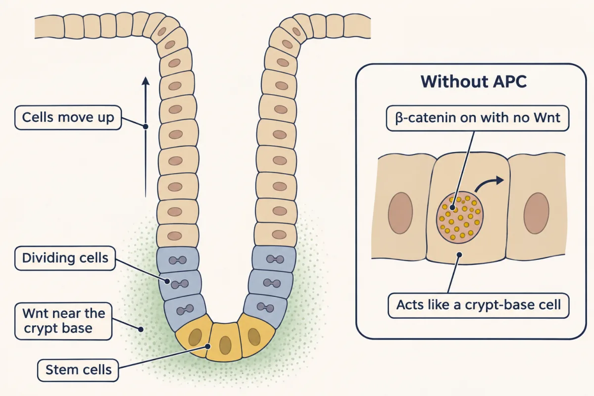
Most people who inherit one faulty copy of the APC gene grow hundreds to thousands of small growths called polyps in the colon, a condition called familial adenomatous polyposis. Nearly all of them get colon cancer unless the colon is removed. APC is also mutated in most colon cancers in people with no family history, about four in five.
Why APC? In 1993 the laboratory of Paul Polakis and the laboratory of Bert Vogelstein and Kenneth Kinzler each reported that the APC protein binds β-catenin. In 1997 Hans Clevers’s laboratory and the Vogelstein and Kinzler laboratory published side by side. Colon cancer cells without working APC had β-catenin and TCF switching genes on all the time, and putting APC back turned that transcription off. Some colon tumours with normal APC carried mutations in β-catenin instead, at the serines and the threonine that casein kinase 1 (CK1) and glycogen synthase kinase 3 (GSK3) phosphorylate.
Both routes end in the same place. Without APC the complex can still phosphorylate β-catenin, but the protein is no longer efficiently ubiquitinated and destroyed. Without the phosphorylation sites β-catenin cannot be marked for destruction. Either way the drain is plugged, and TCF is on with no Wnt. The gut is where this matters most, because the stem cells of the gut lining sit at the base of pits called crypts, and they and the dividing cells above them depend on Wnt: newborn mice made without TCF4 lack the dividing compartment of the small intestine. A cell without APC behaves as if it had never left the base of the crypt.
Rule to keep: the level is the rate of synthesis divided by the rate constant of destruction, so anything that stops the destruction raises it, and a broken destruction complex does so with no Wnt. The numbers 1 and 10 are relative units in this sketch, taken from the lesson's example of slowing destruction ten-fold; they are not measurements, and synthesis is held the same throughout. The picture is a sketch of the logic, and the junction pool of β-catenin is left out. The same 10 is used for Wnt and for each lesion; the lesson does not say they are equally strong.
The same pathway sets bone mass. People with two inactive copies of LRP5 have fragile, low-density bone, and a single change in LRP5, glycine 171 to valine, gives unusually dense bone.
The other Wnt pathway: planar cell polarity tells a cell which side is which
Read the transcript of “Frizzled and the Fly Wing's Hairs”
Film transcript: Frizzled and the Fly Wing's Hairs
Look at a fly wing. Each cell grows one hair, and every hair points toward the wing tip. A direction shared by the cells in the plane of a sheet is called planar cell polarity, or PCP. But no cell can see the whole sheet. So how does each cell know which way to point? At the University of Virginia, in 1987, Charles Vinson and Paul Adler made patches of cells that lack a gene called frizzled. The rest of each wing was normal. Would the normal cells beside a patch notice? Normal cells on the tip side of a patch grew misdirected hairs. Normal cells on the other side did not. So a patch with no frizzled upset its normal neighbours, but on one side only.
Two years later, Adler's laboratory cloned frizzled. It encodes a seven-pass membrane protein. It is the same Frizzled as in the Wnt receptor family, which is named for untidy hairs on a fly. This pathway does not use β-catenin or TCF. In the wing, its output is the place where the cell builds the actin bundle that becomes its hair.
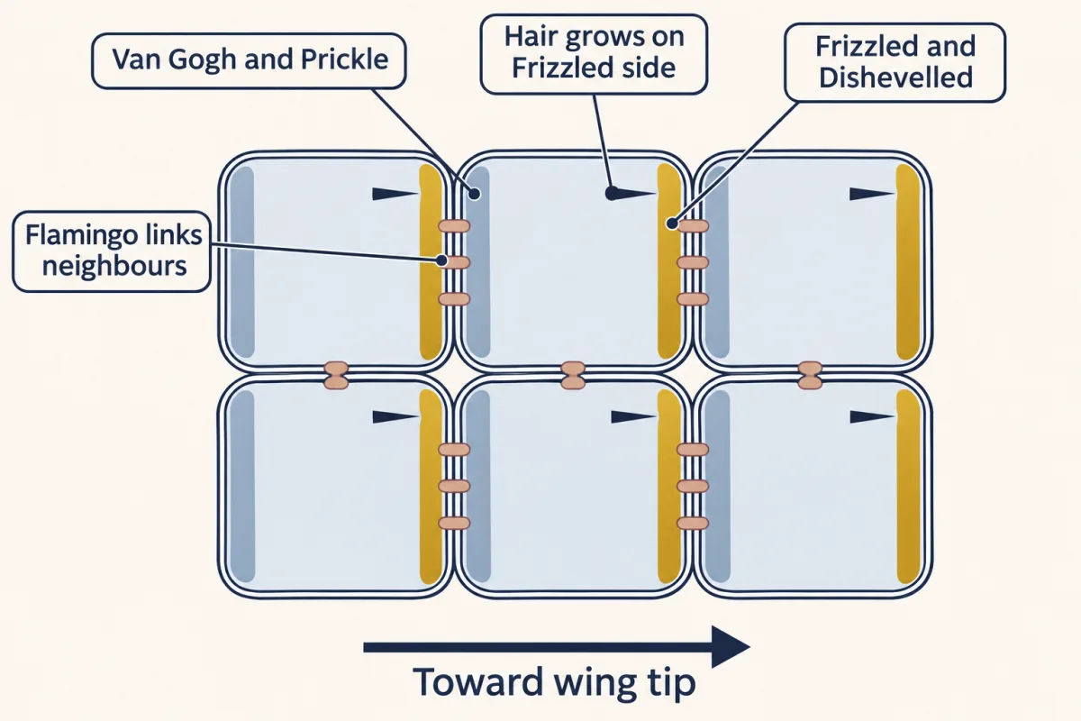
Look at a fly wing. Each cell grows one hair, and every hair points toward the wing tip. A direction shared by the cells within the plane of a sheet is called planar cell polarity, or PCP.
UVa moment: Paul Adler. Every hair must point the same way, though no cell can see the whole sheet. The experiment that showed how the fly solves this, with patches of mutant cells, was done at the University of Virginia. In 1987 Charles Vinson and Paul Adler made patches of cells lacking the gene frizzled in otherwise normal wings. Normal cells on the tip side of a patch grew misdirected hairs. Normal cells on the other side did not. Two years later the laboratory cloned frizzled and found that it encodes a seven-pass membrane protein. It is the same Frizzled: the Wnt receptor family is named for untidy hairs on a fly.
Later work placed the parts. Frizzled and Dishevelled gather on the tip-side edge of each wing cell. A membrane protein called Van Gogh gathers on the opposite edge with its partner Prickle. Flamingo, a cadherin, sits on both edges and links each cell to the next. The hair grows from the Frizzled side. None of this uses β-catenin or TCF. In the wing the output is the place where the cell builds the actin bundle that becomes its hair.
Vertebrates use the same genes to move cells. The neural tube, the future brain and spinal cord, starts as a flat plate that must narrow and lengthen while its edges fold up and meet. Cells narrow the plate by squeezing between their neighbours toward the midline, and to do that each cell must know which of its sides faces the midline. Disrupting PCP causes neural tube closure defects, the group that includes spina bifida. In mice with two mutant copies of Vangl2, a vertebrate Van Gogh, the tube stays open along most of its length, and rare variants of the related human gene VANGL1 have been found in some patients with neural tube defects. Vertebrates need particular Wnts for these movements, Wnt5a and Wnt11 among them, which is one reason the system is counted as a Wnt pathway.
Design problem
Each cell in a sheet grows one hair, and every hair must point the same way. No cell can see the whole sheet. Design a way for each cell to tell its two sides apart using only proteins in its own membrane and in the membranes of the cells it touches. Then predict what a patch of cells that lacks one of your proteins does to the normal cells beside it.
Saved in this browser only.
One way to do it
Use two membrane proteins, A and B, and two rules. Between cells, A on one cell holds B on the cell it touches, so each keeps the other in place across the gap. Within a cell, A and B drive each other away, so no edge keeps both. A weak bias across the tissue is then enough. Each cell ends with A on one side and B on the other, and every boundary has A facing B. Grow the hair on the A side.
Now make a patch of cells with no A. Those cells have B on every edge. Each normal neighbour gathers its A on the edge that touches the patch, so its hair points toward the patch. On one side of the patch that is the normal direction and nothing looks wrong. On the other side the hairs point backwards. Mutant cells have misdirected normal ones, and on one side only.
Going deeper
How int-1 and wingless were matched
Around 1980 Roel Nusse, a postdoctoral fellow in Harold Varmus’s laboratory in San Francisco, began to study mammary tumours in mice infected with mouse mammary tumour virus. This virus makes a DNA copy of its RNA genome and inserts that copy into a mouse chromosome wherever it happens to land, and the inserted copy is called a provirus. If a provirus sits in the same stretch of mouse DNA in tumour after tumour, that stretch probably holds a gene that helps a tumour grow when the virus disturbs it.
In 18 of the 26 tumours they examined, a provirus sat inside one region of about 19 kilobases (19,000 base pairs). In the few tumours tested, an RNA was made from that region, and none was detected in the lactating mammary glands tested. They named the place int-1, for integration site 1. Their 1982 paper proposed only that insertion there strongly favours a tumour, perhaps by raising the expression of a cellular gene.
What the gene does in a healthy animal stayed unknown for five years. Then Frank Rijsewijk, in Nusse’s group in Amsterdam, cloned the fly version. Its protein was 54 percent identical to the mouse protein, with all 23 cysteines in the same positions, and the gene proved to be one that fly geneticists already knew as wingless. A weak wingless mutation, described by R. P. Sharma and V. L. Chopra in 1976, gives adult flies that often lack wings. Stronger ones kill the embryo and scramble the pattern of its segments.
Your notes
Select any text in the lesson to highlight it or add a note to it. Your highlights and notes are saved in this browser and collected in your notebook.
This browser is not letting the site save anything (a private window or strict privacy settings), so highlights and notes will be lost when you leave the page.
How we measure it
Proviral tagging
Infect mice with a retrovirus that inserts its DNA at many places in the genome, collect the tumours, and map where the virus sits in each one. A region hit in many separate tumours probably holds a gene that drives growth when the virus disturbs its expression. This is how int-1 was found.
Genetic mosaics in the fly wing
Make a small patch of mutant cells, marked so that it can be seen, inside a normal wing. If only the mutant cells look abnormal, the gene acts inside the cell that makes it. If normal neighbours change too, the gene helps to pass information between cells, which is what patches lacking frizzled showed.
TCF reporter assay
Place a luciferase gene behind a promoter that carries several TCF binding sites and put it into cells. The light the cells give off reports how much β-catenin is working with TCF, and a control reporter with mutated sites shows what is unrelated to TCF. Colon cancer cells without APC light up with no Wnt added.
Method films · the methods library
- Engineered mutants: site-directed, dominant-negative and constitutively active 4:25
- Family linkage and positional cloning 3:52
- Forward genetic screens 4:07
- Gene cloning: vectors, libraries and screening 4:04
- Gene targeting: knockout and knock-in mice 4:13
- Reporter gene assays 2:42
- X-ray crystallography 4:06
Practice questions on this lesson (3) → MCAT-style, original, each with an explanation
Where this lesson fits
Before thisReview first
- Ubiquitin and Regulated Proteolysis as SignalsThis course · Lesson 8
It gives the general rule that a ubiquitin ligase marks a protein for the proteasome, which the destruction complex uses on β-catenin.
- Adaptors, Docking Proteins and ScaffoldsThis course · Lesson 10
Axin works as a scaffold holding kinases next to their substrate, an idea that lesson teaches.
- PI 3-Kinase, Akt and InsulinThis course · Lesson 20
You need GSK3 as the kinase Akt switches off to see why insulin does not raise β-catenin.
After thisStudy next
- Hedgehog and the Primary CiliumThis course · Lesson 26
It follows another secreted, lipid-modified signal that uses a Frizzled-like receptor to steer embryo pattern.
- Oncogenes: When the Switch SticksThis course · Lesson 41
It takes the stuck-on switch idea further, with mutations that leave a pathway active without its signal.
- The Shape of a Signal: Size, Duration, Frequency and PlaceThis course · Lesson 35
It asks how cells read signal duration and place, including how one pathway gives different outputs.
Elsewhere on the siteFilms, simulations and method films in other courses
- Binding assays: immunoprecipitation, pull-down and gel shiftMethod film
Showing that Axin, APC and β-catenin sit in one complex relies on pulling one partner down with another.
- Gel electrophoresis, SDS-PAGE and western blotMethod film
Reading β-catenin level and its phosphorylated form in cell extracts uses this blot.
Suggested by an AI model from the lessons themselves, then checked. How we use AI
Finish line
Test yourself
8 questions
Practice and MCAT-style, with explained answers.
Next · Lesson 25
Notch: Signaling by Contact and by Cutting
How do two identical cells, side by side, decide which one becomes the nerve cell?
Continue →