Cell Signaling · Unit 4: Receptors with enzymes attached
TGF-β and Smads
Why does the protein that tells healthy cells to stop dividing end up helping late tumours?
Pause when the reading reaches a film, and offer to play it. Saved on this device.
Watch the film now, or keep listening
Builds on:Receptor Tyrosine KinasesCytokine Receptors and JAK–STATUbiquitin and Regulated Proteolysis as SignalsWhy these?
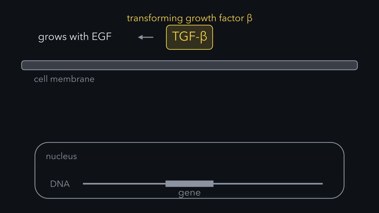 ▶ Watch The Ligand Only Introduces Two Kinases · 3:00
Read the transcript of “The Ligand Only Introduces Two Kinases”
Film transcript: The Ligand Only Introduces Two Kinases
Scientists purified a protein that, with EGF, made normal cells grow like tumour cells. They named it transforming growth factor beta. Then the same protein was caught holding other cells back. What does TGF-β beta do to a cell once it lands? Follow its message from the membrane to a gene. The receptors for TGF-β -beta are kinases of two types. The type 2 kinase is active before any ligand arrives. TGF-β -beta is a dimer, two chains joined together. It binds two type 2 receptors. Then the ligand and type 2 together recruit two type 1 receptors. Four receptor chains now surround one ligand. Type 2 adds phosphate to a stretch of type 1 just inside the membrane, called the GS domain. At rest, the GS domain holds the type 1 kinase in an inactive shape.
Phosphate ends that arrangement. The kinase turns on, and the stretch becomes a docking site for a Smad. Think of type 2 as a lit match, and type 1 as a fuse. The ligand only holds the match to the fuse. The active type 1 kinase phosphorylates a Smad. A Smad is itself a transcription factor, a protein that binds DNA. TGF-β beta receptors phosphorylate Smad2 and Smad3, on two serines at the end of the tail. A phosphorylated tail fits a pocket on a neighbouring Smad. So the Smads leave the receptor, and typically two of them join one Smad4. The complex collects in the nucleus and binds DNA. But it reads a sequence just a few base pairs long, and holds it weakly.
That sequence turns up millions of times in a human genome, so a Smad complex alone cannot pick a gene. It binds firmly mostly next to partner proteins, and each cell type makes its own partners. In many epithelial cells, the partners turn on p15 and p21, and the cell stops dividing. In a fibroblast, the same pathway turns on collagen genes. The ligand brings two kinases together. Type 2 phosphorylates type 1 and type 1 phosphorylates a Smad. The steps run one way. In each cell, the partners decide what the message of the Smad means. Keep this rule: the ligand only introduces two kinases.
Transforming growth factor beta (TGF-β) sends its message through a short relay: the ligand brings two receptor kinases together, the type II kinase phosphorylates the type I kinase, the type I kinase phosphorylates a Smad, and the Smad, joined by Smad4, goes to the nucleus and changes which genes are read. The same relay tells many healthy epithelial cells to stop dividing and tells fibroblasts to make collagen, and in late cancer it can work for the tumour. The name, transforming growth factor β, records a soft-agar assay from around 1980 in which the protein, helped by epidermal growth factor (EGF), made normal cells grow like cancer cells, and it hides the stop-dividing job. This lesson follows the relay first and then asks why one signal gives different answers in different cells.
The idea: the ligand only introduces two kinases
The insulin and epidermal growth factor (EGF) receptors from the receptor field guide are tyrosine kinases. Transforming growth factor beta (TGF-β) receptors are receptor serine/threonine kinases: each crosses the membrane once, and its inner part is a kinase that puts phosphate on serine or threonine. Humans have five type II receptors and seven type I receptors, shared among more than thirty related ligands: the TGF-βs, the activins, the bone morphogenetic proteins (BMPs) and others.
TGF-β itself is a dimer, two chains joined by a disulfide bond. Here is what it does:
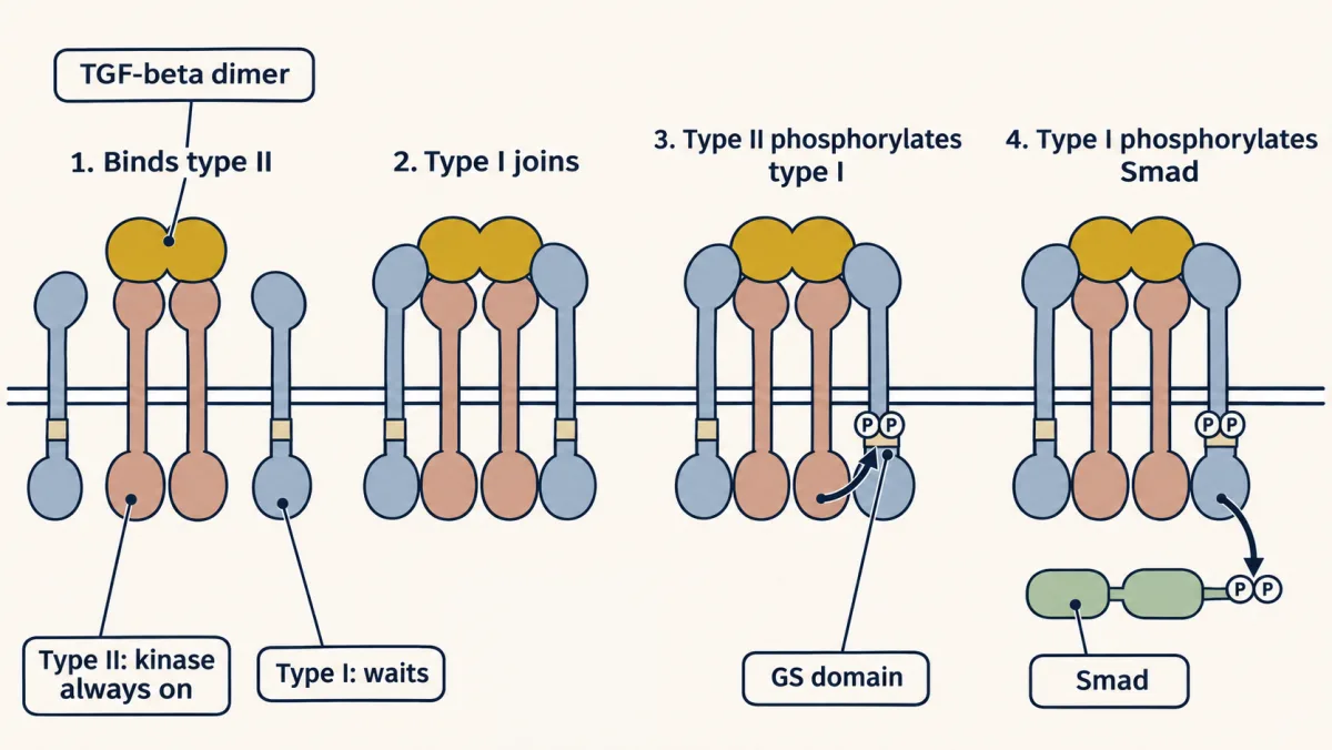
- The dimer binds two type II receptors. (TGF-β2 binds type II weakly and gets help from a co-receptor, betaglycan.) The type II kinase is active before any ligand arrives, and it stays active.
- The ligand and type II together recruit two type I receptors, so four receptor chains surround one ligand.
- Type II phosphorylates type I on a stretch just inside the membrane called the GS domain, named for its glycines and serines.
- The type I kinase, now active, phosphorylates the next protein in line, a Smad.
TGF-β + type II + type I → type II phosphorylates the GS domain of type I → type I phosphorylates a Smad
Think of type II as a lit match and type I as a fuse. The ligand’s only job is to hold the match to the fuse.
Jeffrey Wrana and Liliana Attisano worked this out in Joan Massagué’s laboratory between 1992 and 1994. First they showed that the type II receptor binds TGF-β but cannot signal without type I, and that type I cannot bind TGF-β without type II. Then they showed that phosphorylation runs one way, from the kinase that never turns off to the kinase that cannot start alone.
What keeps the fuse from lighting by accident? In a resting type I receptor the unphosphorylated GS domain holds the kinase in an inactive shape, and a small protein called FKBP12 sits on top of it. Phosphorylation by type II ends that arrangement and turns the GS domain into a docking site for Smads.
The pairing also decides which ligand a cell can hear. Attisano’s 1993 paper found that different pairings of a type I with a type II receptor gave complexes with different signaling capacities, so a cell answers only the ligands for which it displays a working pair. (Some relatives, such as BMP2, grip type I more tightly than type II, but the phosphate still passes from type II to type I.)
The messenger is the transcription factor
In the cAMP and Ras pathways several proteins stand between the receptor and a gene. Here there is one. The protein that the type I receptor phosphorylates is itself a transcription factor, a protein that binds DNA, on its own or in a complex, and changes how often a gene is read.
These proteins are the Smads, named for two genes found in animals: sma in the worm and Mad in the fly.
Humans have eight Smads in three groups:
- Receptor-activated Smads (R-Smads) are the receptor’s substrates. Transforming growth factor beta (TGF-β) and activin receptors phosphorylate Smad2 and Smad3. Bone morphogenetic protein (BMP) receptors phosphorylate Smad1, Smad5 and Smad8.
- Smad4 is the partner that both branches share.
- Smad6 and Smad7 are inhibitory Smads, covered below with the brakes.
An R-Smad has two folded domains. The front one, MH1, binds DNA in most Smads. The back one, MH2, binds other proteins and ends in a short tail. The type I receptor phosphorylates two serines at the very end of that tail. A phosphorylated tail fits a pocket on a neighbouring MH2 domain, so R-Smads leave the receptor and assemble with Smad4, typically two R-Smads to one Smad4. The complex collects in the nucleus and binds DNA.
The signal transducer and activator of transcriptions (STATs) of the last lesson take the same shortcut, except that a STAT is phosphorylated on tyrosine by a separate kinase, a Janus kinase (JAK), while a Smad is phosphorylated on serine by the receptor itself.
The BMP branch does much of its work in shaping the embryo, which belongs to the next unit.
Same signal, different answers
The MH1 domains of Smad3 and Smad4 read very short DNA sequences, such as the four base pairs GTCT, and they hold them weakly. (Smads also read short GC-rich sequences, which the bone morphogenetic protein (BMP) branch favours.) A sequence that short turns up by chance about once every 256 base pairs (4 × 4 × 4 × 4), which means millions of sites in a human genome. A Smad complex alone cannot pick out a gene.
It picks a gene with help. Smads usually bind firmly only next to partner transcription factors that read their own sequences nearby. Each cell type makes its own set of partners and keeps its own set of genes open for reading. The Smad complex carries one message, “transforming growth factor beta (TGF-β) is here.” The partners decide what that means in this cell.
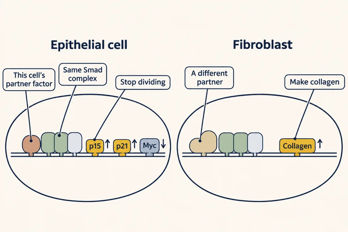
In many healthy epithelial cells it means “stop dividing.” Smad3 and Smad4, with their partners, switch on the genes for p15 and p21, two proteins that inhibit the cyclin-dependent kinases (the enzymes that push a cell through its division cycle). They also switch off the gene for Myc, a transcription factor that drives growth. In a fibroblast the same pathway switches on collagen genes, which is why TGF-β drives wound repair and, in excess, scarring.
This is also why the pathway has two faces in cancer, as Roberts and Lalage Wakefield set out in 2003. Early on, TGF-β acts as a tumour suppressor, because the cells still obey the stop order. Later the tumour cells no longer stop, and the TGF-β around them works in the tumour’s favour: it quiets nearby immune cells and can push tumour cells to loosen from their neighbours and move.
Three brakes, and what breaks
Read the transcript of “Three Brakes on a Strong Signal”
Film transcript: Three Brakes on a Strong Signal
A signal this strong has to be kept off most of the time. First brake: the ligand is stored locked. TGF-β is secreted still wrapped in the front part of its precursor chain. It stays bound and covers the surfaces receptors need. It is usually tethered to the extracellular matrix. For example, a cell's integrins can pull on the wrapper, or a protease can cut it. Active ligand is released locally. Second brake: the signal makes its own inhibitor. One of the genes that Smads switch on encodes Smad7. Smad7 binds the active type I receptor, and keeps the receptor's Smads away. That is negative feedback. Third brake: the parts are destroyed. Smad7 brings ubiquitin ligases called Smurfs to the receptor.
Smurfs tag the receptor, and some Smads, with ubiquitin for destruction. Scott Kern's laboratory found Smad4. It is inactivated in about half of pancreatic cancers. Mice made without the myostatin gene, in Se-Jin Lee's laboratory, have muscles two to three times the normal weight. In fibrodysplasia ossificans progressiva, muscle and connective tissue turn to bone. Nearly all patients with the classic form carry the same one amino acid change. It sits in the GS domain of a BMP type I receptor.
A signal this strong has to be kept off most of the time.
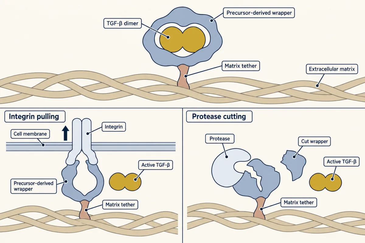
The ligand is stored locked. Transforming growth factor beta (TGF-β) is secreted still wrapped in the front portion of its own precursor chain, which has been cut off but stays bound and covers the surfaces that receptors need. This latent complex is usually tethered to the extracellular matrix, the protein mesh between cells. Active TGF-β is released locally, for example when a cell’s integrins (adhesion receptors) pull on the wrapper or a protease cuts it.
The signal makes its own inhibitor. One of the genes that Smads switch on encodes Smad7, which binds the active type I receptor and keeps R-Smads away from it. This is negative feedback, as in the signaling circuits lesson. Smad6 does a similar job, with a preference for the bone morphogenetic protein (BMP) branch.
The parts are destroyed. Smad7 also brings ubiquitin ligases called Smurfs (Smad ubiquitination regulatory factors) to the receptor. Smurfs tag the receptor, and some Smads, with ubiquitin for destruction, as covered in the lesson on ubiquitin and proteolysis.
Pancreatic cancer. Smad4 was found in 1996, in Scott Kern’s laboratory, as a gene missing from pancreatic tumours, and was first named DPC4, for deleted in pancreatic cancer. It is inactivated in about half of pancreatic cancers.
Muscle size. Myostatin, a TGF-β relative made by muscle, tells muscle to stop growing. Mice made without the gene in Se-Jin Lee’s laboratory in 1997 have muscles two to three times the normal weight.
Bone in the wrong place. In fibrodysplasia ossificans progressiva, bone progressively forms in muscle and connective tissue. Nearly all patients with the classic form carry the same single amino acid change in a BMP type I receptor called ACVR1. The change sits in the GS domain, the part of the receptor that keeps the kinase off until type II phosphorylates it.
Design problem
TGF-β stops a healthy epithelial cell from dividing, yet many late tumours make large amounts of it. Propose two different changes in a tumour cell that would let it ignore the stop signal. For each, say whether the cell would also lose TGF-β’s other effects, and which change you would expect a tumour to gain from more.
Saved in this browser only.
One way to do it
Change 1: cut the relay. Lose the type II receptor or Smad4, and the message never reaches the arrest genes. The cost is that the cell also loses most or all of its other Smad-dependent responses, including any that a tumour could use, such as the programme that lets cells move. Tumours do take this route: the type II receptor gene is inactivated in a subset of colon cancers.
Change 2: cut only the arrest arm. Leave the receptors and Smads intact and remove what they act on: delete the p15 and p21 genes, or drive Myc so hard by another route that the Smads cannot shut it off. The cell still hears TGF-β but no longer stops, and its other responses survive.
Which helps the tumour more? Plausibly the second, because the cell escapes the brake and keeps a signal it can turn to its own use. A cell that took the first route still benefits from the TGF-β around it, which acts on immune cells and other neighbours whose receptors are normal.
Going deeper
How the name was earned: the soft-agar story
Around 1980 the laboratory test for a cancer-like cell was a dish of soft agar. A normal fibroblast (a connective-tissue cell) divides only when it is anchored to a surface. A transformed cell, one with the habits of a tumour cell, needs no anchor and grows into a colony while suspended in the gel. De Larco and Todaro had reported that cells transformed by a sarcoma virus release something that makes normal cells pass this test. The activity was called a transforming growth factor, and the name carried an assumption: this is what cancer cells make.
At the National Cancer Institute, Anita Roberts and Michael Sporn set out to purify it. In 1980 Roberts extracted the activity from cells transformed by a virus or by chemicals. In 1981 the same kind of extraction pulled an activity that needed help from epidermal growth factor (EGF), which the authors called a new class of transforming growth factors, out of tissues of healthy adult mice, among them kidney, liver, heart and brain. Normal organs were making the “cancer” factor. In 1983 the same group showed that the original transforming activity was two proteins: TGF-α, a relative of EGF, and transforming growth factor beta (TGF-β), which made normal cells form large colonies in soft agar only when EGF or TGF-α was also present.
Then the protein was caught doing the opposite job. In 1984 Tucker, Shipley, Moses and Holley reported that a growth inhibitor from kidney epithelial cells (the cells that line surfaces such as skin and gut) was closely related to TGF-β. In 1985 Roberts, Sporn and colleagues called their purified protein “a bifunctional regulator of cellular growth”: it raised colonies in one assay and held cells back in another, depending on the cell and the conditions.
Finding the Smads: a name from a worm and a fly
These proteins are the Smads. The name joins two genes that geneticists found in animals: sma, from the worm C. elegans, whose mutants are small, and Mad, from the fruit fly. In 1996 Macías-Silva, Wrana, Attisano and colleagues, by then in Toronto, reported that the TGF-β receptor phosphorylates a human relative they called MADR2, now Smad2. Other laboratories, Massagué’s among them, reported related Smads the same year.
The ACVR1 change in fibrodysplasia ossificans progressiva
The change is histidine for arginine at position 206 (R206H), as the laboratory of Eileen Shore and Frederick Kaplan reported in 2006.
Your notes
Select any text in the lesson to highlight it or add a note to it. Your highlights and notes are saved in this browser and collected in your notebook.
This browser is not letting the site save anything (a private window or strict privacy settings), so highlights and notes will be lost when you leave the page.
How we measure it
Soft-agar colony assay
Cells are suspended in a soft gel where they cannot attach to a surface. Normal fibroblasts stay single, while transformed cells divide into visible colonies, so counting colonies measures growth without anchorage. This assay defined the transforming growth factors, and it cannot tell a lasting change in a cell from a response that lasts only while a factor is present.
Affinity cross-linking of labelled ligand
TGF-β carrying radioactive iodine is added to cells, and a chemical cross-linker ties it to whatever protein it is touching. On a gel each receptor that held the ligand shows up as a labelled band. Used on mutant cell lines that lack one receptor type, it shows which receptor needs the other in order to bind ligand.
Kinase-dead receptor mutants
Changing the lysine that a kinase uses to hold ATP gives a receptor that usually still folds, pairs and binds ligand but cannot transfer phosphate; experimenters check that it does. Asking which receptor becomes phosphorylated when one partner or the other is kinase-dead shows the direction of the relay.
Genetic screens in flies and worms
Animals are mutated at random and kept if their defects resemble the loss of a known signal, such as small worms or flies with defects like those of dpp mutants. The genes found this way are strong candidates for the pathway before anyone knows their biochemistry. Screens of this kind supplied Mad and the sma genes.
Method films · the methods library
- Engineered mutants: site-directed, dominant-negative and constitutively active 4:25
- Family linkage and positional cloning 3:52
- Forward genetic screens 4:07
- Gene cloning: vectors, libraries and screening 4:04
- Gene targeting: knockout and knock-in mice 4:13
- Protein purification and column chromatography 4:14
- Receptor binding and dose-response analysis 4:18
Practice questions on this lesson (3) → MCAT-style, original, each with an explanation
Where this lesson fits
Before thisReview first
- Receptor Tyrosine KinasesThis course · Lesson 18
You need the receptor kinase idea from there to see how this serine/threonine receptor pair differs.
- Cytokine Receptors and JAK–STATThis course · Lesson 22
The STAT shortcut, where the messenger is itself a transcription factor, is compared directly with Smads here.
- Ubiquitin and Regulated Proteolysis as SignalsThis course · Lesson 8
Smurfs tag the receptor and Smads for destruction, using the ubiquitin tagging described there.
After thisStudy next
- Wnt: β-Catenin and Planar PolarityThis course · Lesson 24
It takes up another signal whose message ends in gene control and that cancer can corrupt.
- Oncogenes: When the Switch SticksThis course · Lesson 41
It takes further the idea that a broken signalling part, like Smad4 loss, drives cancer.
- The Shape of a Signal: Size, Duration, Frequency and PlaceThis course · Lesson 35
It explains how one cell reads a signal's size and duration, extending why one signal gives different answers.
Elsewhere on the siteFilms, simulations and method films in other courses
- One Rise, One FallFilm · Cell Biology
The cell-cycle brake that TGF-β sets in healthy epithelial cells acts on the cyclin-CDK machinery shown there.
- Cre-lox and binary expression systemsMethod film
Deleting a pathway component only in chosen cells is how its early and late roles in tumours are separated.
Suggested by an AI model from the lessons themselves, then checked. How we use AI
Finish line
Test yourself
8 questions
Practice and MCAT-style, with explained answers.
Next · Lesson 24
Wnt: β-Catenin and Planar Polarity
Why would a cell make a protein all day only to destroy it?
Continue →