Physiology · Unit 2: Muscle
Smooth Muscle and the Myogenic Response
Rewritten on : new text, figures and flashcards. Your notes and highlights stay, but the text around them may have moved.
Raise the pressure inside a small artery and it narrows against the stretch instead of simply widening. What is the muscle sensing?
Pause when the reading reaches a film, and offer to play it. Saved on this device.
Watch the film now, or keep listening
Builds on:Muscle, Bone and MovementTransport Across MembranesWhy these?
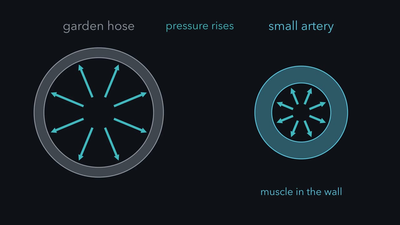 ▶ Watch Smooth muscle force is a balance · 2:42
Read the transcript of “Smooth muscle force is a balance”
Film transcript: Smooth muscle force is a balance
Raise the pressure inside a garden hose, and it bulges. Raise the pressure inside a small artery, and the muscle in its wall pushes back. The artery narrows against the stretch. That answer is built into the muscle, not wired in from the brain. So what is the muscle sensing, and how does it make force? This muscle is smooth muscle. It has no troponin, so calcium cannot act on the thin filament. The switch sits on myosin instead. Calcium binds calmodulin, and calcium and calmodulin together activate myosin light chain kinase. The kinase puts a phosphate on the regulatory light chain, and then the motor can cycle on actin. But the kinase is only half the story.
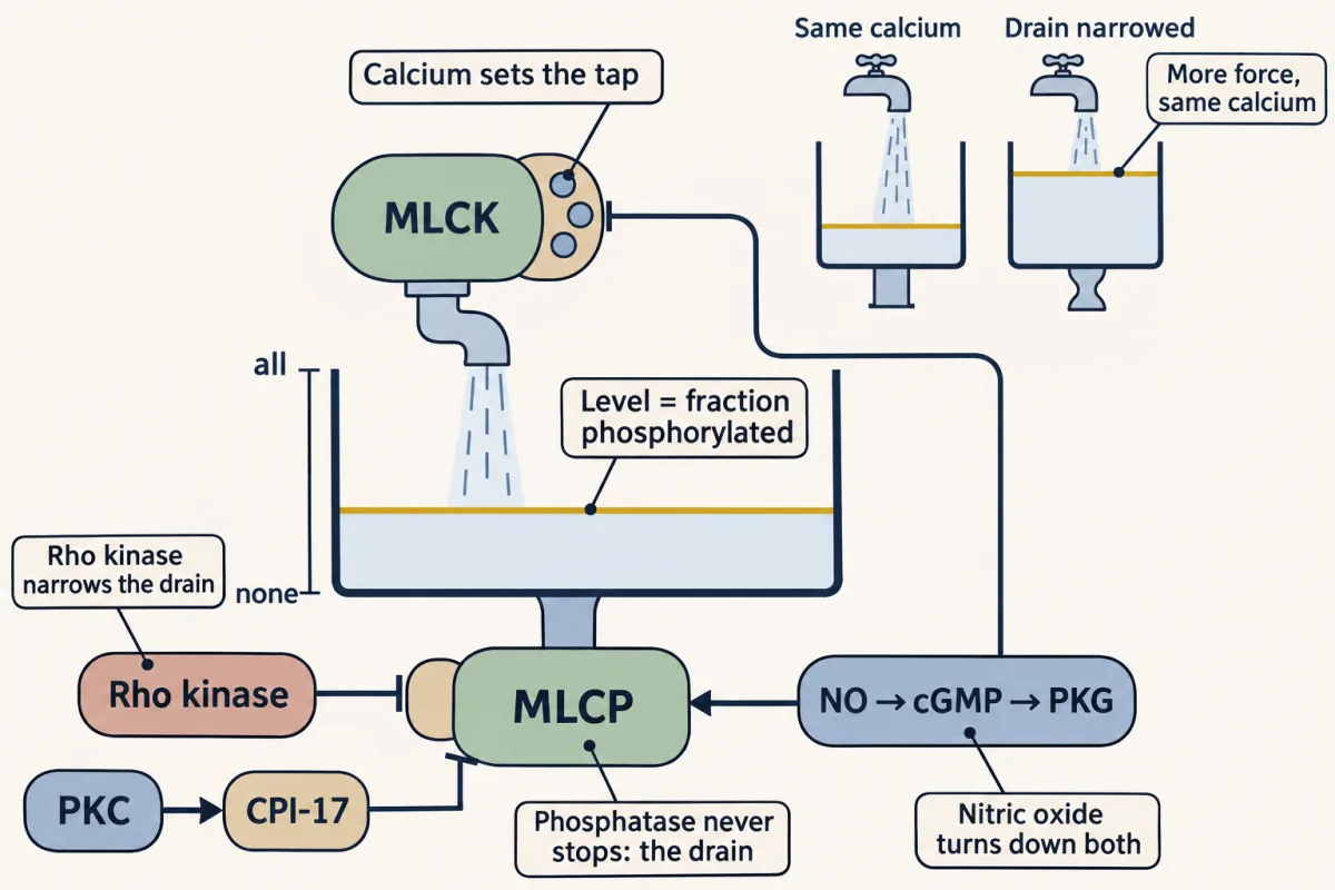
Myosin light chain phosphatase takes the phosphate off, and it works the whole time. So force follows the balance of kinase and phosphatase. Calcium sets the kinase, so anything that changes the phosphatase changes force at the same calcium. That is calcium sensitization: more force at an unchanged calcium concentration. Now move the balance. RhoA activates Rho kinase, and Rho kinase inhibits the phosphatase. More phosphate stays on myosin, so force rises at the same calcium. The opposite arrow runs through nitric oxide, cGMP, and protein kinase G. In rabbit artery, the result is a lower calcium and, the authors read, an indirect activation of the phosphatase.
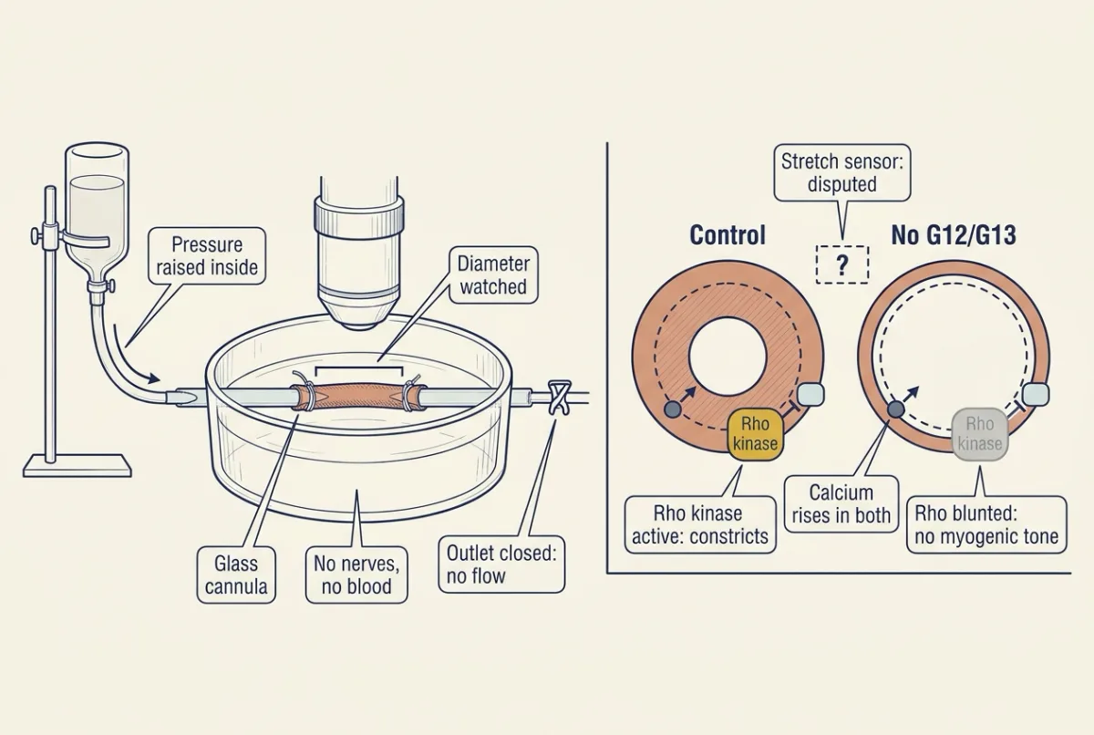
In mouse mesenteric arteries on a pressure myograph, calcium rose with pressure as normal. Arteries lacking G12 and G13 in smooth muscle showed no active constriction and much less active RhoA. In control arteries, a Rho kinase inhibitor blocked the response. So this bed needs two things: a calcium rise, and Rho-dependent sensitization. How the pathways are wired upstream is unsettled. Flow in an idealized rigid arteriole is proportional to the pressure difference times the fourth power of the radius. Take made-up numbers: 60 millimetres of mercury, and a radius of 100 micrometres. Raise the difference to 80 millimetres of mercury, and a passive vessel stretched 5 % carries 62 % more flow.
To hold flow constant, the radius must fall to 93.1 micrometres, a 6.9 % narrowing. Keep one rule: calcium sets the switch, and sensitivity sets the gain.
Push harder on the inside of a small artery and its muscle pushes back. A garden hose does the opposite: raise the pressure and it bulges. The artery’s answer is built into its muscle rather than wired in from the brain. The same machinery runs the gut, airways and bladder.
So what is the muscle sensing? Stretch of its own wall. Which molecule detects the stretch is still disputed; how the muscle turns stretch into force is not. It runs through two enzymes, one that puts a phosphate on myosin and one that takes it off.
The one idea to carry through this lesson. Each myosin light chain is switched on by a kinase (myosin light-chain kinase, MLCK) and off by a phosphatase (myosin light-chain phosphatase, MLCP), over and over. The fraction carrying a phosphate, p, settles at a balance point where the two rates are equal: p = kMLCK / (kMLCK + kMLCP). Calcium sets the kinase; other signals, Rho kinase above all, set the phosphatase. So the same calcium can buy more force or less, and most stimuli, whether a drug, a nerve or a stretched wall, act by moving calcium, the phosphatase, or both (Going deeper lists the exceptions). The rule sets phosphorylation, not force directly: once force is built, latch lets it outlast the phosphate.
The switch on myosin
Smooth muscle has no troponin, so calcium cannot act on the thin filament as it does in skeletal and cardiac muscle. The switch sits on myosin: calcium binds calmodulin, calcium-calmodulin activates myosin light-chain kinase (MLCK), and MLCK puts a phosphate on the 20-kDa regulatory light chain, LC20, after which the motor can cycle on actin. Muscle and movement gave this switch its first look, and calcium signaling covers calmodulin and the stores.
Calcium arrives by two routes. Depolarization opens L-type channels (CaV1.2) and calcium flows in down its electrochemical gradient: electromechanical coupling. Or an agonist on a Gq-coupled receptor makes inositol trisphosphate (IP3), which opens release channels on the sarcoplasmic reticulum (phospholipase C, IP3, DAG and PKC). That route can contract the cell with little change in membrane potential: pharmacomechanical coupling.
Set the three muscle types side by side.
| Muscle | Where the calcium comes from | What calcium acts on |
|---|---|---|
| Skeletal | The store only: a voltage sensor in the T tubule is linked mechanically to the release channel of the sarcoplasmic reticulum, so no calcium from outside is needed | Troponin, on the thin filament |
| Cardiac | Calcium entering during the action potential triggers a larger release from the store (cardiac electrophysiology and the ECG) | Troponin, on the thin filament |
| Smooth | Both: entry through L-type channels and release from the sarcoplasmic reticulum | Calmodulin, which switches on MLCK and so the myosin motor itself |
Smooth muscle’s cross-bridges also cycle more slowly, so it generally shortens slowly and, above all in tonic smooth muscle, holds force cheaply (the latch state, below).
The cell’s build suits that job. A smooth muscle cell is a spindle, a few micrometres across and roughly 30 to 200 micrometres long, with one nucleus, and it has no stripes, because its actin and myosin are not arranged in sarcomeres. Skeletal muscle gets its length-tension curve from that regular overlap; smooth muscle has no such register. Its thin filaments are anchored instead in dense bodies, protein clusters in the cytoplasm, and in dense plaques on the inside of the membrane. Myosin filaments run among them at an angle, and intermediate filaments tie the dense bodies together, so when myosin pulls, the whole cell shortens and bulges. Because the filaments are not stacked in sarcomeres, the cell can develop force over a much wider range of lengths than a sarcomere can, which matters for an organ that fills.
The balance point: kinase against phosphatase
A second enzyme, myosin light-chain phosphatase (MLCP), takes the phosphate back off LC20, and it never stops working; in permeabilized rabbit arterial strips that removal can be followed directly. What you measure at any moment is the fraction of light chains that happens to carry a phosphate.
Picture a sink with the tap running and the drain open. The water settles at the level where it drains out as fast as it pours in. Light-chain phosphorylation settles the same way: at the level where the kinase adds phosphates exactly as fast as the phosphatase removes them.
In words The phosphorylated fraction equals the kinase's activity divided by the kinase's activity plus the phosphatase's activity.
- p“p”
- the fraction of LC20 carrying a phosphate0 to 1
- kMLCK“k sub M L C K”
- how fast the kinase phosphorylates a light chain that has no phosphate; calcium-calmodulin sets itper s
- kMLCP“k sub M L C P”
- how fast the phosphatase removes the phosphate; Rho kinase, PKC and nitric oxide set itper s
Sanity check Only the ratio of the two activities matters: double both and p does not change. In the model, the kinase off means p = 0, and the phosphatase stopped means p climbs toward 1 at any kinase activity above zero. Real muscle keeps a little phosphorylation at rest, from kinases that do not need calcium (Going deeper).
Try one number
Illustrative numbers, not measurements. Let the kinase run three times as fast as the phosphatase: kMLCK = 3 and kMLCP = 1 per second.
p = 3 / (3 + 1) = 0.75, so three light chains in four carry a phosphate at any moment.
Where it comes from optional
Step 1, phosphates go on at a rate kMLCK × (1 − p), because only light chains without a phosphate can receive one, and come off at a rate kMLCP × p.
Step 2, at the balance point the two rates are equal: kMLCK × (1 − p) = kMLCP × p.
Step 3, collect the p terms, p × (kMLCK + kMLCP) = kMLCK, and divide.
Need a refresher? rates and “per”
Predict. Start with the kinase and the phosphatase equally active, so p = 0.5. A signal halves the phosphatase and leaves calcium alone. How much would calcium have to raise the kinase to reach the same p?
Check your prediction
It would have to double it. Halving the phosphatase gives p = 1 / (1 + 0.5) = 0.67. Doubling the kinase gives p = 2 / (2 + 1) = 0.67. Halving the drain does what doubling the tap does. That is why a signal that never touches calcium can still raise force.
Calcium sensitization is any change that buys more force, or more phosphorylation, at an unchanged calcium concentration; desensitization buys less. The film puts it as “calcium sets the switch, and sensitivity sets the gain”: gain here means how much force each level of calcium buys. On a graph of force against calcium, sensitization mainly slides the curve to the left, so less calcium gives the same force.
Predict. Permeabilize a strip of artery so that the bath, not the cell, sets the calcium inside, and hold it at a moderate level. Add a drug that activates the GTPase RhoA. What happens to LC20 phosphorylation and to force, and how do you know calcium did not do it?
Check your prediction
Both rise. RhoA activates Rho kinase (ROCK), which phosphorylates MYPT1, the targeting subunit of the phosphatase, and inhibits MLCP. In the ratio, kMLCP falls while kMLCK stays put, so p rises. Calcium cannot have risen: the bath holds it constant. That is the experiment that defines sensitization.
The clearest early evidence came from the Somlyo laboratory, which ran the mirror image of that experiment in permeabilized rabbit portal vein. With the kinase stopped, so that only the phosphatase could act, Kitazawa, Masuo and Somlyo found that GTP-gamma-S, which keeps G proteins active, slowed dephosphorylation and relaxation to about half the control rates: a G protein can raise force by slowing the drain. The route is now drawn through RhoA, a GTPase switch (GTPase switches covers the cycle and its exchange factors, and integrins and Rho GTPases the same kinase elsewhere), and Rho kinase. Protein kinase C gives a second brake on the phosphatase: it phosphorylates CPI-17, a protein that inhibits MLCP once it is itself phosphorylated.
Relaxation moves both sides of the ratio at once. Nitric oxide from the endothelium activates soluble guanylate cyclase (through the enzyme’s heme group, shown with purified bovine enzyme), and the cGMP that follows activates protein kinase G (nitric oxide and cGMP). In muscle that lowers calcium, which turns the tap down, and raises phosphatase activity, which opens the drain. In permeabilized rabbit artery with calcium clamped, the cGMP analog 8-bromo-cGMP still relaxed the muscle, which the authors read as indirect activation of the phosphatase.
In the Hai-Murphy model below, force rises steeply as phosphorylation rises from zero and then levels off, because of latch, so a muscle can hold most of its force with only a modest fraction of its light chains phosphorylated.
Latch: force outlasts the phosphate
Predict. Stimulate a strip of arterial muscle and keep the stimulus on. LC20 phosphorylation rises first, then falls back toward low values. What would you expect force to do?
Check your prediction
If force simply tracked phosphorylation, it would follow it down. It does not: force stays near its peak. Something holds the load after the phosphates come off, and that state is called latch.
Hai and Murphy proposed a kinetic model in 1988. Myosin heads move among four states: phosphorylated or not, attached or not. Kinase and phosphatase act on heads whether or not they are bound to actin, so the balance-point rule still sets p. But a head that loses its phosphate while attached does not release at once. It becomes a latch bridge, whose slow detachment is the rate-limiting step, so attached heads keep bearing load while the cycling rate, and ATP cost, fall. The slow release is an assumption of a model fitted to phosphorylation and stress data, not a structure anyone has imaged.
Latch is not a state without ATP: phosphorylated cross-bridges keep cycling, latch bridges let go slowly, and calcium pumps keep working, so latch is economy, not shutdown. Nor is it rigor, the stiff state of a muscle whose ATP has run out.
In 2022 Hammell and colleagues tested the prediction on a few purified myosin molecules in a laser trap, injecting the phosphatase once force had reached a plateau. Force was held for a median of 32 seconds with tonic myosin (pig stomach, from muscle that holds tone) and 21 seconds with phasic myosin (chicken gizzard, from muscle that contracts in bursts). Dephosphorylation was timed indirectly, in a parallel motility assay rather than on the trapped heads, with a midpoint near 27 seconds; only the tonic hold outlasted that clock, so it is the more convincing. A further limit: purified non-human proteins, not an intact vessel.
The film says force follows the balance of kinase and phosphatase. That holds while force is being built; after that, latch bridges hold it as the phosphates come off.
Arterioles answer stretch
In his 1902 paper in the Journal of Physiology, William Bayliss reported that the muscular coat of an artery contracts when stretched and relaxes when tension falls, independently of the central nervous system. He saw it first in a rabbit hind limb with its nerves cut, where a passive tube should simply have followed the pressure, then in dogs and cats. Limb volume sums arteries, veins and tissue, so his tracings could not say which vessels responded, and the tracing from an excised dog carotid he described was spoilt and not reproduced. Bayliss appears again, with Starling, in signals and distance.
The clean answer to the opening question came later, from isolated arteries on a pressure myograph: tied between two glass cannulas and pressurized, with no nerves and no flow, they still narrow. The myogenic response belongs above all to small resistance arteries and arterioles, and stretch moves both sides of the ratio.
The modern version adds a mouse. Chennupati, Wirth and colleagues deleted G-protein alpha subunits in the smooth muscle of adult mice, mounted mesenteric arteries and raised the pressure inside them from 20 to 140 mmHg, measuring diameter, a calcium dye signal and active RhoA.
Predict. In arteries lacking G12 and G13, calcium rises with pressure as normal. Does myogenic tone appear?
Check your prediction
No. Arteries lacking G12 and G13 lost their active myogenic constriction and had strongly reduced RhoA activity, yet calcium rose with pressure as normal. In control arteries a Rho kinase inhibitor blocked the response. In the rule’s terms, the tap opened but the drain was never narrowed, and calcium alone was not enough.
Two requirements, then, in this bed: a calcium rise and Rho-dependent sensitization. What senses the stretch, and which G proteins carry the signal, is disputed between mouse studies (see Going deeper).
Why a small narrowing matters
Treat an arteriole as a rigid cylinder of fixed length carrying steady laminar flow of a Newtonian fluid, assumptions blood and arterioles both violate, and Poiseuille’s law applies (blood and vessels). The example shows what a passive vessel would do; the Predict asks what the muscle must do instead.
In words The new flow over the old equals the new pressure difference over the old, times the ratio of the radii raised to the fourth power.
- Q“Q”
- flow through the segment; from Poiseuille's law, Q = (π × ΔP × r⁴) / (8 × viscosity × length)volume per s
- ΔP“delta P”
- the pressure difference driving flow along the segmentmmHg
- r“r”
- the radius of the lumenµm
Sanity check Viscosity and length cancel in the ratio because they do not change. Resistance scales as 1 / r⁴, so flow equals ΔP divided by resistance, as Q = ΔP / R requires.
Try one number
Made-up but convenient numbers: ΔP1 = 60 mmHg across the segment and r1 = 100 µm, with downstream and outside pressures fixed, so a rise in inlet pressure raises both the driving gradient and the transmural pressure (inside minus outside) that stretches the wall.
Raise the gradient to 80 mmHg. If the vessel were passive and distended by 5 percent: Q2 / Q1 = (80 / 60) × 1.05⁴ = 1.62, a 62 percent rise in flow.
Need a refresher? rates and “per”
Predict. The pressure difference has risen by a third, from 60 to 80 mmHg. How much must the radius shrink to hold flow at its old value?
Check your prediction
Only 6.9 percent. For flow to stay constant, (r2 / r1)⁴ = 60 / 80 = 0.75, so r2 = 100 × 0.751/4 = 93.1 µm. Resistance rises as 1 / r⁴, so R2 / R1 = 1 / 0.75 = 1.33, the same one-third increase as the pressure gradient. The fourth power turns a small narrowing into a large change in resistance.
Steady flow is a result of answering stretch, not the signal the muscle reads: a pressure myograph has no flow at all.
Muscle side: the tap and the drain on the light chain
Illustrative rates in made-up units; the preset values are made up too, and only their directions come from the lesson. Latch, calcium-independent kinases and thin-filament regulation are left out (Going deeper).
- Flow relative to baseline, Q / Q₁
- Resistance relative to baseline, R / R₁
- Change in radius from 100 micrometres
In this sketch: idealized rigid tube with Poiseuille flow. The tube has fixed length and viscosity, steady laminar flow of a Newtonian fluid, and downstream and outside pressures held fixed, so the pressure difference is the only driver; blood and arterioles both violate these assumptions, so the numbers are a sketch of the logic, not a prediction for a real vessel. The baselines (60 mmHg, 100 micrometres) are the lesson’s made-up but convenient numbers; the slider ranges are made up for the sketch; the toggle simply sets whatever radius keeps flow at baseline.
Rule to keep: flow scales with the fourth power of radius, so a small change in radius can cancel a large change in pressure. Holding flow while the difference rises from 60 to 120 mmHg takes only a 16 percent fall in radius. The muscle reaches a narrower radius by moving calcium and the phosphatase, not by measuring flow.
Holding flow steady across a range of pressures is autoregulation, produced by several mechanisms together; its metabolic and endothelial parts belong to resistance, compliance and local flow, and the renal version, with tubuloglomerular feedback, to glomerular filtration. Three inputs act on the same kinase-phosphatase balance: pressure raises calcium and sensitization, endothelial nitric oxide lowers both, sympathetic alpha-1 input raises both. Net diameter reflects their combined effect.
Use that to reason about raised pressure. In hypertensive rodents small arteries often show more myogenic tone at a given pressure, and in people with essential hypertension small arteries show inward remodelling of the wall (how much myogenic tone contributes in people is less settled), so the lumen is narrower at the same transmural pressure; by the fourth-power law that raises segment resistance steeply, and higher resistance at a given cardiac output holds mean arterial pressure up. Practise the directions: tone up, lumen down, resistance up.
A fall in pressure runs the loop the other way: after blood loss, pressure in a small artery falls, wall tension falls, the muscle tends to relax and lower segment resistance, while the baroreflex raises sympathetic outflow (blood pressure regulation) and alpha-1 input pulls the diameter back down.
Hollow organs and nerves
A hollow organ that fills is stretched steadily, and visceral smooth muscle can relax after being stretched, a behaviour called stress relaxation: tension falls back after the stretch, so pressure inside rises much less during filling than in a stiff container. A small resistance artery does the opposite when transmural pressure rises: it contracts.
In single-unit (visceral) smooth muscle, gap junctions couple neighbouring cells electrically, so excitation spreads and the cells contract as one mass; the gut and uterus are the usual examples. Some of these tissues also carry pacemaker activity, and the gut’s slow waves belong to gut motility and secretion. In multi-unit smooth muscle, cells have little electrical coupling and each is driven by its own nerve terminals, allowing finely graded control; the iris and the piloerector muscles are the usual examples. Real tissues fall between the two types: bladder and arterial muscle cells are less tightly coupled, and in arteries the endothelium carries much of the electrical spread.
Receptors, not nerves in general, set the direction, so “sympathetic input contracts smooth muscle” is wrong as a rule. Norepinephrine on alpha-1 adrenoceptors contracted isolated rat aorta and mesenteric resistance vessels, while in isolated human detrusor strips noradrenaline relaxed the muscle, mainly through beta-3 adrenoceptors as the authors suggested. The receptor families are mapped in the hypothalamus and the autonomic nervous system. Name the receptor and the preparation, and the contradictions go away.
Design problem
Does nitric oxide relax arterial smooth muscle by lowering calcium, by lowering calcium sensitivity, or both? You have endothelium-denuded rabbit femoral arteries, a nitric oxide donor, an alpha-1 agonist, the calcium dye Fura-2, alpha-toxin to permeabilize strips, and antibodies reporting LC20, MYPT1 and CPI-17 phosphorylation. Design the experiments.
Saved in this browser only.
One way to do it
Start with intact, denuded strips, confirming the denudation (acetylcholine no longer relaxes them). Contract them with the alpha-1 agonist, then add the donor while recording force and Fura-2 together. A calcium-force curve in the same strips says what a given fall in calcium should cost in force; more relaxation than that points to lost sensitivity too.
Then clamp calcium: in alpha-toxin-permeabilized strips the bath sets calcium, so if the donor or cGMP still relaxes them, desensitization is part of the mechanism at fixed calcium. Lee, Li and Kitazawa used that design in 1997 (the 8-bromo-cGMP result above).
To ask whether the phosphatase speeds up, reverse the 1991 logic: phosphorylate LC20, remove calcium and ATP, block MLCK, and follow dephosphorylation with and without cGMP. Faster dephosphorylation means more phosphatase activity. Follow MYPT1 and CPI-17 too; the 2009 study found CPI-17 phosphorylation at a minimum within 15 seconds of the donor in this preparation, in parallel with the fall in calcium, which the authors read as a consequence of suppressed calcium release.
Controls: time-matched strips with no donor, a guanylate cyclase inhibitor to confirm the cGMP route, the same starting force each time. Limits: a donor on a denuded artery is not endogenous nitric oxide in a flowing arteriole, rabbit is not human, and permeabilization removes ion gradients and voltage control, though receptors and G proteins remain coupled. Expect both arrows at once.
Going deeper
What the balance-point rule leaves out
The rule treats calcium-calmodulin as what sets the kinase and the phosphate on LC20 as the only switch. It leaves out thin-filament proteins such as caldesmon and calponin, and kinases that phosphorylate LC20 without calcium. It also treats each enzyme’s rate as proportional to its substrate, which a saturated enzyme does not obey.
The Somlyo preparation
The portal vein strips were permeabilized with staphylococcal alpha-toxin. After letting the light chain become phosphorylated, Kitazawa, Masuo and Somlyo removed calcium and ATP and blocked myosin light-chain kinase (MLCK) with the inhibitor ML-9, so that any change in dephosphorylation had to come from the phosphatase.
Latch in numbers
The trap buffer held 200 micromolar MgATP, and the tonic myosin came from pig stomach fundus. The interquartile ranges of the force holds were 27 to 48 seconds for the tonic and 15 to 27 seconds for the phasic myosin, five assays each. In the motility assay, the midpoint of the loss of actin movement came at about 27 seconds for the tonic and 26 seconds for the phasic myosin, so the authors read the tonic hold as force kept during dephosphorylation and the phasic hold less convincingly.
In one illustrative run of the Hai-Murphy model (phosphatase 0.5, attachment 0.4, detachment 0.1 and latch detachment 0.01 per second), a steady 17 percent phosphorylation held about 86 percent of the force held at 67 percent, most of it on latch bridges. The figure depends on the constants: halving or doubling latch detachment moves it to about 92 or 76 percent.
Bayliss’s experiments in detail
In the rabbit, Bayliss lowered systemic pressure by stimulating the central end of the depressor nerve. In dogs and cats he raised pressure by stimulating the splanchnic nerves or by asphyxia, and lowered it by compressing the aorta. A footnote in the 1902 paper points to a preliminary report of March 1901.
The mouse study and a conflicting result
Chennupati, Wirth and colleagues raised pressure in 20 mmHg steps, reading active diameter in calcium-containing and passive diameter in calcium-free solution. Control arteries peaked at about 80 to 120 mmHg. Arteries lacking Gq and G11 behaved like controls, and cerebral arteries kept about half their response. L-type plus TRP channel blockers together cut the calcium rise by about half.
In 2022 Cui and colleagues found that deleting the angiotensin II type 1a receptor in smooth muscle left about 35 percent of wild-type constriction in mouse mesenteric arteries, which still constricted to a depolarizing potassium solution, and that a Gq/11 inhibitor abolished the response in the beds they tested. Deletion and inhibition, and different vessel orders and beds, are not interchangeable, so the upstream wiring is unsettled.
More Poiseuille arithmetic
Overshoot and flow falls: at r2 = 90 µm with the gradient at 80 mmHg, Q2 / Q1 = 1.333 × 0.6561 = 0.875, 12.5 percent below baseline. Tone is a separate quantity: 200 µm passive and 160 µm active at one pressure give tone = 100 × (200 − 160) / 200 = 20 percent of diameter, while flow at the same gradient would be 0.8⁴ = 0.41 of passive.
Receptors by preparation
In the rat vessels, alpha-1 activation mobilized calcium from inside and outside the cell; in the human detrusor strips, relaxation by noradrenaline was concentration-dependent. Beta receptors classically signal through cAMP and protein kinase A (cAMP and PKA), though in rat bladder strips cAMP played only a minor part in relaxation by the beta agonist isoproterenol.
Your notes
Select any text in the lesson to highlight it or add a note to it. Your highlights and notes are saved in this browser and collected in your notebook.
This browser is not letting the site save anything (a private window or strict privacy settings), so highlights and notes will be lost when you leave the page.
How we measure it
Pressure myography
A small artery is tied between two glass cannulas in a warmed bath, pressurized from one side with no flow, and its diameter is tracked by a camera. Comparing the diameter in calcium-containing solution (active) with that in calcium-free solution (passive) at the same pressure gives myogenic tone as 100 x (passive - active) / passive.
Permeabilized (skinned) smooth muscle
A toxin such as staphylococcal alpha-toxin punches pores in the membrane of a muscle strip, so the bath sets calcium inside the cells while receptors and G proteins stay attached. Force and light-chain phosphorylation can then be measured at a clamped calcium, which separates calcium sensitivity from calcium concentration.
Laser trap with an in vitro motility assay
A focused laser holds a bead attached to an actin filament over a few purified myosin molecules, and the bead's displacement reports their force. A parallel motility assay, in which myosin on a surface moves fluorescent actin, reports how phosphorylation changes the motor's activity over time.
Limb plethysmography
A limb is sealed in a rigid volume recorder, so changes in its blood content move a recording lever. With arterial pressure recorded at the same time, it shows whether the vessels of the limb widen or narrow, but it sums arteries, veins and tissue and does not measure one vessel.
Practice questions on this lesson (3) → MCAT-style, original, each with an explanation
Where this lesson fits
Before thisReview first
- Muscle, Bone and MovementOrganismal Biology · Starting college
It first showed the calcium switch and filament sliding in skeletal muscle, which you compare with smooth muscle here.
- Transport Across MembranesCell Biology · College: the core
Calcium entering through L-type channels down its electrochemical gradient is used here without being re-taught.
After thisStudy next
- Resistance, Compliance and Local Blood FlowThis course · Lesson 8
It uses the myogenic response and Poiseuille's fourth-power law to explain how arteriole radius sets local flow and resistance.
- Regulating Arterial PressureThis course · Lesson 10
Arteriole tone set by stretch, nitric oxide and sympathetic input here feeds into how resistance holds arterial pressure.
- Glomerular Filtration and Renal Blood FlowThis course · Lesson 15
The renal arteriole's myogenic response applies this stretch-contraction idea to keep filtration steady.
Suggested by an AI model from the lessons themselves, then checked. How we use AI
Finish line
Test yourself
9 questions
Practice and MCAT-style, with explained answers.
Next · Lesson 5
Cardiac Action Potentials and the ECG
A ventricular cell stays depolarised for about a fifth of a second. Why can't your heart cramp into a sustained contraction the way a skeletal muscle can?
Continue →