Cell Signaling · Unit 3: G-protein-coupled receptors and second messengers
PLC, IP3, DAG and Protein Kinase C
Rewritten on : new text, flashcards and film. Your notes and highlights stay, but the text around them may have moved.
How does a kinase know that a receptor has just cut a lipid, here and now?
Pause when the reading reaches a film, and offer to play it. Saved on this device.
Watch the film now, or keep listening
Builds on:GPCRs and Trimeric G ProteinsProtein Modules: SH2, SH3, PTB, PDZ and PHLipids as Anchors and AddressesWhy these?
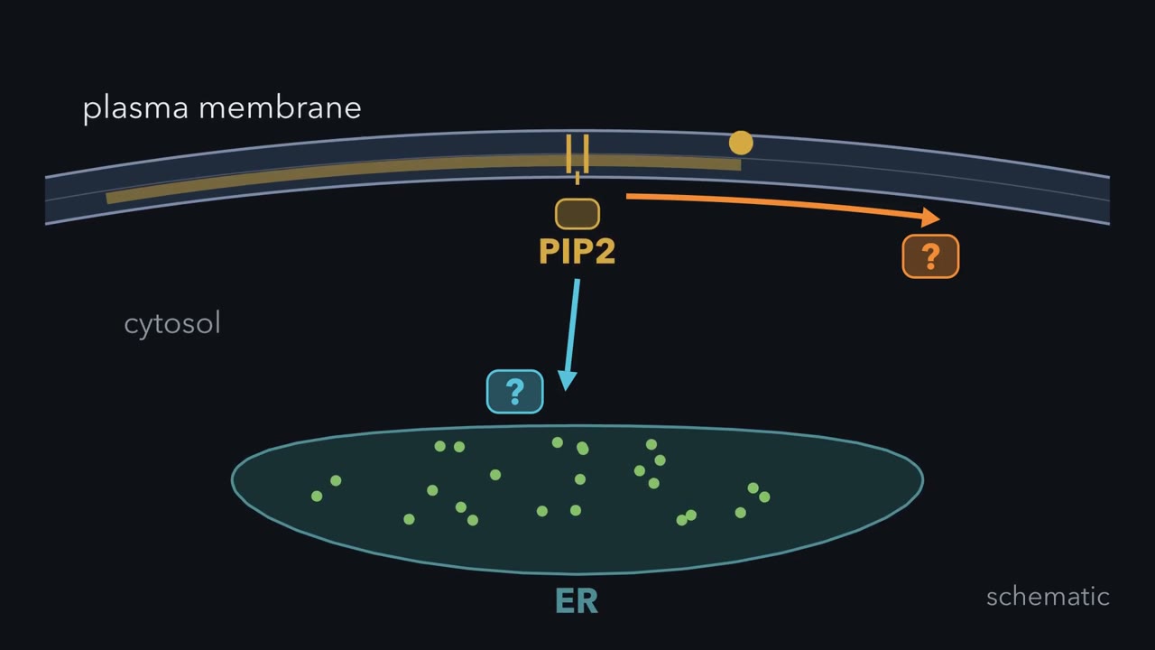 ▶ Watch One Cut, Two Destinations · 3:16
Read the transcript of “One Cut, Two Destinations”
Film transcript: One Cut, Two Destinations
How can one cut at a membrane send one signal inward while keeping another at the surface? Follow a single plasma membrane closely around one cell. The answer depends on where each product can go after the cut. The starting block is PIP2, a lipid in the inner leaflet of the plasma membrane. Its oily tails stay buried, while its charged head faces the watery cytosol. The same lipid serves as an address label and a stored signal. Phospholipase C approaches from inside the cell. The enzyme cuts between the head and the glycerol carrying the tails. Both products begin at the same tiny patch of membrane. Which product can leave the bilayer? Choose the phosphate-rich head or the oily pair of tails.
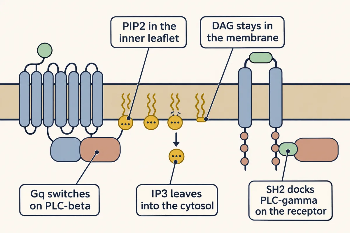
IP3 carries three phosphates, so it dissolves in cytosol and moves away. DAG keeps two oily tails, so it stays in the inner leaflet. Their different chemistry creates a moving branch and a local membrane branch. One cut has created two second messengers in different places. Different receptors can start the same cut. A G-protein-coupled receptor activates Gq, which activates PLC-beta. A receptor tyrosine kinase displays phosphorylated tyrosines, where SH2 domains dock PLC-gamma. Docking places PLC-gamma beside the receptor kinase. The kinase phosphorylates it and releases its internal brake. Both enzymes then make the same cut, producing IP3 and DAG.
Now follow IP3. It crosses the cytosol and binds an IP3 receptor in the endoplasmic reticulum. That receptor is a calcium channel. IP3 does not carry calcium; it opens the route out. The channel opens, and green calcium dots flow from the store into cytosol. Researchers tested whether IP3 was sufficient without a hormone. They made a cell surface leaky while internal stores stayed sealed. No outside hormone was present. Added IP3 released calcium from a nonmitochondrial store. That separation of cause from hormone made IP3 a true messenger. Later work identified the store as endoplasmic reticulum. Return to the original cell. The moving branch is IP3, which opens the calcium channel.
The local branch is DAG, still marking the membrane. Keep this rule: one PIP2 cut sends IP3 away and leaves DAG at the membrane.
From the film
- One PIP2 cut sends IP3 away and leaves DAG at the membrane.
- IP3 is water-soluble, while DAG remains in the inner membrane leaflet.
- Gq activates PLC-beta; receptor phosphotyrosines dock PLC-gamma, and the receptor kinase then phosphorylates it.
- IP3 opens an ER calcium channel; it does not carry calcium itself.
The one idea. One phosphatidylinositol 4,5-bisphosphate (PIP2) cut sends inositol trisphosphate (IP3) away and leaves diacylglycerol (DAG) at the membrane. Conventional protein kinase C (PKC) integrates calcium targeting with DAG activation at membranes.
Scope: this rule covers phospholipase C (PLC) cleavage of PIP2 and conventional PKC. IP3 opens a calcium channel; it does not carry calcium. Other routes can raise calcium or make DAG, and novel and atypical PKCs use different inputs. “Integrates” means a stronger, longer response where the two inputs overlap, not an all-or-none switch.
One cut makes two messengers
The first film showed where each product goes; here is the chemistry behind it. The substrate is PIP2 (phosphatidylinositol 4,5-bisphosphate), the inner-leaflet label read by PH domains in lipid anchors and addresses. Two fatty acid tails on a glycerol backbone sit in the membrane. The cytosolic head is a six-carbon inositol ring, with a hydroxyl on each carbon, joined to glycerol through one phosphate and carrying two more at positions 4 and 5.
Phospholipase C (PLC) hydrolyzes the bond between glycerol and the linking phosphate. The phosphate leaves with the ring, so a lipid named for two phosphates yields a soluble product named for three:
PIP2 + H2O → inositol trisphosphate (IP3) (inositol 1,4,5-trisphosphate) + DAG (diacylglycerol)
Each product is a second messenger, a small molecule made inside a cell in response to an external signal.
Two receptor routes converge on the cut
The first film showed Gq switching on PLC-beta, and PLC-gamma docking on a receptor and then being phosphorylated by its kinase. Both fold their X and Y regions into one catalytic barrel; their regulatory parts read different receptors.
A receptor coupled to Gq loads GTP onto Gq alpha, as in GTPase switches, and Gq-GTP activates PLC-beta at the membrane. Some forms, especially PLC-beta2 and PLC-beta3, also respond to beta-gamma subunits released from G proteins such as Gi. PLC-beta accelerates GTP hydrolysis by Gq, helping reset its own activator; cutting continues only while receptors reload Gq.
PLC-gamma carries two SH2 domains, which recognize phosphorylated tyrosines in particular sequence contexts, and an SH3 domain, which binds proline-rich protein segments. For the platelet-derived growth factor (PDGF) receptor, the growth factor pairs two receptors, and each phosphorylates tyrosines on the other’s tail. Docking alone is not activation: a PLC-gamma lacking the tyrosines the receptor kinase phosphorylates would still dock but cut little phosphatidylinositol 4,5-bisphosphate (PIP2), because the brake on its catalytic barrel stays on.
Thus acetylcholine at a Gq-coupled muscarinic receptor can use PLC-beta, and PDGF at its receptor tyrosine kinase can use PLC-gamma.
How IP3 earned the name messenger
By 1983 Michael Berridge, working on blowfly salivary glands, had shown that inositol trisphosphate (IP3) appears within seconds of hormone arrival. Robert Michell had proposed eight years earlier that breaking down inositol lipids is how receptors raise calcium. Correlation was not enough: a hormone starts many events, so IP3 and calcium could both follow a third cause.
The first film showed the 1983 test that closed the gap. In it H. Streb permeabilized pancreatic acinar cells in Irene Schulz’s laboratory, making the plasma membrane admit small molecules while internal compartments stayed sealed. IP3 is too charged to cross an intact membrane, which is why the cells had to be opened. Robin Irvine prepared the IP3 from human red blood cells.
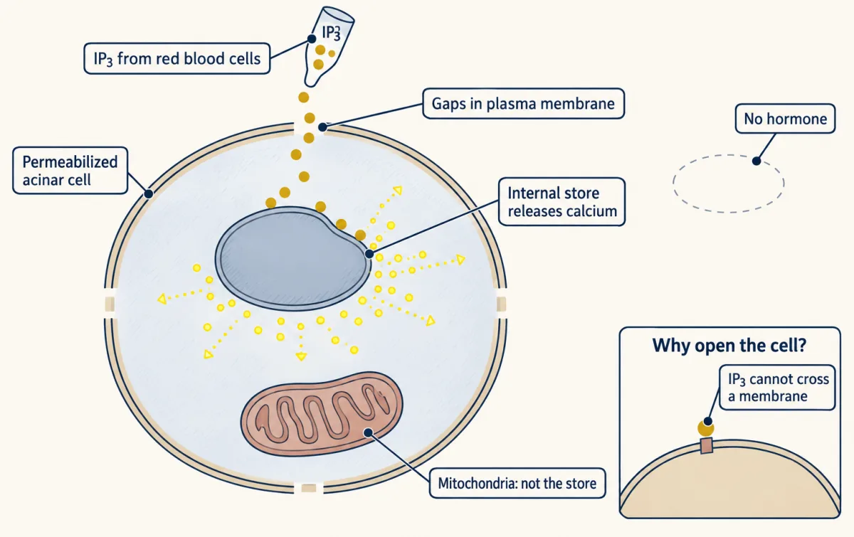
The store, later identified as the endoplasmic reticulum (ER), holds free calcium at a thousand times or more the resting cytosolic level of about 100 nM. Its IP3 receptor is a four-subunit calcium channel, so opening it produces a fast rise. The next lesson follows how cells read and clear that rise.
The second film asks how the calcium it releases and the diacylglycerol (DAG) left in the membrane meet at one enzyme.
Read the transcript of “How Conventional PKC Reads Both Branches”
Film transcript: How Conventional PKC Reads Both Branches
The first film left IP3 traveling and DAG in the membrane. How can conventional protein kinase C use both branches? The branches reach the same cell but keep different locations. Return to the same cell and follow one kinase. Follow its movement and timing.
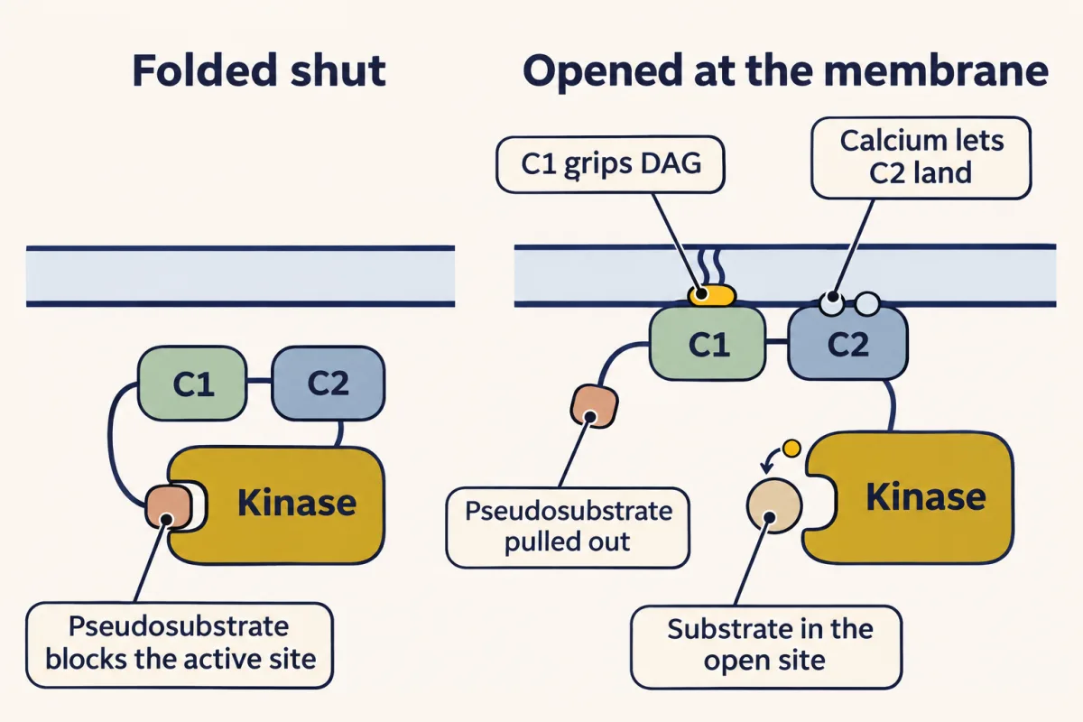
At rest, conventional PKC is folded shut. Its pseudosubstrate occupies the kinase active site, blocking real substrates. All four parts are connected in one protein chain. A C2 domain can respond to calcium. A C1 domain can recognize DAG. IP3 opened the endoplasmic reticulum channel, raising cytosolic calcium. Calcium binds C2 and changes its membrane affinity. C2 then engages anionic lipids, bringing conventional PKC toward the plasma membrane. The kinase is now close enough to sample the local membrane. At the membrane, C1 can search for DAG. DAG binding strengthens membrane engagement and helps displace the pseudosubstrate. The active site opens, so the kinase can phosphorylate protein substrates.
Now compare three schematic conditions: calcium without DAG, DAG without raised calcium, and both branches together. Which condition should retain conventional PKC most strongly at this membrane? Each contact alone can be weak under physiological conditions. Together, their binding energies add, so membrane preference can rise sharply. Conventional PKC gives a stronger, longer response where calcium targeting and DAG activation overlap. This is not a perfectly simultaneous digital switch. Calcium can recruit the kinase rapidly, before DAG engages C1. The response is graded, not all or none. The detector integrates timing as well as place. The rule belongs to conventional PKC.
Novel PKC has a C1 module suited to DAG-driven recruitment without calcium. Atypical PKC responds to neither messenger and instead uses protein partners. A phorbol ester can occupy the DAG site and activate PKC directly. Unlike DAG, it is cleared slowly. Prolonged activation can lead to kinase degradation, so signal duration changes the outcome. Return once more to the membrane. Calcium changes C2-dependent targeting.
DAG changes C1-dependent activation and retention. Keep this rule: conventional PKC integrates calcium targeting with DAG activation at membranes.
From the film
- Conventional PKC integrates calcium targeting with DAG activation at membranes.
- Calcium-bound C2 recruits conventional PKC to anionic membrane lipids.
- DAG-bound C1 strengthens membrane engagement and helps displace the pseudosubstrate.
- The response is graded: stronger and longer where calcium and DAG overlap, not an all-or-none switch.
Conventional PKC joins the branches
The second film showed calcium bringing conventional protein kinase C (PKC) to the membrane and diacylglycerol (DAG) helping displace its pseudosubstrate. The evidence came from assays with purified kinase and defined lipids: by 1980 Yasutomi Nishizuka’s laboratory had traced the extra activity of impure lipid preparations to DAG, which lets the kinase work at much lower calcium.
Protein kinase C (PKC) phosphorylates serines and threonines. Its pseudosubstrate resembles a substrate but has alanine where the target serine or threonine would be, so it occupies the active site and blocks it. The calcium-loaded C2 domain binds anionic membrane lipids such as phosphatidylserine, and conventional and novel PKCs carry two DAG-binding C1 domains, C1A and C1B.
Energies add, preferences multiply
Predict. A purified conventional PKC reads 1 unit of activity with no additions, 4 with calcium and 3 with DAG. What would simple addition predict with both, and what would a measured 40 units imply?
Check your prediction
Calcium adds 3 above baseline and DAG adds 2, so simple addition predicts 1 + 3 + 2 = 6. A value of 40 is about sevenfold above that expectation, evidence that the inputs act synergistically under these assay conditions.
Synergy comes from two membrane contacts, each weak alone. Binding energies add, which means preferences multiply:
ΔG(both) ≈ ΔG(C1) + ΔG(C2), so the combined membrane preference ≈ preference(C1) × preference(C2)
A membrane preference is a plain ratio: how many times more the kinase favors the membrane with that contact than without it. If each contact alone favors the membrane thirtyfold, say, the pair favors it about 30 × 30 = 900-fold. The numbers are illustrative, not measurements, and assume the two contacts act independently.
Calcium also rises when a voltage-gated channel opens, and other lipid reactions make DAG, so a kinase that answered either input alone would fire for many unrelated reasons. Calcium and DAG appear together at the inner face of the plasma membrane mainly when phospholipase C (PLC) has just cut phosphatidylinositol 4,5-bisphosphate (PIP2) there. Conventional PKC is therefore a coincidence detector in a graded sense: it gives a stronger, longer response where calcium targeting and DAG activation overlap. It recognizes the cut by sensing both halves of it.
Keep the rule: neither signal alone is enough, because only the pair holds a conventional PKC at the membrane firmly enough to free its active site. Thirtyfold is the lesson's example, not a measured value, and this is true of conventional PKC, not of every PKC. This is a sketch of the logic. The slider's range, 2 to 100, is this sketch's own, and the lesson states no threshold.
Not every PKC asks for both signals.
| Class | Members | Regulatory parts | Needs |
|---|---|---|---|
| Conventional | alpha, beta, gamma | Two C1 domains; C2 that binds calcium | DAG and calcium |
| Novel | delta, epsilon, eta, theta | Two C1 domains; C2-like domain that does not bind calcium | DAG only |
| Atypical | zeta, iota | C1-like domain that does not bind DAG; PB1 domain for protein partners | neither |
A DAG mimic shows why duration matters
Phorbol esters from croton oil promote tumors in animal skin without mutating DNA. In 1982 Monique Castagna and Nishizuka’s group showed that a phorbol ester can substitute for diacylglycerol (DAG) and activate protein kinase C (PKC) directly; later work showed that it binds the same C1 site. As the second film showed, cells clear DAG quickly but cannot efficiently break down a phorbol ester, and PKC held on for hours is degraded. A long exposure therefore ends with less enzyme than the cell started with, so a brief signal and a prolonged one can have opposite outcomes.
Predict. If PKC mainly drove tumors, what kind of PKC mutations would you expect to find in human cancers? What would it mean if most of them disabled the kinase?
Check your prediction
An enzyme that drives tumors should pick up activating mutations, as oncogenes do. If most tumor mutations disable it instead, the working enzyme behaves more like a brake, at least for the isozymes tested.
Alexandra Newton’s laboratory tested 46 PKC mutations from human tumors: most disabled the kinase and none activated it. For many isozymes that points to a brake on tumors, although the direction depends on isozyme and context; PKC-iota in lung cancer can act as an oncogene.
Ending and rebuilding the signal
Both branches are reversible. Phosphatases dephosphorylate inositol trisphosphate (IP3), or a kinase adds a fourth phosphate. Diacylglycerol (DAG) kinase converts DAG to phosphatidic acid. The inositol and lipid backbone are recycled into phosphatidylinositol 4,5-bisphosphate (PIP2). Meanwhile Gq hydrolyzes GTP and calcium is pumped away, so conventional protein kinase C (PKC) returns toward its closed cytosolic state.
Predict. Suppose IP3 breakdown is blocked but DAG kinase remains active. Which messenger should outlast the other, and why should conventional PKC output still fall?
Check your prediction
IP3 should outlast DAG, because its breakdown is blocked. That does not guarantee high cytosolic calcium for as long: calcium feeds back on the IP3 receptor, the ER store can run down, and pumps keep removing calcium. Conventional PKC output can fall either way as DAG is converted to phosphatidic acid, because calcium targeting without the DAG contact is a weaker input.
Design problem
Design a detector that responds strongly only when two of these signals overlap: calcium, DAG, phosphorylation, cAMP, an active small G protein or a protein partner. Name its sensor domains and explain why either contact alone should be weak.
Saved in this browser only.
One way to do it
Choose DAG and an active small G protein; copy the PKC plan and swap one sensor.
- Output: a kinase domain blocked by a tethered pseudosubstrate.
- First sensor: a C1 domain for DAG.
- Second sensor: replace C2 with a domain that binds a small G protein only in its GTP form. Raf has such a domain for Ras, which is membrane-anchored by lipid.
- Tuning: make each contact weak enough that either signal alone gives only brief membrane visits.
DAG alone says a lipid reaction occurred; Ras-GTP alone says a receptor activated Ras; together they report both events at one membrane patch. Each alone is ambiguous: if either could switch the kinase on fully, you would have built an OR gate, whose output cannot tell the pair of events from a single one. Disabling either sensor should weaken the response; DAG alone should not strongly activate the detector while Ras holds GDP.
Going deeper
Domain architecture
The fuller maps are PLC-beta: PH · EF hands · X · Y · C2 · long tail and PLC-gamma: PH · EF hands · X · SH2 · SH2 · SH3 · Y · C2. PLC-gamma also contains a split PH domain in the insert. The shared X and Y regions make the catalytic barrel; the inserted modules regulate access and recruitment.
Proteolysis bypasses the switch
Cutting the tether between a protein kinase C (PKC) pseudosubstrate and kinase domain creates activity that no longer needs the normal signal, explaining how Nishizuka’s laboratory first activated the kinase with a protease in 1977. A related event occurs in programmed cell death, where a caspase clips PKC-delta and frees its kinase domain.
Two routes from environment to cancer
Soot has been tied to cancer since Percivall Pott’s 1775 report on chimney sweeps, and hydrocarbons later isolated from coal tar damage DNA. A phorbol ester can contribute by impersonating a second messenger instead.
Your notes
Select any text in the lesson to highlight it or add a note to it. Your highlights and notes are saved in this browser and collected in your notebook.
This browser is not letting the site save anything (a private window or strict privacy settings), so highlights and notes will be lost when you leave the page.
How we measure it
Permeabilized cells
Make the plasma membrane leaky to small molecules while the compartments inside the cell stay sealed. A charged compound that cannot cross a membrane, such as IP3, can then be added straight to the cytosol, and you can ask whether that one molecule is enough to produce a response with no hormone present.
Radiolabeled inositol tracing
Feed cells inositol that carries tritium so their inositol lipids become radioactive, stimulate them, then separate the water-soluble inositol phosphates and count each fraction in a scintillation counter. Adding lithium blocks the last step of their breakdown, so the products pile up and are easier to measure.
Reconstituted kinase assay with defined lipids
Mix purified kinase, a substrate protein, ATP labeled with phosphorus-32 and lipid vesicles of known composition, then measure the radioactivity transferred to the substrate. Leaving out calcium, phosphatidylserine or DAG one at a time shows which cofactors the enzyme needs and whether they act together.
Correcting a tumor mutation by genome editing
Use CRISPR to change one mutated base in a cancer cell line back to the normal sequence, then compare the growth of corrected and uncorrected cells. If the corrected cells grow less well, the mutation was helping the tumor.
Method films · the methods library
- Cell-free extracts and reconstitution 4:01
- CRISPR-Cas9 genome editing 2:46
- Engineered mutants: site-directed, dominant-negative and constitutively active 4:25
- Pharmacological blockade and inhibitors 4:10
- Protein purification and column chromatography 4:14
- Radioactive and stable isotope tracers 4:03
- X-ray crystallography 4:06
Practice questions on this lesson (3) → MCAT-style, original, each with an explanation
Where this lesson fits
Before thisReview first
- GPCRs and Trimeric G ProteinsThis course · Lesson 12
You need to know how a receptor loads GTP onto Gq alpha, since that is what switches on PLC-beta.
- Protein Modules: SH2, SH3, PTB, PDZ and PHThis course · Lesson 9
PLC-gamma docks on receptor phosphotyrosines through its SH2 domains, so you need how SH2 domains read marks.
- Lipids as Anchors and AddressesThis course · Lesson 11
PIP2 is the lipid that PLC cuts, and this lesson assumes you know it as a membrane label.
After thisStudy next
- Calcium: The Messenger Kept at Arm's LengthThis course · Lesson 15
It takes up the calcium that IP3 releases from the ER and shows how the cell reads and clears it.
- Turning the Signal Off: Arrestins, RGS Proteins and PhosphodiesterasesThis course · Lesson 16
It shows how the receptor and G-protein side of this pathway is switched off.
- Receptor Tyrosine KinasesThis course · Lesson 18
It explains how receptor tyrosine kinases are activated and how they recruit PLC-gamma.
Suggested by an AI model from the lessons themselves, then checked. How we use AI
Finish line
Test yourself
8 questions
Practice and MCAT-style, with explained answers.
Next · Lesson 15
Calcium: The Messenger Kept at Arm's Length
How can the same calcium ion trigger contraction, secretion or gene expression without making the cell respond everywhere at once?
Continue →