Organismal Biology · Unit 3: Supply and exchange
Blood Vessels, Blood and Lymph
Rewritten on : new text, flashcards and film. Your notes and highlights stay, but the text around them may have moved.
Blood moves slowest in the narrowest vessels in your body. How can narrower mean slower?
Pause when the reading reaches a film, and offer to play it. Saved on this device.
Watch the film now, or keep listening
Builds on:Circulation and the HeartSize, Scale and SurfaceTissues, Organs and HomeostasisWhy these?
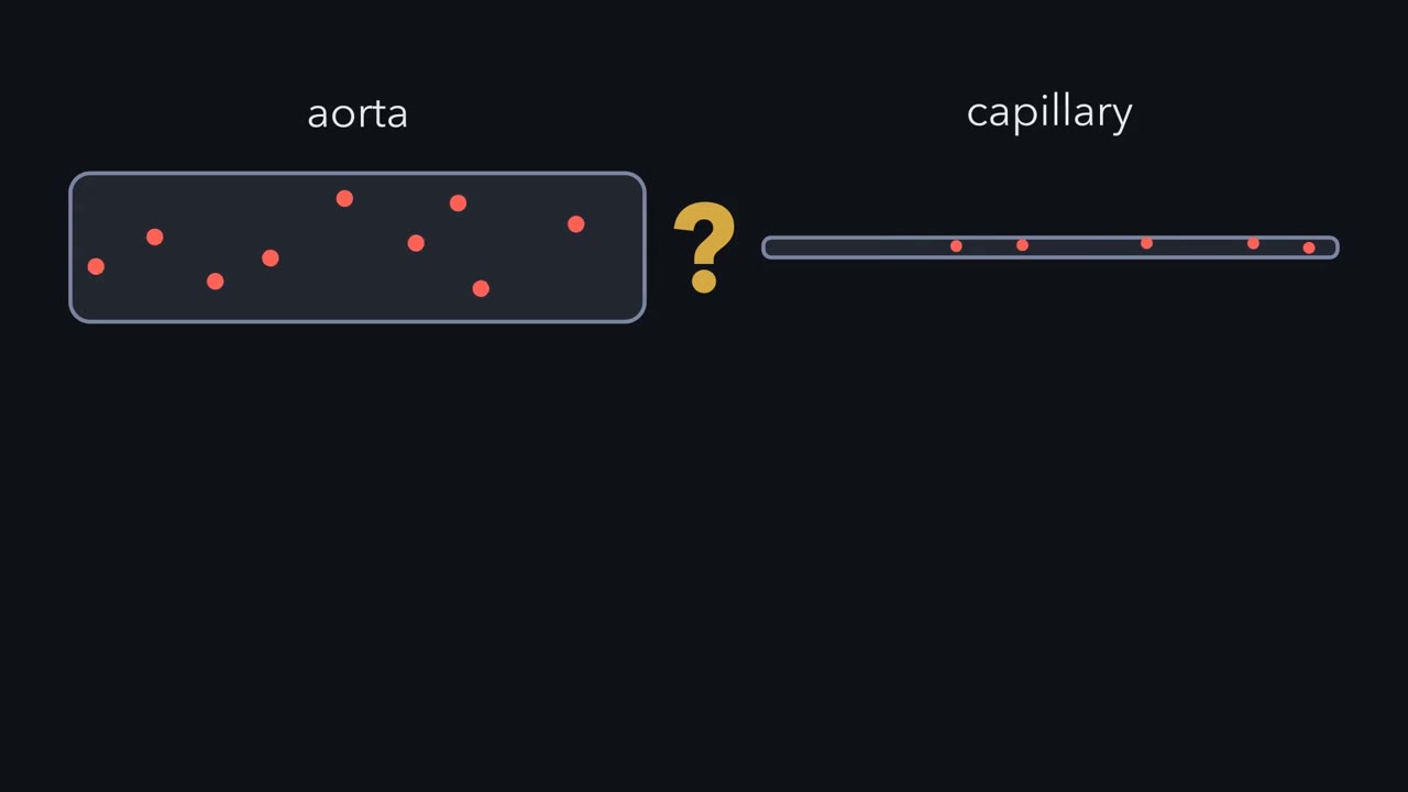 ▶ Watch Downhill in Pressure · 3:21
Read the transcript of “Downhill in Pressure”
Film transcript: Downhill in Pressure
Blood moves fastest in your widest artery and slowest in your narrowest vessels. How can narrower mean slower? A single river channel does the opposite. Where its cross-section shrinks, the same water has to speed up. Start with what makes blood flow along a vessel. In a level tube, fluid flows only from higher pressure to lower pressure. Equal pressure at both ends drives no flow, however high that pressure is. How much flows depends on two things. One is the pressure difference between the ends. The other is the resistance in the way. Flow equals the pressure difference divided by the resistance. Now lay out the whole circulation as one branching tree.
The aorta splits into arteries, then arterioles, then capillaries. The capillaries gather again into veins that return to the heart. Above the tree, follow the pressure. It falls all the way from the aorta to the heart's right atrium. It falls most steeply across the arterioles. Arterioles have walls of smooth muscle. When the muscle tightens, the arteriole narrows. In an ideal tube, resistance rises with the fourth power of a falling radius. A small squeeze makes a large change. So arterioles act like taps.
Nerves, hormones and local signals set their tone. Tightening one organ's arterioles cuts that organ's share of the blood. But a capillary is narrower than a typical arteriole. On its own, it can resist more. So which level sets most of the resistance?
The arterioles. Most capillaries are not lined up in a chain. They lie side by side, in huge numbers. Side by side, paths share the flow, so together they resist less. Now return to speed. In this simplified tree at steady state, no blood bypasses the capillaries. So the same flow crosses every level.
Speed is that flow divided by the total cross-section at each level. Each capillary is tiny. But together their cross-section far exceeds the aorta's. Spread the same flow over that huge area, and it has to move slowly. That is why blood creeps through the capillaries, the narrowest vessels of all. So narrow does not, by itself, mean slow. A single narrow tube carrying the whole flow would speed blood up. What matters is the total area at each level. That slow passage gives blood time to trade with the tissues. And fluid crossing the capillary wall obeys the same rule: a pressure difference over a resistance.
From the film
- Along a level vessel with no pump inside, flow equals the pressure difference divided by the resistance in the way.
- Equal pressure at both ends, however high, drives no flow.
- The arterioles hold most of the resistance, and their smooth muscle lets them change it: in an ideal tube, resistance rises with the fourth power of a falling radius.
- A single capillary can resist more than an arteriole, but so many lie side by side that together they resist less.
- In a simplified steady-state tree where no blood bypasses the capillaries, the same flow crosses every level, so blood is slowest where the total cross-section is largest: the capillaries.
The first film showed why blood is slowest in the narrowest vessels: the same flow spreads over the largest total area. It left a second puzzle: capillary walls leak water, so why doesn’t the plasma drain into the tissues? One rule answers both.
The one idea. Fluid flows down a pressure difference, and how much flows is that difference divided by the resistance in the way: Q = (Pin − Pout) / R. Across a capillary wall the driving pressure is the blood’s push outward minus the plasma proteins’ pull inward, each a difference between inside and outside the wall.
Scope: steady flow through a simplified vessel tree at one height, with no pump inside and no blood bypassing the capillaries (gravity, pumps and shunts add terms of their own), and fluid crossing capillary walls; not the diffusion of gases and solutes, the pulse, or what blood is made of. Later mentions of the rule assume these conditions rather than repeat them.
Each wall fits its job
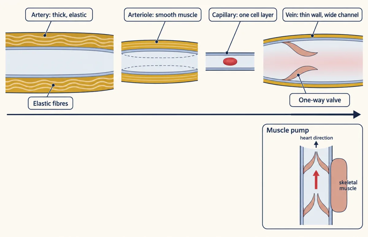
In a closed circulation (see circulation and the heart) blood leaves the heart in arteries and returns in veins. The names describe direction, not oxygen: in mammals the pulmonary artery carries oxygen-poor blood away from the heart, and the pulmonary veins carry oxygen-rich blood toward it. Between them lie the arterioles, the smallest arteries, and the capillaries, each with a wall fit for its work (tissues and homeostasis).
- Arteries have thick walls with smooth muscle and elastic fibres. Their elastic recoil between beats keeps pushing blood forward, so arterial pressure never falls to zero.
- Arterioles have walls dominated by smooth muscle. When it contracts (vasoconstriction) the arteriole narrows; when it relaxes (vasodilation) it widens. These are the film’s taps, holding most of the circulation’s resistance. Besides nerves (the hypothalamus and autonomic system) and hormones, local signals set their tone: stretch of the wall, chemicals from active tissue, and nitric oxide from the endothelium.
- Capillaries have a wall of a single layer of endothelial cells, the flat cells that line every vessel, on a thin basement membrane. Little else separates blood from tissue fluid, so exchange happens here (how the cells join: epithelia, adhesion and the extracellular matrix).
- Veins have thin walls and wide channels. They are more compliant than arteries: compliance is the change in volume per change in pressure, so they hold much more blood for a small rise in pressure. In adult humans at rest, most of the blood is in the veins. Many limb veins have one-way valves; contracting muscles squeeze them and push blood toward the heart: the muscle pump.
Numbers on the 1/r4 lever
For steady flow of a simple fluid through a rigid tube, Poiseuille’s law gives resistance from the fluid’s viscosity (its thickness: honey resists more than water), the tube’s length and its radius:
R = 8 × viscosity × length / (π × radius^4)
Blood is not a simple fluid and vessels are not rigid, so this is an idealization.
Worked problem. Illustrative numbers for an adult human at rest: mean arterial pressure (the average pressure in the aorta) 93 mmHg, right atrial pressure (where blood re-enters the heart) 3 mmHg, cardiac output (the heart’s flow) 5.0 L per min.
- The whole body as one resistance. Total peripheral resistance = (93 − 3) / 5.0 = 90 / 5.0 = 18 mmHg min per L. That lumps every vessel together.
- One arteriole narrows. Its radius halves, all else fixed. Its resistance rises 24 = 16 times, so its flow falls to 1/16, or 6.25 percent. If you got one half, you treated resistance as proportional to 1/r.
Predict. Now every arteriole in one organ halves its radius. Resistances in a chain (in series) add. If the arterioles held half the organ’s resistance and the pressure across the organ is fixed, does the organ’s flow also fall to 1/16?
Check your prediction
No. The arterioles’ half becomes 0.5 × 16 = 8 and the rest stays 0.5, so the organ’s resistance becomes 8.5 times the old and its flow falls to 1/8.5, about 12 percent. One sixteenth describes the narrowed vessels, not the organ. Narrowing a single arteriole among many side by side would change the organ’s flow very little.
Narrowest, but not the main resistance
Paths side by side (in parallel) work the other way: the flow divides, so n equal paths resist 1/n as much as one. With a made-up figure, if one capillary resisted 50 times as much as one arteriole, 100 side by side would resist half as much. Real beds also branch and rejoin, so this sketches the logic only.
Behind the film’s speeds, in made-up round numbers: 5 L per min of volume flow (how much passes a point) is about 83 cm3 per s; 83 / 4 cm2 of aorta is about 21 cm per s, and 83 / 3,000 cm2 of capillaries is about 0.03 cm per s.
Slow blood in a leaky tube brings back the second puzzle, and the second film follows the first measurement of fluid crossing one capillary’s wall.
Read the transcript of “Landis's Blocked Capillary”
Film transcript: Landis's Blocked Capillary
Capillary walls let water and small solutes through. So why doesn't all your plasma end up in your tissues? Something must hold most of it in. The first film showed that blood flows down a pressure difference, divided by a resistance. Across the capillary wall, the same rule decides which way fluid moves. Two pressures act across the wall. The first is blood pressure inside the capillary, minus the tissue fluid's pressure outside. It pushes fluid outward. Physiologists call it hydrostatic pressure. The second comes from plasma proteins, mostly albumin. Few cross the wall, so they pull water back in. Protein outside pulls the other way, so the difference counts.
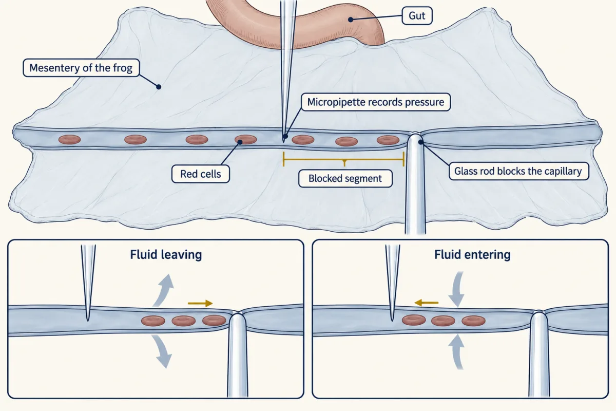
That pull is the oncotic pressure. In 1896, Ernest Starling, working with dogs, argued that these two pressures set the direction of flow. But neither had been measured inside a single capillary. In 1927, Eugene Landis took up the test in frogs. He spread out the mesentery, the thin sheet that holds the gut, and watched single capillaries under a microscope. A fine glass pipette slid into one capillary to record its pressure. Then he pressed the capillary shut with a fine glass rod. With the flow stopped, the trapped red cells drifted slowly. They moved toward the block when fluid was leaving through the wall. They moved away when fluid entered.
From the cells' speed and the vessel's size, he worked out the fluid crossing each patch of wall. Then he plotted that rate against the pressure he had measured. If Starling was right, what should happen when capillary pressure falls below the pull of the plasma proteins? Which way should the fluid go?
Fluid should come in, and it did. Filtration rose roughly in proportion to pressure. It fell to zero near the oncotic pressure of frog plasma, then reversed. So across the wall, the driving pressure is the push minus the pull. The more the push wins, the faster fluid leaves. When the pull wins, fluid comes in. But Landis measured single capillaries in one frog tissue. His result ruled in the balance there. It did not show what every capillary does over hours. Later reviews concluded that a lasting return at the venous end lacks reproducible support. In most tissues, capillaries filter slowly, and lymph vessels carry the fluid back.
From the film
- Across a capillary wall, the driving pressure is the blood's push outward minus the plasma proteins' pull inward, each a difference between inside and outside the wall.
- Landis blocked single frog capillaries and timed the drift of trapped red cells to measure fluid crossing the wall.
- Filtration rose roughly in proportion to capillary pressure and reversed near the oncotic pressure of frog plasma.
- His experiment did not show that fluid keeps returning at the venous end; in most tissues, lymph returns it.
Starling’s balance in numbers
The film weighed push against pull without sizes. In symbols, with P for hydrostatic pressure (the pressure of the fluid itself), π for oncotic pressure (the osmotic pull of proteins that cross the wall poorly, in mammals mostly albumin), c for capillary, i for interstitial (the fluid between cells) and outward positive:
net pressure = (Pc − Pi) − (πc − πi)
This form assumes protein cannot cross the wall at all (Going deeper gives the full relation). Flow across the wall is this net pressure divided by the wall’s resistance to water: the one idea again.
The classic model, worked
Illustrative adult human numbers: capillary pressure 35 mmHg at the arterial end, falling to 15 mmHg at the venous end as pressure drops along the capillary’s own resistance; interstitial pressure 0; plasma oncotic pressure 25 mmHg; interstitial oncotic pressure 3 mmHg. The oncotic difference is 25 − 3 = 22 mmHg. Arterial end: 35 − 22 = +13 mmHg, outward. Venous end: 15 − 22 = −7 mmHg, inward.
- Oncotic difference (πc − πi, with πi = 3)
- Net, arterial end (Pc − 0) − difference
- Net, venous end
- Net crosses zero at
In this sketch: illustrative adult human numbers, reflection coefficient 1, interstitial pressure 0, interstitial oncotic pressure 3 mmHg, capillary pressure falling linearly between the two ends (an assumption; the lesson gives only the ends).
Net pressure = (Pc − Pi) − (πc − πi), outward positive. The classic arithmetic is a teaching model, not a measured steady state.
What survives, and what does not
The film’s verdict: the outward push survives, but a steady venous-end return does not hold as a general account for most tissues. The reason is the oncotic term. The difference that matters lies across the glycocalyx, a sugar-rich coat on the inner surface of the endothelium that holds back protein: between plasma and the fluid just beneath the coat (πsub, which replaces πi in the equation). That fluid has less protein than average tissue fluid, so tissue protein does not cancel the pull. In perfused rat mesenteric microvessels (fed with a fluid of set make-up), the effective oncotic opposition stayed near 70 percent of the plasma oncotic pressure inside the vessel, even with equal albumin in plasma and tissue (Adamson and colleagues, 2004). Applied, for illustration only, to 25 mmHg: 17.5 mmHg.
Predict. With that 17.5 mmHg pull and interstitial pressure 0, which way does fluid move at a capillary pressure of 35 mmHg, and at 15 mmHg?
Check your prediction
At 35 mmHg, net pressure is 35 − 17.5 = +17.5 mmHg, outward. At 15 mmHg it is 15 − 17.5 = −2.5 mmHg, inward, but only for a while. Water drawn in leaves the protein beneath the coat more concentrated, the pull shrinks, and the absorption fades.
Some beds do absorb steadily: those where an epithelium, a sheet of lining cells, keeps supplying fluid for the capillaries to take up.
The giraffe stretches these forces: its feet are far below its heart, and its arterial pressure is much higher than ours. Hargens and colleagues (1987) reported three defences against fluid build-up in its legs: a tight skin layer that, with pressure changes during exercise, helps move fluid upward against gravity; narrowing of the small vessels that feed the capillaries; and capillary walls that let little plasma protein through.
Lymph: the return route
Filtered fluid enters blind-ended lymphatic capillaries and is then called lymph. In mouse trachea their cells are joined by short “buttons” with loose, overlapping flaps between them, thought (from structure alone) to let fluid in but not out; larger collecting vessels have continuous “zipper” junctions and valves.
Predict. Frogs and other amphibians have lymph hearts, small pulsating sacs that pump lymph into veins. Mammals have none. What moves theirs?
Check your prediction
The walls of mammalian collecting lymphatics contract, body movements squeeze them, and their valves keep lymph moving one way, much as the muscle pump moves venous blood.
Lymph passes through nodes, where white cells sample it (see innate immunity), and in humans it re-enters the blood where the jugular and subclavian veins join in the neck. Leaked protein comes back mainly this way. In the small intestine, lymphatics called lacteals carry absorbed fat (see digestive systems).
Blood and stopping a leak, compared across animals
A cut opens a new path out of the vessel; the blood carries what closes it.
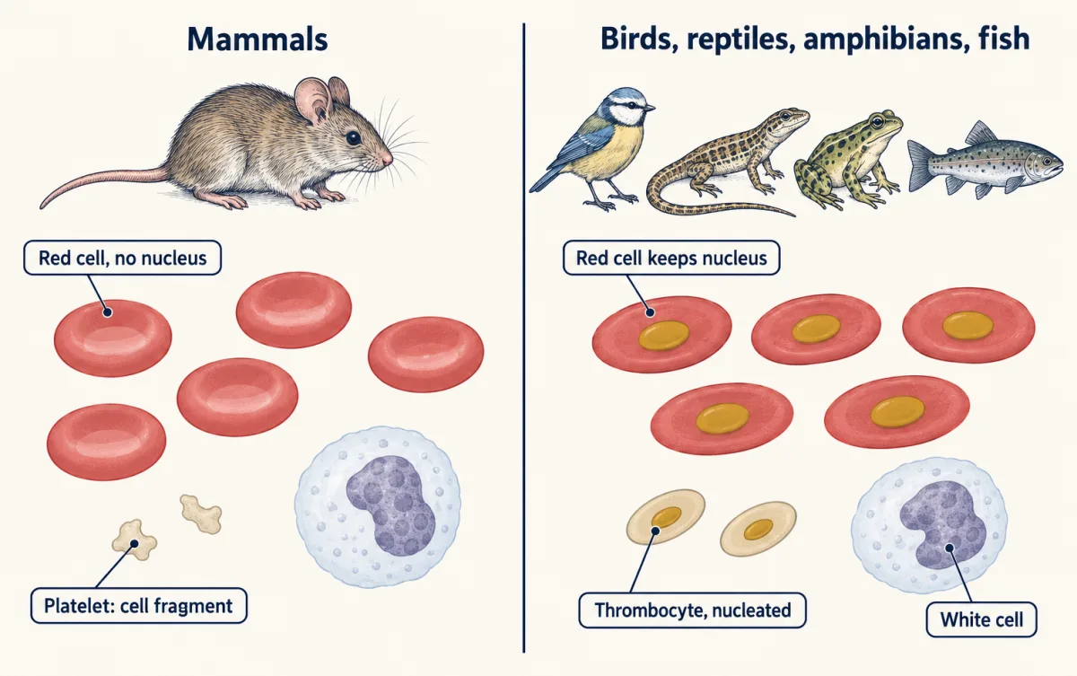
Blood is plasma, the liquid, carrying cells. Human plasma is more than 90 percent water, with proteins including albumin, globulins and fibrinogen. Red cells (erythrocytes) carry hemoglobin, the oxygen-binding protein (see oxygen transport). White cells (leukocytes) defend the body. Platelets are cell fragments shed by large cells called megakaryocytes. The fraction of blood volume made up of red cells is the hematocrit (Going deeper gives the human range).
Mammalian red cells lose their nucleus as they mature; those of birds, reptiles, amphibians and fish keep it, with rare exceptions such as a few salamanders. Platelets are a mammal feature: other vertebrates have nucleated thrombocytes that do a similar job.
Hemostasis, stopping blood loss, runs in overlapping steps in mammals:
- Vascular spasm: the cut vessel’s smooth muscle contracts. Narrowing steeply raises the resistance of the path out (the 1/r4 lever, roughly: a wound is no ideal tube).
- Platelet plug: platelets stick to the damaged wall and recruit more platelets.
- Coagulation: tissue factor, a protein exposed at the injury, starts a chain of enzymes. It ends with the enzyme thrombin converting soluble fibrinogen into insoluble fibrin strands. A cascade amplifies a small start into a large output, the logic of signaling circuits.
- Clot contraction: in human plasma clots in vitro (outside the body), platelet extensions pull on fibrin fibres and compact the clot.
- Fibrinolysis: the enzyme plasmin later breaks the fibrin down.
The horseshoe crab, Limulus, has an open circulation and a clot built another way. Its blood, called hemolymph, is rich in amebocytes, cells that store clotting factors in granules. Levin and Bang (1964) reported that bacterial endotoxin makes amebocyte material clot; later work traced a cascade that ends in a gel protein, coagulin, not fibrin.
Design problem
Do one tissue’s capillaries reabsorb fluid at their venous ends in steady state, or filter along their length while lymph returns it? Design the measurements.
Saved in this browser only.
One way to do it
In single microvessels (Landis’s occlusion method, or perfused vessels as in Adamson and colleagues’ rat mesentery), measure filtration in the first seconds after a pressure step and again after many minutes. The revised model predicts that absorption after a drop fades as protein beneath the glycocalyx evens out again; the classic model predicts it lasts. Measure oncotic pressure as close to the outside of the endothelium as you can, not only in bulk tissue fluid. Blocking the lymphatics is weaker: swelling shows net filtration, but the classic model also leaves a small filtrate for lymph, so swelling cannot tell the models apart. Repeat in another tissue; one bed in one species does not speak for all.
Going deeper
The full Starling relation
In full, Jv = K × [(Pc − Pi) − σ(πc − πsub)]: Jv is the fluid flux, K the hydraulic conductance of the wall (how easily water passes; the inverse of its resistance), and σ is the reflection coefficient for protein: 1 if protein cannot cross at all, 0 if it crosses freely. The worked problems above set σ = 1, and the classic model also sets πsub = πi.
Sources for the revised view
The reviews are Levick and Michel (2010) and Woodcock and Michel (2021): sustained venous-end reabsorption lacks reproducible experimental support as a general account.
Blood numbers and clotting factors
In adult humans red cells make up roughly 36 to 54 percent of blood volume, a hematocrit that is lower in women than in men. In the platelet plug, von Willebrand factor helps platelets stick to the damaged wall, and the platelets release ADP, which recruits more platelets. Much of the coagulation chain of enzymes and cofactors is assembled on the surface of activated platelets; it ends when prothrombinase converts prothrombin to thrombin, and Factor XIII stabilizes the fibrin mesh.
Your notes
Select any text in the lesson to highlight it or add a note to it. Your highlights and notes are saved in this browser and collected in your notebook.
This browser is not letting the site save anything (a private window or strict privacy settings), so highlights and notes will be lost when you leave the page.
How we measure it
Micropipette pressure measurement and single-capillary occlusion
Insert a fine glass pipette into one capillary to record its pressure, then block the vessel and follow the drift of red cells in the blocked segment. The drift gives the rate of fluid filtration or absorption per unit wall area at a known pressure.
Blood-pressure and flow measurement
Measure the pressure at the two ends of a vascular bed and the flow through it, and divide the pressure difference by the flow to obtain resistance. Repeating the measurement before and after a change in vessel tone shows how the bed's total resistance changes; pressures measured at points along the bed show which vessels set it.
Light, confocal and intravital microscopy
Inject a marker such as India ink, or label cell junctions with fluorescent antibodies, and image vessels in fixed tissue or in a living animal. It shows which capillaries carry blood (ink counts in fixed tissue can miss vessels that were flowing), how lymphatic cells join, and how platelets act inside a clot.
Method films · the methods library
Practice questions on this lesson (3) → MCAT-style, original, each with an explanation
Where this lesson fits
Before thisReview first
- Circulation and the HeartThis course · Lesson 10
You need the closed circuit and the heart's output from there to follow flow through arteries, capillaries and veins.
- Size, Scale and SurfaceThis course · Lesson 2
Surface area and volume reasoning from there explains why a huge total capillary area suits exchange.
- Tissues, Organs and HomeostasisThis course · Lesson 6
The tissue types, including smooth muscle and endothelium, come from there and are used in each vessel wall here.
After thisStudy next
- Gas Exchange and BreathingThis course · Lesson 12
Gases cross thin capillary walls in the lungs, using the slow flow and large area shown here.
- The KidneyThis course · Lesson 16
The Starling forces here reappear as the filtration pressure across the glomerular capillary wall.
- Carrying Oxygen and Carbon DioxideThis course · Lesson 13
It picks up the red cells and hemoglobin introduced here and shows how blood carries oxygen.
Suggested by an AI model from the lessons themselves, then checked. How we use AI
Finish line
Test yourself
8 questions
Practice and MCAT-style, with explained answers.
Next · Lesson 12
Gas Exchange and Breathing
Water holds far less oxygen than air, yet a trout's gill can send its blood out at a higher oxygen partial pressure than the water leaving it. Your lung cannot do that with air. Why not?
Continue →