Cell Signaling · Unit 5: Signals that build and defend an animal
Integrins and Rho GTPases
How does a cell hold on to what it stands on, feel how hard it is pulling, and use that grip to crawl?
Pause when the reading reaches a film, and offer to play it. Saved on this device.
Watch the film now, or keep listening
Builds on:GTPase Switches: The TimerProtein Modules: SH2, SH3, PTB, PDZ and PHAdaptors, Docking Proteins and ScaffoldsWhy these?
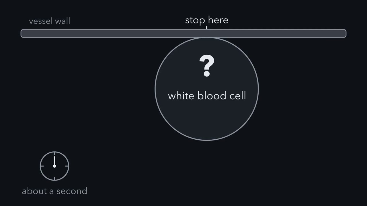 ▶ Watch A Grip Switched On From Inside · 2:05
Read the transcript of “A Grip Switched On From Inside”
Film transcript: A Grip Switched On From Inside
A white blood cell is carried along in flowing blood, and it must stop where it is needed. There, it can stop on the vessel wall in about a second. What switches its grip on? A second is too short to make a new protein. So the receptor that grips must already sit on the cell surface, switched off. The receptor that grips the wall is an integrin. It is two different chains, called α and β, and each chain crosses the membrane once. Their large outer parts together form a head that grips its ligand. Inside the cell, each chain has only a short tail. There is no kinase domain and no channel. In integrins studied most closely, those of platelets and white blood cells, the outer part can fold at a knee.
The head then tips back toward the membrane. Bent, with the head closed, the integrin holds its ligand weakly. A white blood cell flowing in the blood keeps its integrins in this form. So the grip is switched off. A signal on the vessel wall starts a relay inside the cell. The relay helps bring a protein called talin to the integrin tail. Talin binds the tail of the β chain and separates the two tails. The change passes through the membrane, the outer part straightens, and the head opens. Extended like this, the integrin holds its ligand tightly. The extended integrin grabs its ligand on the vessel wall and holds it tightly. The cell stops. Nothing had to be made, so the relay can run in about a second.
The message started inside the cell and changed the grip outside. So this is called inside-out signalling. A hormone receptor waits for its ligand. An integrin is often surrounded by ligand, so the cell controls the receptor instead. So ligand can be all around, and the cell can still keep the grip weak. The control is a change of shape, thrown from inside the cell. Keep this rule: signals inside the cell can strengthen an integrin's grip.
Integrins tie the material a cell stands on to the actin inside it, and they signal in both directions. Together with three Rho-family switches that build actin, they let a cell grip, feel a pull and crawl. A white blood cell carried along in your blood can stop on the wall of a vessel in about a second, and the receptor that stops it has no enzyme activity of its own.
Most receptors in this course read a signal that drifts in from somewhere else. This one reads something solid: the material the cell is standing on. So it has to answer two questions that a hormone receptor never faces. Am I holding on to anything? How hard is it pulling back? The answers help decide whether a cell stays where it is or crawls away. A crawling cell pushes its front edge forward, grips, pulls its body forward against the grip, and lets go at the back. The integrin supplies the grip, and the Rho-family switches supply the push and the pull.
The idea: a receptor for something solid
Most cells in your body sit on or in the extracellular matrix, a mesh of protein fibres and sugar chains that cells secrete around themselves. Collagen is its rope. Fibronectin is one of its glues, a long protein that sticks to collagen with one part and to cells with another.
Which part do cells hold? In 1984 Michael Pierschbacher and Erkki Ruoslahti tested short synthetic pieces of fibronectin’s cell-binding region and found that a peptide of four amino acids, Arg-Gly-Asp-Ser, could stand in for it. The first three, RGD in one-letter code, are the handle. Add enough free RGD peptide to a dish and fewer cells hold on to fibronectin, because the peptide fills the site that the matrix would have filled.
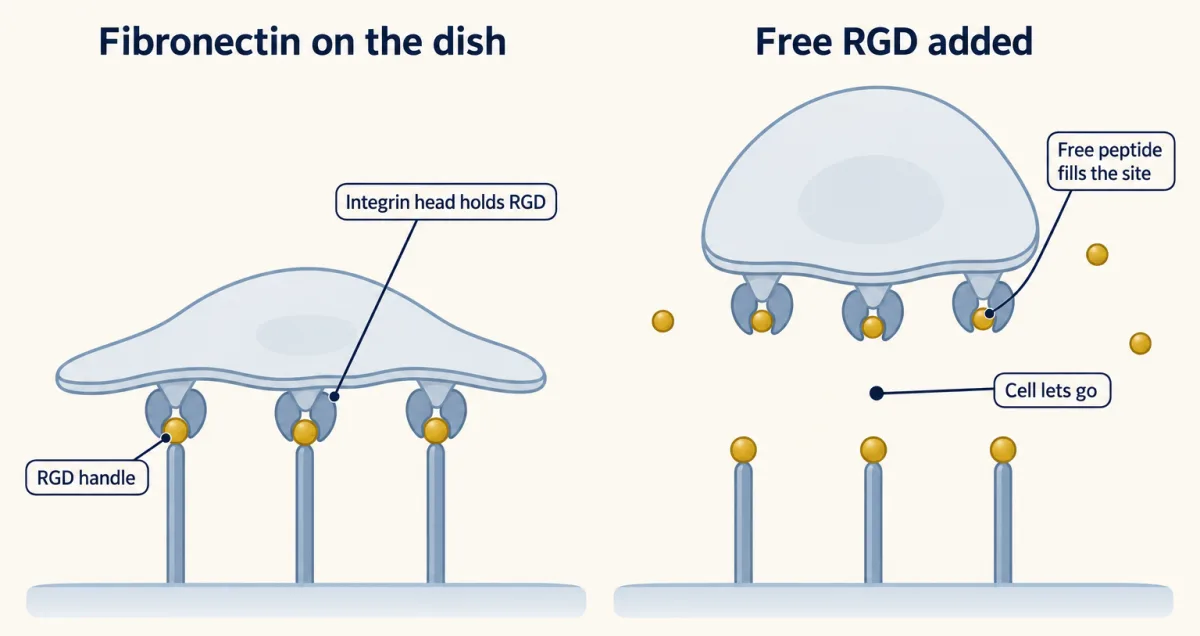
The receptor that holds the handle is an integrin. The name was proposed in 1986 by the laboratories of Richard Hynes, Clayton Buck and Alan Horwitz, for an integral membrane protein that ties the matrix outside to the cytoskeleton inside. An integrin is two different chains, α and β, and each crosses the membrane once. Their large outer parts together form a head that grips the matrix. Inside the cell each chain has only a short tail, a few dozen amino acids in nearly every case, and linker proteins join the tails to actin filaments (one integrin, α6β4, joins keratin filaments instead). There is no kinase domain and no channel.
Integrins come as a family of α and β pairs with different ligand preferences. Some recognise RGD. Others grip collagen or laminin at different sites, and some integrins of white blood cells grip proteins on the surface of other cells.
So an integrin is a mechanical link first. Its signalling comes from the proteins that gather on its tails, and it runs in both directions.
A grip that is switched on from inside
A hormone receptor waits for its ligand. An integrin is often surrounded by ligand, so the cell controls the receptor instead.
In the integrins studied most closely, those of platelets and white blood cells, the outer part can fold at a knee so that the head tips back toward the membrane. Bent, with the head closed, it holds its ligand weakly. Extended, with the head open, it holds tightly.
The switch is thrown from the cytoplasm. A protein called talin binds the tail of the β chain. That separates the two tails, the change passes through the membrane, and the outer part straightens. A second tail-binding protein, kindlin, is needed as well. The message starts inside the cell and changes the grip outside, so this is called inside-out signalling.
A platelet shows why it matters. It carries its integrin, αIIbβ3, on its surface all the time, switched off, while blood full of the integrin’s ligand, fibrinogen, flows past. When a vessel is cut, signals from the wound switch the integrin on within seconds. Fibrinogen has two ends, so it bridges one platelet to the next, and a plug forms. A white blood cell faces a harder version of the same problem, and the design problem at the end of this lesson works it through.
The focal adhesion: a signalling machine that can feel a pull
Once integrins hold the matrix they cluster, and their tails collect a crowd of proteins. Talin joins the β tail to actin. Vinculin binds both talin and actin and reinforces the join. Paxillin is a scaffold, with many docking sites and no enzyme activity. Focal adhesion kinase (FAK) is a tyrosine kinase that is recruited from the cytoplasm when an adhesion forms; it also has other jobs, some in the nucleus. The whole assembly is a focal adhesion.
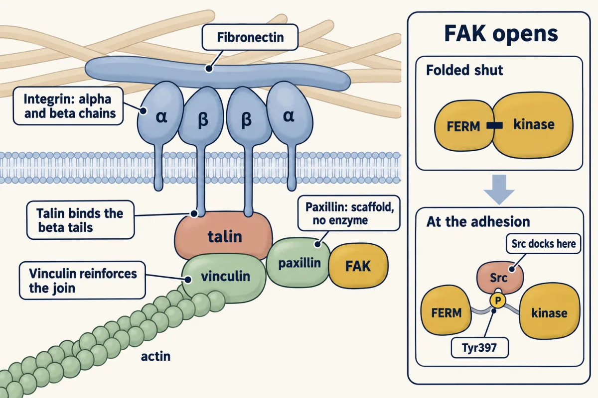
This is how a receptor with no enzyme signals inward: it borrows one, as the cytokine receptors in the Janus kinase (JAK) and signal transducer and activator of transcription (STAT) lesson do. Resting FAK is folded shut, with one of its own domains lying against the kinase. At a cluster of integrins it opens and phosphorylates itself on one tyrosine, Tyr397. How clustering opens it is not fully worked out. That phosphotyrosine is a docking site for the SH2 domain of Src. Docked Src phosphorylates FAK further, and the pair then phosphorylates its neighbours, paxillin among them, which makes more docking sites.
One site on FAK binds Grb2, the adaptor that leads to Ras.
The adhesion also measures force. A cell pulls on its adhesions with myosin, and a stiff surface resists more than a soft one.
Laser-trap experiments showed that the harder a fibronectin-coated bead on the cell surface was held, the more firmly the cell tied that patch of integrins to its actin. At the level of a single molecule, the long rod of talin is a string of helix bundles, and helices that vinculin binds are folded away inside them. When a piece of the rod was pulled, it bound more vinculin than when it was not.
No phosphate was added and no GTP was exchanged. Force alone uncovered binding sites. In a cell, full-length vinculin that binds this way can also hold actin, so each one adds a link and an adhesion that is pulled can get stronger. The experiment used purified protein. Later work saw single talin molecules stretch and relax in living cells, pulled by myosin. Whether stretch uncovers sites in many other proteins is still open.
Three switches for the actin skeleton
Read the transcript of “Three Switches, Three Shapes”
Film transcript: Three Switches, Three Shapes
A cell that can grip still needs to change shape. Shape is the work of actin filaments. Three small GTPases of the Rho family decide what the filaments build. They work as Ras does. Bound to GTP, each one is on. Bound to GDP, it is off. GEFs and GAPs set the balance. In 1992, Anne Ridley and Alan Hall injected purified proteins into fibroblasts. The cells had been starved of serum, which leaves them with little organised actin. Active Rho filled the cells with thick actin cables. Active Rac made their edges ruffle.
In 1995, Catherine Nobes and Alan Hall showed that Cdc42 produces thin spikes. Separately, the laboratory of Louis Lim showed the same. So three switches gave three different shapes. Rho builds stress fibres. These are cables of actin and myosin II that contract. Rac builds lamellipodia. These are broad, thin sheets, pushed forward by a branching mesh of filaments. Cdc42 builds filopodia. These are thin fingers with a core of parallel filaments. Keep this rule: each Rho family switch decides which shape the filaments build.
A grip is no use to a cell that cannot change shape. Shape is the work of actin filaments, and three small GTPases of the Rho family decide what the filaments build. They work as Ras does: GTP-bound, “turn on”; GDP-bound, “turn off”; with guanine-nucleotide exchange factors (GEFs) and GAPs setting the balance.
In 1992 Anne Ridley and Alan Hall injected purified proteins into fibroblasts that had been starved of serum, which leaves them with few stress fibres. Active Rho filled the cells with thick actin cables. Active Rac made their edges ruffle. In 1995 Catherine Nobes and Hall, and separately the laboratory of Louis Lim, showed that Cdc42 produces thin spikes.
- Rho builds stress fibres: cables of actin and myosin II that end in focal adhesions and contract. One effector is Rho kinase (ROCK), which raises the activity of myosin II. Rho is also the switch that G12/13-coupled receptors turn on.
- Rac builds lamellipodia: broad, thin sheets pushed forward by a branching mesh of filaments. Rac-GTP turns on the WAVE complex, and WAVE turns on Arp2/3, which starts each new filament as a branch on the side of an old one.
- Cdc42 is best known for triggering filopodia: thin fingers with a core of parallel filaments. The bundles themselves are built mainly by formins and Ena/VASP proteins, and some cells make filopodia without Cdc42 or N-WASP. Cdc42 also turns on WASP-family proteins (WASP in blood cells, N-WASP in most other cells), which are folded shut until Cdc42-GTP binds them and frees the part that turns on Arp2/3.
The adhesion and the switches are wired together: proteins phosphorylated by focal adhesion kinase (FAK) and Src recruit GEFs for Rac, so a new grip at the front calls for more protrusion there.
How a cell crawls, and what happens when it cannot
Put the parts in order and the cell moves.
- Push. At the front, Rac drives a lamellipodium forward.
- Grip. Under the new sheet, integrins catch the matrix and small adhesions form.
- Pull. Rho, through ROCK and myosin II, contracts the cell body against those adhesions and hauls it forward.
- Let go. At the back, old adhesions come apart and the tail is drawn in.
Rac in front and Rho behind is the short version. It is a simplification: sensors that report Rho activity in living cells find some at the front edge too.
The child in the design problem at the end of this lesson is real. In leukocyte adhesion deficiency type I, the gene for β2 (ITGB2), the chain shared by the β2 integrins of white cells, is mutated. White cells cannot stop on the vessel wall, and affected children have severe bacterial infections starting in infancy. A bone marrow transplant can cure it.
Platelets have a matching disease. People without working αIIbβ3 bleed easily, a condition called Glanzmann thrombasthenia. A drug can copy that state on purpose. Abciximab, a fragment of an antibody that bound αIIbβ3 and blocked it, is no longer supplied. Eptifibatide and tirofiban are small molecules that sit in its ligand pocket. They are given by vein, in hospital, to some patients with a heart attack or a threatened one, most often around the catheter procedure that reopens the blocked artery, so that platelets cannot bridge. The main risk is the one the genetics predicts: bleeding.
Design problem
A white blood cell is carried along in flowing blood. It must stop and crawl out of the vessel where it is needed, such as at an infection, and not elsewhere. So its integrins must be off while it flows and on within about a second at the right place. Design the control, using a signal on the vessel wall and parts from earlier lessons. Then predict what happens to a child whose white cells cannot make this integrin.
Saved in this browser only.
One way to do it
A second is too short to make a new protein, so the integrin must already be on the surface, held in its bent form. The “on” signal must sit on the vessel wall, and only at the inflamed place.
The address. Cells lining a vessel next to an infection put two things on their surface. Selectins are sticky proteins whose short-lived bonds make a passing white cell slow down and roll. Chemokines are small signal proteins that stay stuck to the wall.
The receptor. The white cell carries a G-protein-coupled receptor for the chemokine, a fast architecture whose parts are already at the membrane.
The relay. The G protein turns on phospholipase C, so calcium and diacylglycerol rise. Calcium turns on a GEF for Rap1, a GTPase of the Ras family (CalDAG-GEFI in platelets and mouse neutrophils; other blood cells use other GEFs). Rap1 with GTP bound (its “on” state) helps bring talin to the integrin tail. The details differ between cell types.
The grip. Talin binds the β tail, the integrin extends, and it grabs its partner on the vessel wall, a protein called ICAM-1. The cell stops. Then it crawls out between the cells of the wall.
Nothing has to be made, so the relay can run in about a second.
The child. White cells without this integrin roll but cannot stop. They stay in the blood, so the blood count of white cells, mostly neutrophils, is very high while infected tissue has very few neutrophils. You would predict repeated bacterial infections that form little pus, since pus is mostly dead white cells. That is what is seen.
Going deeper
The integrin family in numbers
Humans have 18 α chains and 8 β chains, which pair to make 24 known integrins. About a third of them recognise RGD.
A scaffold for the MAP kinase cascade
From Grb2 and Ras the next step follows a rule you met with Ste5 and KSR: “Scaffolds can localize ras-mapk.” In cells responding to hepatocyte growth factor through its receptor c-Met, paxillin holds Raf, MAPK/ERK kinase (MEK) and extracellular signal-regulated kinase (ERK) next to focal adhesion kinase (FAK) at the focal adhesion. The cascade is the one you know, running at one address.
Two experiments on force at adhesions
Michael Sheetz’s laboratory spent years asking where pull becomes chemistry. In 1997 Daniel Choquet, Dan Felsenfeld and Sheetz held fibronectin-coated beads on fibroblasts with a laser trap. The harder the trap resisted, the more firmly the cell tied that patch of integrins to its actin.
In 2009 Armando del Rio and colleagues went down to one molecule. They fixed a piece of the talin rod to glass at one end and to a magnetic bead at the other, and pulled. Unpulled, a rod bound at most one vinculin head, the part of vinculin that grips talin. Pulled with about 12 piconewtons, it bound up to three. The later work on single talin molecules in living cells also came from Sheetz’s laboratory.
Your notes
Select any text in the lesson to highlight it or add a note to it. Your highlights and notes are saved in this browser and collected in your notebook.
This browser is not letting the site save anything (a private window or strict privacy settings), so highlights and notes will be lost when you leave the page.
How we measure it
Microinjection with actin staining
A purified protein is injected into one cultured cell through a fine glass needle. The cell is later fixed and stained with fluorescent phalloidin, a mushroom toxin that binds actin filaments, so the microscope shows what the injected protein made the actin do. Ridley and Hall tested Rho and Rac this way, one protein at a time.
Peptide competition
Short synthetic peptides that copy part of a matrix protein are added to cells in solution. If a peptide stops cells from attaching to the whole protein, and a control peptide with one residue changed does not, then its sequence is what the receptor recognises. This is how the RGD sequence was pinned down.
Optical trap (laser tweezers)
A focused laser beam holds a small bead and pulls it back toward the focus with a force the experimenter sets, in the piconewton range. With a matrix-coated bead resting on a cell, the trap reports how the cell responds when the thing it is holding resists.
Magnetic tweezers with single-molecule counting
One protein molecule is tethered between a glass surface and a magnetic bead, and a magnet pulls on the bead with a chosen force. Fluorescent partner proteins that bind the stretched molecule are counted one at a time, because each dye goes dark in a single step.
Practice questions on this lesson (3) → MCAT-style, original, each with an explanation
Where this lesson fits
Before thisReview first
- GTPase Switches: The TimerThis course · Lesson 7
Rho, Rac and Cdc42 work as GTP-on, GDP-off switches with GEFs and GAPs, as taught there.
- Protein Modules: SH2, SH3, PTB, PDZ and PHThis course · Lesson 9
Src docks on phosphorylated FAK through its SH2 domain, and paxillin works as a docking scaffold.
- Adaptors, Docking Proteins and ScaffoldsThis course · Lesson 10
Paxillin and Grb2 act as adaptors and scaffolds at the focal adhesion, linking an enzyme-free receptor to kinases.
After thisStudy next
- Endocytosis and Signaling EndosomesThis course · Lesson 36
Integrins and their adhesion signals are also taken into the cell and recycled, which moves where they signal.
- Antigen Receptors and CheckpointsThis course · Lesson 29
White-cell integrins are switched on from inside by receptor signals, as in your design problem, to hold other cells.
Elsewhere on the siteFilms, simulations and method films in other courses
- One Chain, Four LinksFilm · Cell Biology
It shows the chain from matrix to receptor to adaptors to a cable inside, the same link integrins make.
- How Internal Motion Becomes MovementFilm · Cell Biology
It shows how actin polymerization and integrin coupling drive forward movement at a cell's leading edge.
- Polymerization minus flowSimulation · Cell Biology
It lets you vary polymerization and rearward flow to see how adhesion coupling sets crawling speed.
- Tight Ends, Loose EndsFilm · Cell Biology
It shows how actin filament ends grow and shrink, the structures that Rho-family switches shape.
Suggested by an AI model from the lessons themselves, then checked. How we use AI
Finish line
Test yourself
8 questions
Practice and MCAT-style, with explained answers.
Next · Lesson 28
Innate Immunity: Toll-like Receptors and NF-κB
How does a cell recognize an enemy it has never met, and answer within minutes?
Continue →