Organismal Biology · Unit 5: Defense, movement and reproduction
Muscle, Bone and Movement
Rewritten on : new text, flashcards and film. Your notes and highlights stay, but the text around them may have moved.
In a skeletal muscle fibre, none of the filaments gets shorter, yet the whole fibre shortens. How can that be?
Pause when the reading reaches a film, and offer to play it. Saved on this device.
Watch the film now, or keep listening
Builds on:Nervous Control: A First LookRespiration and ATPTissues, Organs and HomeostasisWhy these?
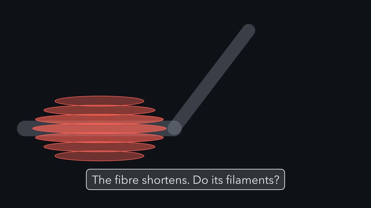 ▶ Watch Sliding, Not Shrinking · 3:03
Read the transcript of “Sliding, Not Shrinking”
Film transcript: Sliding, Not Shrinking
A muscle fibre shortens, yet none of its filaments gets any shorter. How can that be? Let's zoom in and watch one contract. A skeletal muscle fibre is one long cell. It is packed with chains of repeating units called sarcomeres. Each sarcomere runs from one Z disc to the next. Thin filaments of actin hang from each Z disc. A thick filament of myosin sits in the middle. Its heads stick out toward the actin on both sides. At rest, a strand called tropomyosin covers the sites where myosin binds.
Calcium is the switch. In skeletal and heart muscle, it binds troponin, and troponin moves tropomyosin aside. Now the myosin heads grab actin and swing. Heads on each half pull toward the middle. So both sets of thin filaments slide inward. The Z discs draw together, and the sarcomere shortens. But the thick filament keeps its length, and so do the thin ones. Only their overlap changes. Each head pulls only a tiny distance. Then it must let go and grab again further along. ATP binds, and the head lets go. ATP is split, and the head re-cocks. Then it grabs actin again and swings once more. Here is a question for you. Take away all the ATP. Do the heads stay stuck to actin, or do they fall off?
They stay stuck. Without ATP, an attached head cannot let go, so the fibre locks stiff. That is why muscles stiffen after death, the state called rigor mortis. One sarcomere moves only a hair. So how does that become a pull you can see? The answer is in how sarcomeres are arranged. A long fibre holds tens of thousands of sarcomeres in a row. Each one shortens a little. Their small shortenings add up along the chain into one visible pull. Notice what this machine can do. It can pull its ends together. It cannot push them apart. So every muscle needs something to stretch it back. In your arm, one muscle bends the elbow and another straightens it.
Each takes a turn pulling. Filaments slide; they never shrink. Many small slides in a row add up to one pull. Every muscle in this lesson pulls with this same machine. They differ in how calcium throws the switch.
From the film
- Myosin heads grab actin and pull the thin filaments toward the middle of each sarcomere; no filament gets shorter, only their overlap changes.
- Calcium is the switch: in skeletal and heart muscle it binds troponin, which moves tropomyosin off the sites where myosin binds.
- ATP binding lets a myosin head let go, and splitting ATP re-cocks it; without ATP the heads stay stuck, as in rigor mortis.
- A fibre's shortening is the sum of many tiny sarcomere shortenings in a row; a long fibre holds tens of thousands of them.
- Muscles can only pull, so something else must stretch each one back, such as a partner muscle on the other side of a joint.
The first film built the machine. This text follows its switches and what it pulls against.
The one idea. Every muscle pulls the same way: myosin heads use ATP to drag actin filaments past them, and calcium switches the sliding on. A muscle can only pull. In striated muscle, many tiny sarcomere shortenings in a row add up to one visible pull. Muscles differ in how calcium throws the switch and how they make ATP; skeletons differ in what the pull works against.
Scope: smooth muscle also slides actin past myosin, but it has no sarcomeres, and its switch is on myosin, not on troponin. The lever equation later is for an idealized static joint.
From nerve to calcium to cross-bridge
The film’s sarcomeres sit end to end in myofibrils, which pack each skeletal muscle fibre: one long cell with many nuclei, formed when embryonic muscle cells (myoblasts) fuse. Their regular overlap makes the stripes that give striated muscle its name. The A band is the length of the thick filaments. The I band, the light stripe straddling each Z disc, is thin filament with no thick filament beside it. The H zone, in the middle, is thick filament with no thin filament beside it. As overlap grows, the I band and H zone narrow and the A band keeps its width: the sliding filament model.
Predict. Two muscles have the same fibres, the same cross-section and the same load, and both are fully switched on. One is twice as long, with twice as many sarcomeres in a row. Which pulls harder? Which shortens farther, and faster? (Hint: in a chain, each link pulls on the next.)
Check your prediction
Equally hard: like links in a chain, sarcomeres in a row pass the same force along. But each one shortens the same amount at the same rate, so the longer muscle shortens twice as far in the same time, twice as fast. Force depends instead on how many filaments lie side by side, set by the cross-section across the fibres.
At the neuromuscular junction, the synapse between a motor neuron and a muscle fibre, the neuron releases acetylcholine, which opens channels in the fibre’s membrane and depolarizes it (synaptic transmission and the neuromuscular junction cover the details). That starts an action potential, an electrical signal that runs along the membrane (action potential) and down T tubules, infoldings that reach deep into the fibre.
Next to each T tubule lies the sarcoplasmic reticulum (SR), the fibre’s internal calcium store. In vertebrate skeletal muscle, a voltage sensor in the T-tubule membrane (a channel called CaV1.1) is linked to the SR’s calcium release channel (RyR1). When the voltage changes, the sensor opens the release channel, and calcium floods out. Calcium entering from outside the cell is not required for this step; that is where the heart differs (below). The film’s switch, troponin, is a complex of three proteins on the thin filament, with calcium binding at troponin C; the attached heads are cross-bridges.
ATP’s first job, releasing and re-cocking each head, is in the film. Its second is relaxation: SR pumps use ATP to move calcium back into the store, tropomyosin covers the sites again, and the muscle relaxes. The cross-bridge cycle and the 1954 sliding filament papers are in molecular motors; calcium as a signal is in calcium signaling.
Three muscles, three triggers
Vertebrates have three kinds of muscle, all built from actin and myosin (each type makes its own versions) but switched on differently.
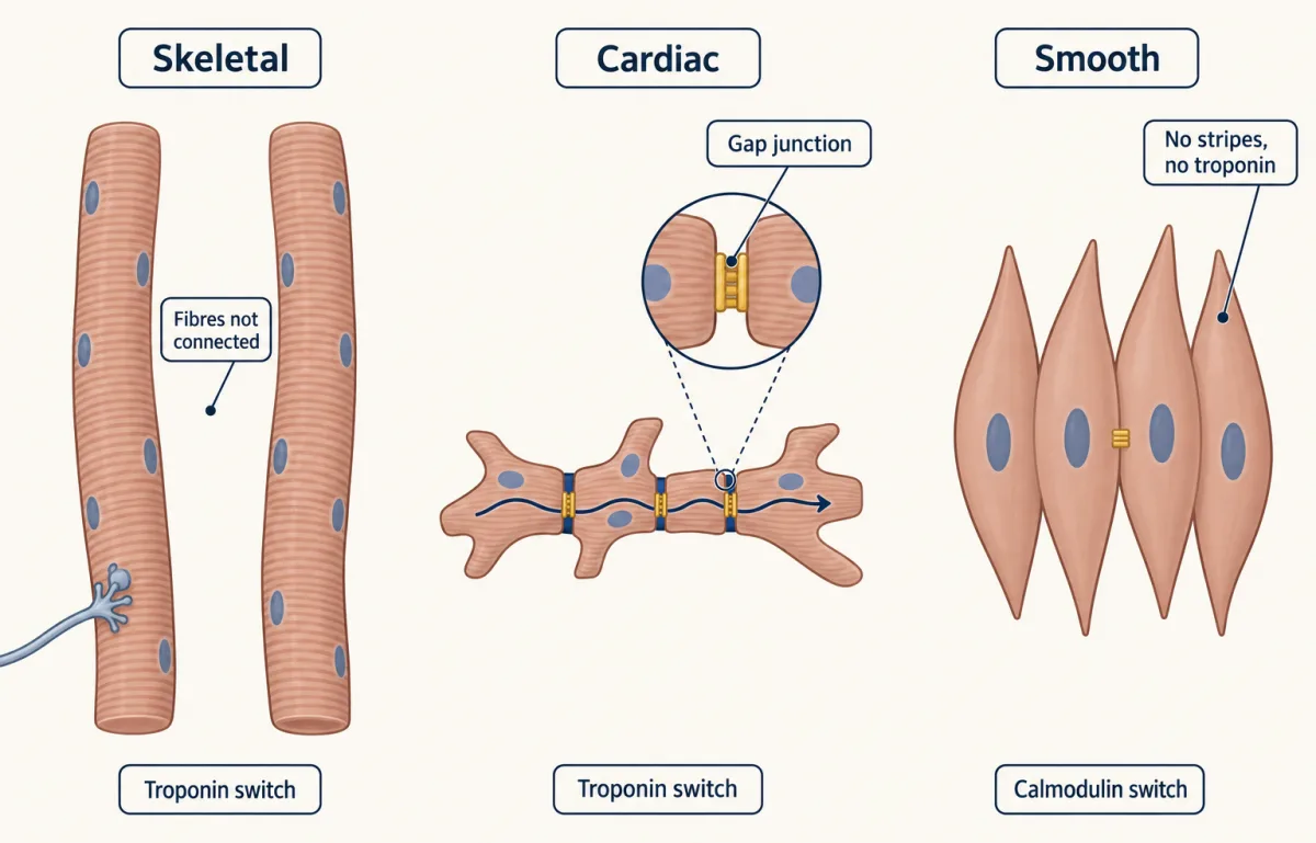
Skeletal muscle is striated and voluntary. Each fibre contracts when its own motor neuron fires, and fibres are not electrically connected to each other. Graded force comes from how many motor units (a motor neuron and the fibres it drives) fire and how often (motor units and reflexes).
Cardiac muscle is also striated, but its cells are short, branched and joined end to end by gap junctions, channels that let electrical current pass from cell to cell, so a wave of excitation spreads through the heart. No motor neuron starts the beat: pacemaker cells generate action potentials on their own, and nerves adjust their rate and the force of each beat (see circulation and the heart). The calcium trigger differs too. In mammalian heart cells, calcium entering through membrane channels during the action potential triggers a much larger release from the sarcoplasmic reticulum (SR). Alexandre and Françoise Fabiato gave key evidence for this calcium-induced calcium release in 1975: in skinned cells (surface membrane removed) from adult rat ventricles, a small controlled rise in bath calcium triggered calcium release from the SR. Frog hearts and many fish hearts rely mostly on the entering calcium itself.
Smooth muscle lines the gut, blood vessels, airways and ducts. Its actin and myosin are not organized into sarcomeres, so it has no stripes. It is not under voluntary control; autonomic nerves, hormones and stretch act on it. In many smooth muscles, such as those of the gut wall, gap junctions couple neighbouring cells. Smooth muscle has no troponin switch; its switch is on myosin. Calcium binds calmodulin, and calcium-calmodulin activates myosin light-chain kinase, an enzyme that adds a phosphate to the myosin light chain and turns myosin on. Chacko, Conti and Adelstein showed in 1977, with purified guinea-pig smooth muscle myosin, that this phosphate raised the myosin’s actin-activated ATPase activity (its rate of splitting ATP with actin present) and removing it lowered that activity. As calcium falls, the kinase slows and a phosphatase (an enzyme that removes phosphates) takes the phosphate off, so the muscle generally relaxes.
Fast and slow: fibres, tuna and chickens
Skeletal fibres also differ in speed and in how they make ATP. Oxidative fibres make most of their ATP with oxygen, by aerobic respiration in mitochondria. Glycolytic fibres make ATP fast by glycolysis (glycolysis), which does not need oxygen, but they have less capacity for aerobic respiration.
- Slow oxidative fibres contract slowly, are rich in mitochondria and in myoglobin (an oxygen-binding protein, see oxygen transport), look dark red, and resist fatigue.
- Fast oxidative fibres contract quickly but still rely mainly on aerobic respiration.
- Fast glycolytic fibres contract quickly, look pale, and fatigue sooner.
In humans the three main types are I, IIa and IIx; older papers use other labels (see Going deeper).
Predict. A domestic chicken stands and walks all day but flaps only in brief bursts. Which would you expect to be pale, its leg muscles or its breast muscles?
Check your prediction
The breast. The leg muscles used in standing and walking are darker, and the breast muscles that power brief bursts of flapping are pale.
Whole animals show the same split. In many fishes a thin band of dark red muscle runs along each side of the body under the skin and powers steady swimming, while the bulk of white muscle is used in bursts. In tunas the red muscle lies deeper, close to the backbone, and stays warmer than the water (thermoregulation). In each of these animals, chickens included, the muscles used for steady work are red and oxidative and those used for bursts are pale and glycolytic, although species also differ in which fibre types they have.
Fast or slow, each contraction of a vertebrate skeletal fibre waits for its own nerve impulse. The second film asks whether a fly’s far faster wings obey that rule.
Read the transcript of “Wings Faster Than Nerves”
Film transcript: Wings Faster Than Nerves
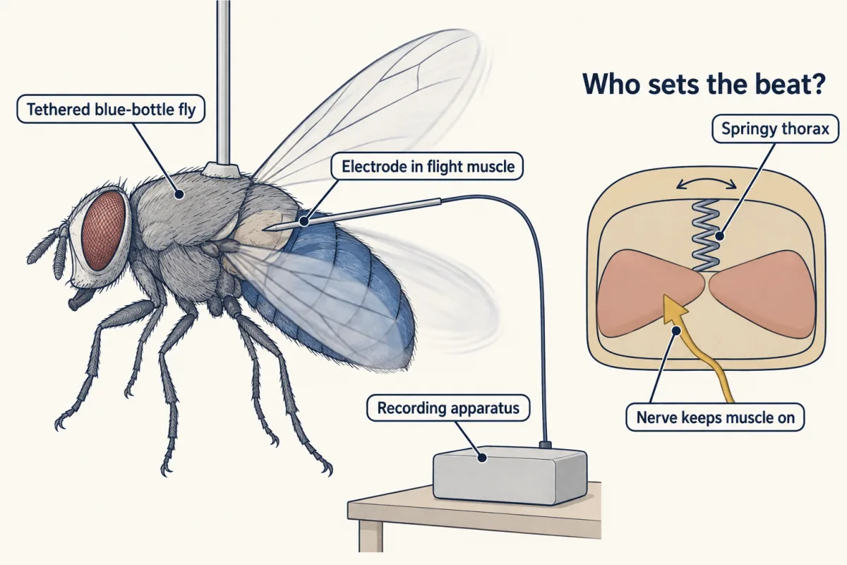
Does every wingbeat need its own nerve impulse? A fly beats its wings so fast that the answer seemed in doubt. One recording put it to the test. The first film built one machine. Calcium switches it on, and myosin heads pull actin. Now the question is how a fly's flight muscle throws that switch. In vertebrate skeletal muscle, each contraction follows its own nerve impulse. So does the flight muscle of a locust or a dragonfly. This is called synchronous muscle. In nineteen forty-nine, John Pringle tested this rule in blue-bottle flies. He tethered each fly so that it beat its wings in place. Then he put a fine electrode into its flight muscle.
If every beat needs its own impulse, the trace should show one large spike under every wingbeat. What do you think he saw? The large spikes, which he linked to nerve impulses, came far less often than the wings beat. And they were not timed to particular beats. The one-to-one rule failed. So what sets the beat?
Picture the fly's thorax as a springy box. Two sets of power muscles pull on it in different directions. When one set shortens, the box bends, and the other set is stretched. These muscles are already switched on. In them, a stretch raises force, after a short delay. So the stretched set builds force and shortens. That stretches the first set in turn. The muscles and the springy thorax keep the rhythm going between them. This is asynchronous muscle. It powers the wings of flies, bees and wasps, beetles and some other insects. In nineteen fifty-nine, Machin and Pringle reported stretch activation in beetle flight muscle. Does that make the nerve useless?
No. Its few impulses keep the muscle's calcium raised, so it stays switched on. Without them, the wings stop. The pulling machine is the same as in the first film. What changed is the switch. The nerve holds it on, and stretch times each beat. Pringle's recordings left questions open. And how stretch raises force, at the level of the molecules, is still being worked out. So here is the rule for these insects. In asynchronous muscle, stretch times each beat, and the nerve keeps it on.
From the film
- In a locust, each flight-muscle contraction follows its own nerve impulse; in Pringle's blue-bottle flies, the large spikes came far less often than the wingbeats.
- In asynchronous flight muscle that the nerve keeps switched on, a stretch raises force after a short delay, so two muscle sets pulling on a springy thorax take turns stretching each other.
- The nerve still matters: its few impulses keep the muscle's calcium raised, so it stays switched on; without them the wings stop.
- Pringle's 1949 recordings showed the mismatch; how stretch raises force at the molecular level is still being worked out.
A fly’s wings outrun its nerves
Which signal counts. Pringle’s trace also held a small signal with every wingbeat. The one-to-one rule is about the large spikes he linked to nerve impulses, so they are what break it.
Predict. Suppose instead that the force rose the instant the switched-on muscle was stretched. Would the two muscle sets keep the wings beating?
Check your prediction
No. Force that rose in step with the stretch would act like a stiffer spring: it would add no net work, so the beat would die away. The delay lets force peak while a set is shortening, so each set does work on the thorax and stretches its partner in turn.
How firm the evidence was. In 1949 Pringle only suggested that the muscle responds to stretch; the 1959 beetle work used isolated muscle (Going deeper).
Where it is found. Even in flies, the small steering muscles at the wing base are synchronous.
Skeletons: water, armour and bone, with levers
A hydrostatic skeleton is fluid under pressure in a closed cavity. An earthworm contracts the muscles of its body wall against fluid in its body cavity, so each segment can lengthen or shorten (animal body plans). A muscular hydrostat has no fluid cavity: octopus arms and mammalian tongues are mostly muscle at constant volume, so muscle in one direction acts against muscle in another.
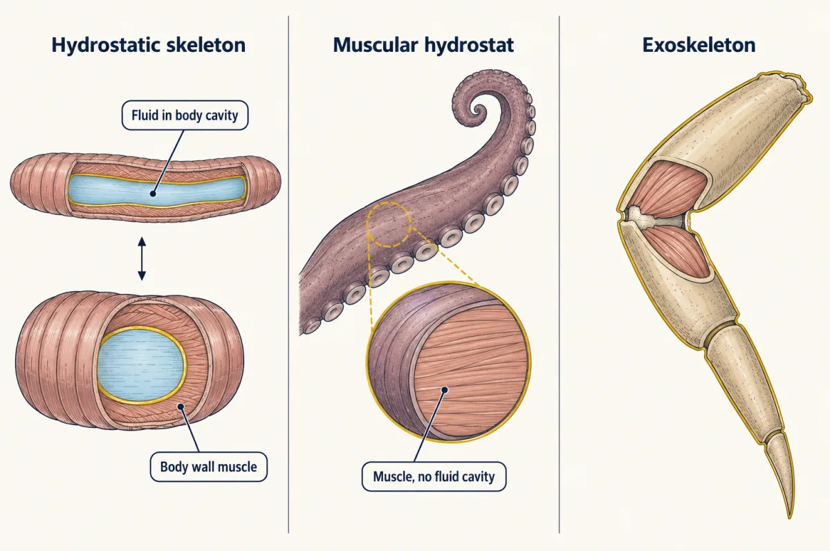
An exoskeleton is a hard outer case with muscles attached inside, as in insects and crabs. It cannot grow, so arthropods shed it and make a larger one at each moult (endocrine system). An endoskeleton lies inside the soft tissues, as in vertebrates.
Bone is living tissue. Its matrix combines collagen fibres with a calcium phosphate mineral, hydroxyapatite. Compact bone forms the dense outer wall and is built of osteons, cylinders of matrix around a central canal. Spongy bone, inside, is a lattice of thin struts called trabeculae. Osteoblasts lay down new bone, osteoclasts dissolve it, and osteocytes are former osteoblasts trapped in the matrix. Because bone is resorbed and rebuilt throughout life, its shape changes with the loads it carries (on how bone thickness scales with body size, see size and scaling).
Bones meet at joints. In humans, joints are grouped by structure as fibrous (bones joined by connective tissue, as in skull sutures), cartilaginous (joined by cartilage) or synovial (a lubricated, fluid-filled cavity, as at the elbow and knee). At a synovial joint, muscles work in antagonistic groups: flexors such as the biceps and brachialis bend the elbow, and the triceps straightens it. Tendons carry the pull to bone, and the bone acts as a lever.
Worked problem. These numbers are invented for an idealized model. A forearm is held horizontal and treated as weightless, and the biceps is treated as the only muscle bending the elbow; in a real arm the brachialis and brachioradialis share the load. A 20 N load (about 2 kg) is held in the hand 0.30 m from the elbow. The biceps pulls straight up with a moment arm of 0.040 m, the perpendicular distance from the elbow to the line of the biceps’ pull. Assume static equilibrium, with the elbow as the pivot. Each torque is a force times its moment arm, and the torques must balance:
muscle force x muscle moment arm = load x load arm
- Torque from the load. 20 N x 0.30 m = 6.0 N m.
- Muscle force. F x 0.040 m = 6.0 N m, so F = 6.0 / 0.040 = 150 N, which is 7.5 times the load.
- Check. 150 N x 0.040 m = 6.0 N m, matching the load’s torque.
- What the trade buys. The same geometry means the hand moves 0.30 / 0.040 = 7.5 times as far, and as fast, as the point where the biceps attaches. If the biceps shortens by 1.0 cm, the hand moves about 7.5 cm (for a small angle). This is a third-class lever: the muscle pulls between the pivot and the load, and gives up force for distance and speed.
- Load torque
- Muscle force
- Muscle force as a multiple of the load
- Hand movement per 1.0 cm of muscle shortening
In this sketch: invented numbers, an idealized weightless forearm held horizontal, static equilibrium, the biceps pulling straight up, the elbow as the pivot; not anatomy.
Rule to keep: muscle force x muscle moment arm = load x load arm. A short moment arm means a large muscle force, and the same ratio means the hand moves that many times as far and as fast as the muscle’s attachment (for a small angle).
The one idea, once more: one actin-myosin machine, switched on in different ways, and what it pulls against decides what the pull does.
Design problem
Does a muscle require calcium from outside the cell to contract? You have frog skeletal muscle fibres and isolated cardiac cells, a calcium-free bathing solution with a calcium binder, and a drug that blocks the L-type calcium channels in the cell membrane. Design an experiment that tells the two triggers apart.
Saved in this browser only.
One way to do it
Stimulate each preparation electrically and record force in normal solution, then in the calcium-free solution, and in a separate run with the channel blocker added to normal solution.
The prediction for skeletal fibres: twitches continue for a time without calcium entry, because the voltage sensor is linked mechanically to the SR release channel. For mammalian ventricular cells: force falls quickly once the calcium-free solution reaches the cells and drops with the blocker, because entering calcium triggers SR release, as the Fabiatos’ work on skinned rat ventricular cells suggested. The calcium-free bath is the cleaner test (Going deeper explains why skeletal twitches eventually fade and cautions about the blocker).
Controls matter. Record action potentials in every condition, so that a loss of force cannot be blamed on a membrane that no longer fires. Return each preparation to normal solution to show that the effect reverses and the cells were undamaged. Keep the calcium-free period short, because heart cells can be injured when calcium is restored after a long time without it. Keep the temperature the same, and take the cardiac cells from a mammal such as the rat: frog ventricle cells rely almost entirely on the entering calcium itself and have little SR release, so they would not test calcium-triggered release. Name the species in your conclusion: the result holds for frog skeletal muscle and the heart cells you used. The older story of Ringer’s frog heart and bath calcium is told in calcium signaling.
Going deeper
Fibre names depend on the species
Human fibres long labelled IIb by staining express mostly IIx myosin, not the rodent IIb isoform (Smerdu and colleagues, 1994), so rodent IIb results cannot be read straight across to people.
What Pringle’s recordings did not settle
Pringle left open whether the large spikes were true propagated action potentials, and the recordings did not show how the muscle oscillates. How stretch raises the force of an already calcium-activated fibre is still being worked out, with proposals involving troponin isoforms specific to these muscles and the geometry of thick and thin filaments.
Reading the calcium-free test with care
Twitches may still fade after some minutes, but not because the store has run down: the voltage sensor itself needs calcium bound to the outside of the membrane. Read the blocker run with care for skeletal fibres: there the L-type channel (CaV1.1) is itself the voltage sensor, so some blockers of this kind reduce skeletal force without any need for calcium entry.
Your notes
Select any text in the lesson to highlight it or add a note to it. Your highlights and notes are saved in this browser and collected in your notebook.
This browser is not letting the site save anything (a private window or strict privacy settings), so highlights and notes will be lost when you leave the page.
How we measure it
Electrical recording from working muscle
Place fine electrodes in or on a muscle and record its electrical activity while the animal moves, here a tethered fly beating its wings. Counting impulses against movements tells you whether each contraction follows its own impulse.
Skinned muscle cells with calcium buffers
Remove the surface membrane from a muscle cell, mechanically or with detergent, so that the experimenter sets the calcium, ATP and other contents of the bath. The Fabiatos used this in rat ventricular cells to separate calcium acting on the filaments from calcium released by the sarcoplasmic reticulum.
Myosin typing by histochemistry and in situ hybridization
Stain muscle sections for myosin ATPase activity, which differs among fibre types, and probe neighbouring sections for the messenger RNA of each myosin heavy chain. Smerdu and colleagues used both on human muscle and found that fibres stained as IIb carry IIx myosin transcripts.
Protein purification and enzyme assays
Purify a protein from a tissue, change it (here by adding or removing phosphate), and measure its activity. Chacko, Conti and Adelstein used this on guinea-pig smooth muscle myosin.
Practice questions on this lesson (3) → MCAT-style, original, each with an explanation
Where this lesson fits
Before thisReview first
- Nervous Control: A First LookThis course · Lesson 7
You need the motor neuron's signal and the neuromuscular junction from there to follow how a nerve impulse starts contraction.
- Respiration and ATPBiology · High school
The two jobs of ATP in the cross-bridge cycle build on how cells make and use ATP.
- Tissues, Organs and HomeostasisThis course · Lesson 6
The lesson compares muscle types as tissues, using what you learned there about tissue types.
Elsewhere on the siteFilms, simulations and method films in other courses
- Grip, Swing, Let GoFilm · Cell Biology
It shows how myosin and other motors turn ATP into steps along a filament.
- Reading an optical trapSimulation · Cell Biology
It lets you read single-motor force and steps, the molecular basis of the sliding you study here.
Suggested by an AI model from the lessons themselves, then checked. How we use AI
Finish line
Test yourself
8 questions
Practice and MCAT-style, with explained answers.
Next · Lesson 20
Animal Reproduction
A monkey's pituitary, cut off from the brain's drive, was given its missing hormone either as a steady drip or once an hour, the drip giving at least as much. Only one schedule kept it working. Why would timing matter?
Continue →