Physiology · Unit 7: Digestion and absorption
Gut Motility, Secretion and Gut Hormones
Rewritten on : new text, figures and flashcards. Your notes and highlights stay, but the text around them may have moved.
A glass of water is usually half gone from your stomach in about 10 minutes. A rich, high-calorie drink may take about 2 hours to be half gone. What holds the rich drink back?
Pause when the reading reaches a film, and offer to play it. Saved on this device.
Watch the film now, or keep listening
Builds on:Digestive Systems and NutritionTransport Across MembranesSignal-Transduction Pathways: How Cells Compute With Molecular SwitchesWhy these?
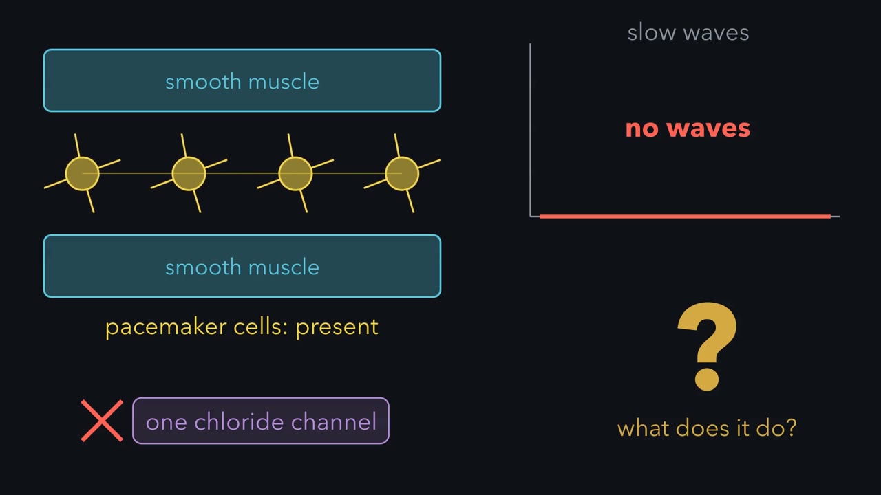 ▶ Watch The gut keeps its own time · 2:35
Read the transcript of “The gut keeps its own time”
Film transcript: The gut keeps its own time
Mice born without one chloride channel have normal-looking pacemaker cells in the gut but no electrical slow waves. What does that channel do? The gut wall keeps its own time. The beat comes from branched cells between the muscle layers, the interstitial cells of Cajal, or ICC. Record from a gut smooth muscle cell and you see slow waves, which are rhythmic depolarizations that last seconds. In the human stomach, pacemaker cells generate about 3 a minute. Isolated ICC show this rhythm, but isolated smooth muscle cells do not typically show it. In paired recordings from mouse gut muscle, the waves were larger in ICC. They reached the muscle passively, shrinking as they spread, so smooth muscle cannot regenerate them.
A slow wave is not an action potential, and it sets when the muscle can contract. A contraction follows only when a wave carries the muscle past threshold for calcium entry. Think of a metronome and a pianist. The metronome fixes the beat, and the pianist decides how loud each note is. Excitatory and inhibitory neurons, and hormones, set how strongly each wave couples to contraction. Small-intestinal muscle from Kit-mutant mice, with few myenteric ICC, lacked the slow waves of wild-type siblings. Mice lacking ANO1 failed to develop slow waves, yet their ICC networks looked normal. So a calcium-activated chloride current is necessary for the rhythm, separately from building the network.
It does not show that ANO1 is the whole pacemaker. It describes neonatal mice, not adults or any human disorder. The enteric nervous system is the network of neurons in the gut wall. The myenteric plexus, between the circular and longitudinal muscle layers, mainly controls movement. In 1899, Bayliss and Starling recorded loops of small intestine in anesthetized dogs. Distension of the wall caused contraction on the oral side and relaxation on the anal side, driving contents anally. The response persisted after the extrinsic nerves were cut, but was lost when conduction within the wall was blocked. So the gut splits the work. ICC keep the beat, and the beat sets when the muscle can contract.
Neurons and hormones set how strongly each wave couples to contraction. Keep this rule. Slow waves mainly set the timing of contraction.
Drink a glass of water and half of it has usually left your stomach in about 10 minutes. Drink a rich, high-calorie liquid such as a milkshake, and half may still be there after about 2 hours.
Predict. Why is the milkshake held back? Is it simply thicker, or is something telling the stomach to slow down?
Check your prediction
Something is telling it: calories reaching the duodenum slow emptying, beyond what thickness alone would do. When calories reach the duodenum, the first part of the small intestine, it releases hormones and starts nerve reflexes that slow the stomach. Water carries no calories, meets no calorie brake, and pours through.
The one idea to carry through this lesson. Food arriving in a part of the gut turns that part on, and what it makes or passes downstream turns it back down. Nerves and hormones carry both signals, but they adjust machinery that the gut wall runs on its own. In the muscle, pacemaker slow waves set when a contraction can happen; the signals decide whether it does, and how strongly.
The on-then-brake idea covers the fed stomach and duodenum, with two exceptions; the clock and the reflex circuits work along the whole tract.
- Anticipation. The vagus can act on a part before food arrives: the cephalic phase of acid secretion, the stomach’s relaxation as you swallow, and relaxation of the lower esophageal sphincter. Only the trigger comes early.
- Fasting. Between meals the gut runs a separate program, the migrating motor complex, and eating switches it off.
The timing half applies to muscle that contracts in phases, such as the lower stomach and small intestine; the upper stomach and tonic sphincters such as the lower esophageal sphincter hold a steady tone with no comparable beat.
The gut wall runs three machines itself: a clock that times the muscle, reflex circuits of nerves, and the stomach’s acid pump. The film covers only the first two; the acid pump is in the text and the simulation. The first look at the gut and the first hormone table are in digestive systems; here they get cells, receptors and numbers.
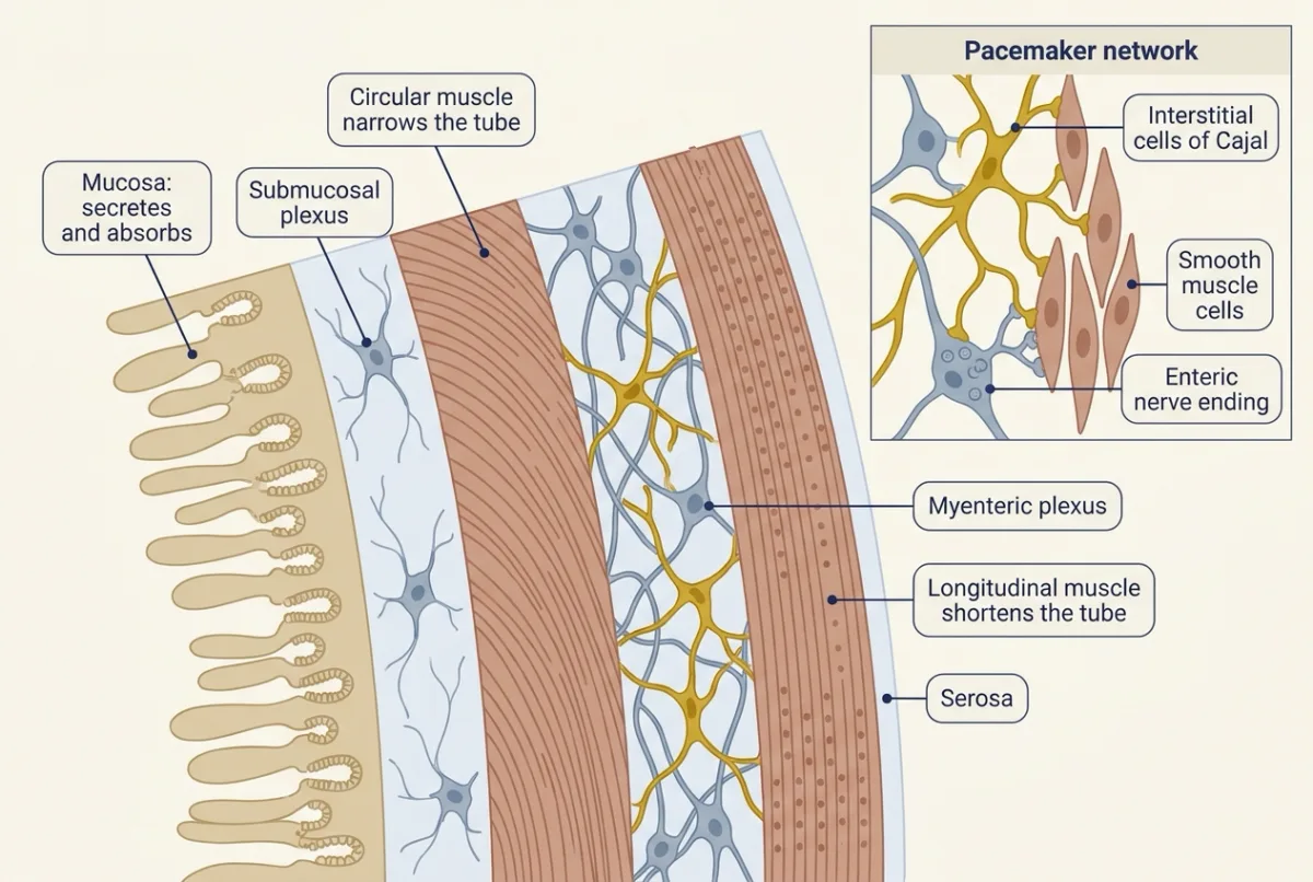
The wall, from the lumen outward
Picture a cross-section of the gut and read it from the inside out.
- Mucosa, the lining that faces the lumen (the inside of the tube), secretes and absorbs; in the stomach it holds the acid-making and hormone-making cells.
- Submucosa, carrying the submucosal plexus of neurons.
- Circular muscle, which narrows the tube.
- Myenteric plexus, a second mesh of neurons. Here and within the muscle layers sit the pacemaker cells of the next section.
- Longitudinal muscle, which shortens the tube.
- Serosa, the outer covering.
The clock: Cajal’s cells keep the beat; nerves decide how hard
Reading a slow-wave recording
Picture a voltage recording from a smooth muscle cell of the human antrum, the lower stomach, above a force recording of the same muscle.
- The voltage jumps up quickly, holds a raised plateau for some seconds, then falls back, about every 20 seconds. These are slow waves: rhythmic depolarizations lasting seconds, about 3 per minute here.
- On some waves the plateau rises higher, sometimes with fast spikes on top. Under exactly those waves the force trace shows a contraction.
- On other waves the force trace stays flat.
A contraction follows only when a wave carries the muscle past threshold for Ca2+ entry, the voltage at which its Ca2+ channels open. A slow wave is not an action potential: it sets when the muscle can contract.
In the human stomach the waves start high on the corpus, the body of the stomach, and spread toward the pylorus, its outlet. In humans, other regions keep other rates: about 11 to 12 per minute in the duodenum and about 8 to 9 in the ileum.
The metronome and the pianist
Slow waves are a metronome, fixing the beat. Excitatory and inhibitory neurons and hormones are the pianist, deciding whether to play on each beat and how loud: they set whether each wave carries the muscle past threshold, and how far, which physiologists call how strongly each wave couples to contraction. So in the antrum and small intestine the slow-wave rate is the fastest rate at which a region can repeat a phasic contraction, about 3 per minute in the antrum, and the same 3 waves can give no contractions or three. Force generation itself belongs to smooth muscle.
Where the beat comes from
The rhythm comes from the gut wall, but not from its smooth muscle: it comes from the interstitial cells of Cajal (ICC), branched cells that form networks between and within the muscle layers. Isolated smooth muscle cells do not typically show the rhythm; isolated ICC do. In paired recordings from mouse gut muscle, slow waves were larger in myenteric ICC than in the smooth muscle beside them; they reach the muscle passively, shrinking as they spread, and the muscle does not regenerate them.
ICC generate the beat inside a network: they are electrically coupled to the muscle and to PDGFRα-positive cells, together sometimes called the SIP syncytium.
W/Wv mice, with mutations in Kit, a receptor ICC need to develop, have few myenteric ICC.
Predict. In W/Wv muscle the smooth muscle and enteric neurons are still present. Could the neurons stand in for the missing clock?
Check your prediction
No. Their small-intestinal muscle lacked the slow waves always present in normal siblings, and a second laboratory independently found the same, so ICC are necessary. Neurons are the pianist, not the metronome: they adjust whether each wave gives a contraction, but they do not make the waves.
The channel in the film
The film opens by asking what a missing chloride channel does. The channel is ANO1 (TMEM16A), a Ca2+-activated Cl- channel. Mice born without it had normal-looking ICC networks, yet their gut muscle failed to develop slow waves, so ANO1 is needed to run the clock, separately from building it. Periodic Ca2+ release from stores in each ICC is the proposed upstream trigger, and ANO1 turns that Ca2+ into the depolarizing current of the slow wave. Current reviews put the Ca2+ channel CaV3.2 beside ANO1 as a second main pacemaker conductance. The knockout results come from neonatal mice, not adults or any human disorder.
The gut wall has its own nervous system
The enteric nervous system is the network of neurons in two plexuses of the gut wall: the myenteric (Auerbach’s) plexus mainly for movement and the submucosal (Meissner’s) plexus mainly for secretion and local blood flow. Together they hold sensory and motor neurons, with interneurons linking them, enough to run reflexes without the brain. Extrinsic nerves adjust these circuits, and some functions, such as normal defecation, also need spinal pathways (the hypothalamus and autonomic system; vagal sensory neurons).
The signature reflex is peristalsis. In 1899 William Bayliss and Ernest Starling, three years before their secretin work (signals and distance), studied it in loops of small intestine in anesthetized dogs. A local stimulus, pinching the wall or a bolus distending it, produced contraction on the oral side and relaxation on the anal side, so contents were driven anally. Their summary became the “law of the intestine”.
Predict. If the brain and spinal cord organize peristalsis, cutting the extrinsic nerves, the vagi and splanchnics, should abolish it. Bayliss and Starling cut them. What happened?
Check your prediction
The reflex survived, and was lost only when nervous conduction within the wall was blocked: nerves in the gut wall carry it without the central nervous system. The experiment did not show which neurons or transmitters carry it, or where the rhythm comes from.
The same intrinsic wiring ends the tract. The rectum stores feces. When it is distended, an enteric reflex relaxes the internal anal sphincter, which is smooth muscle and not under voluntary control. The external anal sphincter is skeletal muscle supplied by the pudendal nerve; it keeps up a resting contraction and can be squeezed voluntarily to hold defecation off; sacral parasympathetic outflow through the pelvic nerves strengthens colonic and rectal contraction.
Mouth to stomach: wet, swallow, store, grind, meter
An adult makes roughly 1 to 1.5 L of saliva a day, in two steps. Acinar cells secrete a fluid close to plasma in composition; the ducts, poorly permeable to water, then reabsorb Na+ and Cl- and secrete K+ and HCO3-, so the saliva ends hypotonic. At high flow the ducts have less time per volume and the saliva comes out closer to plasma tonicity. Both autonomic divisions stimulate secretion, with parasympathetic input dominant.
Swallowing starts voluntarily; the pharyngeal phase is a reflex patterned in the brainstem, and esophageal peristalsis carries the bolus down while vagal pathways activate inhibitory enteric neurons that relax the lower esophageal sphincter.
When you swallow, the vagus relaxes the upper stomach before the food gets there (receptive relaxation), the anticipation exception at work. As the stomach fills, distension keeps it relaxed through vagal and local reflexes (accommodation). Either way, volume rises with little rise in pressure.
The antrum grinds. Its contractions, timed by the slow waves, push chyme, the soupy mix of food and juice, against a pylorus that is closing, and most of the chyme is thrown back, until particles are small enough to pass.
Why the milkshake waits: the duodenal brake
Nutrients reaching the duodenum release cholecystokinin (CCK) and, further down, glucagon-like peptide 1 (GLP-1), and these slow emptying, partly through vagal reflexes that slow the antrum and tighten the pylorus. The stomach hands calories on at roughly 1 to 4 kcal per minute.
The opening’s numbers are one review’s example for healthy humans; real times depend on the meal’s volume and calories. They fit the brake: if the drink held, say, 400 kcal, emptying half of it in 2 hours would be about 1.7 kcal per minute, inside the 1 to 4 kcal per minute range. Because the brake meters calories, a caloric liquid tends to empty at a more nearly steady rate, while water empties fastest at first and then slows, roughly losing half of what remains every 10 minutes.
Digestible solids leave only after a lag, while they are ground small, and emptying a solid meal takes hours. In fasting, motilin speeds emptying, and so does ghrelin given as a drug (see the ledger below).
Between meals: the migrating motor complex
Fasting brings the migrating motor complex (MMC), a cycle interrupted by feeding.
- Phase I lasts about 45 to 60 minutes, with slow waves but few or no contractions.
- Phase II has irregular contractions.
- Phase III is a 5 to 15 minute front of strong contractions that sweeps toward the pylorus and down the small bowel.
Phase III, thought to sweep undigested residue onward between meals, shows the clock’s limit. Its contractions are the strongest of the fasting cycle and come as fast as they can, yet each rides on a slow wave, so the front contracts at the local slow-wave rate, about 3 per minute in the antrum. Enteric neurons, with vagal and hormonal input, decide when the front starts and how it moves; plasma motilin rises in cycles, and in humans intravenous motilin or ghrelin can start antral phase III. Isolated mouse small intestine can make migrating complexes without slow waves, which has not been shown for the human stomach.
Acid from a pump, protection from a gel
At rest the parietal cell keeps its H,K-ATPase in cytoplasmic tubulovesicles. Stimulation moves the pumps into the membrane of the secretory canaliculus, a deep fold of the apical surface. Each cycle exchanges cytoplasmic H+ for luminal K+ using ATP, electroneutrally (one charge in for one out, so no net current); K+ returns to the lumen through apical channels and Cl- exits with it, so HCl accumulates. The H+ comes from carbonic anhydrase, and the matching HCO3- leaves across the basolateral membrane in exchange for Cl-, the alkaline tide of acid-base balance. The canaliculus of a stimulated cell reaches about pH 1.
The pump has three switches. Acetylcholine acts on Gq-coupled M3 receptors and gastrin on Gq-coupled CCK2 receptors, both raising Ca2+; histamine acts on Gs-coupled H2 receptors and raises cAMP (GPCRs and G proteins; cAMP and PKA). The H2 receptor is James Black’s story (drugs against a part).
In humans, gastrin probably acts mainly by releasing histamine from enterochromaffin-like (ECL) cells beside the parietal cells: in volunteers given gastrin, blocking H2 receptors abolished the acid response, and muscarinic block cut the response to the lower dose sharply, so acetylcholine is a needed partner.
Switching acid on and off
Before the food arrives: the cephalic phase. Sight, smell, taste and chewing signal through the vagus. Vagal fibres excite enteric neurons in the stomach wall; some release acetylcholine onto parietal cells, and others release gastrin-releasing peptide (GRP), which makes G cells in the antrum release gastrin. Acid starts before there is food in the stomach, as Pavlov’s sham-fed dogs showed (digestive systems).
While food is in the stomach: the gastric phase. Distension excites vagal and local reflexes that release more acetylcholine and, through GRP-containing neurons, more gastrin. Digested peptides and amino acids act on G cells directly, sensed through receptors such as the calcium-sensing receptor. Gastrin travels in the blood to the body of the stomach.
The brake inside the stomach. Acid made in the body of the stomach flows down to the antrum. There, acid makes D cells release somatostatin, and somatostatin holds back the G cells beside them: negative feedback, the more acid, the less gastrin.
Predict. A patient takes a proton pump inhibitor, a drug that blocks the H,K-ATPase, every day for months. What happens to the gastrin level in the blood?
Check your prediction
It rises. Little acid reaches the antrum, so D cells release less somatostatin, and the G cells, no longer held back, release more gastrin. The extra gastrin cannot bring the acid back to normal, because the block sits downstream, at the pump; it shows up as rebound acid when the drug is stopped.
Once chyme reaches the duodenum: the intestinal phase. Acid entering the duodenum releases secretin from S cells, and fat and digested protein release cholecystokinin (CCK) from I cells. Both inhibit gastric acid; CCK acts at CCK1 receptors, probably via somatostatin. CCK also slows emptying, so less acid arrives: the intestinal phase is mostly a brake.
So the meal switches acid on in the stomach and off from two places downstream of the pump: the antrum and the duodenum.
Why the stomach does not digest itself. A layer of mucus gel traps the bicarbonate that surface cells secrete and holds it against the lining, so the surface stays far less acidic than the lumen. Prostaglandins keep that mucus and bicarbonate coming, and tight junctions between the cells limit how much H+ leaks back in (epithelia).
Worked problem: what does it cost to make pH 1?
In this illustrative example, stimulated parietal cells secrete 1.0 L of juice at pH 1.0. Assume cytosolic pH 7.0, body temperature 310 K, R = 8.314 J per mol per K, and electroneutral exchange, so the membrane potential drops out.
- Luminal [H+] = 10−1.0 = 0.10 mol per L, so 1.0 L holds 0.10 mol H+. Assume 1 H+ per ATP (step 4 checks that this is the most the budget allows at pH 1); that takes at least 0.10 mol ATP.
- The concentration ratio is 10−1 / 10−7 = 106.
- Free energy per mol H+:
delta-G = 2.303 x R x T x (pH cytosol - pH lumen)
2.303 x 8.314 x 310 = 5,936 J per mol per pH unit; x 6 = 35,600 J, about 35.6 kJ per mol H+. Ignoring the uphill K+ step makes this a minimum. 4. ATP hydrolysis in cells releases roughly 50 to 60 kJ per mol (take 55). Two H+ at pH 1 would cost 71.2 kJ, too much. Two per ATP stays affordable while 2 x 5.94 x delta-pH is at most 55, so delta-pH is at most 4.6, a luminal pH of about 2.4.
Whether the pump ever moves 2 H+ per ATP at milder pH is debated (Going deeper, “One proton or two”). The budget does not set the stomach’s floor near pH 1, since one proton per ATP could in principle go below pH 0. In practice the floor is set by the juice being close to isotonic: about 150 to 160 mM HCl, which is pH of roughly 0.8 to 1.
- pH difference, cytosol − lumen
- Free energy per mol H+ = 5.936 × pH difference
- Work per cycle = n × free energy per H+
- Energy released by one ATP
- Luminal pH at which 2 H+ per ATP stops being affordable = pH cytosol − E ÷ (2 × 5.936)
Illustrative model, not a measurement. In this sketch (made up for the sketch unless the lesson gives it): each cycle spends one ATP; the cost is 2.303 × 8.314 × 310 × pH difference, at 310 K with electroneutral exchange so the membrane potential drops out, which is the lesson’s rule; the K+ step is ignored, so every cost shown is a minimum; the starting values, cytosolic pH 7.0, luminal pH 1.0 and 55 kJ per mol per ATP, are the lesson’s illustrative worked-problem values (ATP hydrolysis in cells releases roughly 50 to 60 kJ per mol); the slider ranges for pH are made up (the energy range is the lesson’s roughly 50 to 60), and the luminal slider goes down to 0 only to show that one ATP could still pay for one proton below the stomach’s real floor, which the lesson puts at pH roughly 0.8 to 1 because the juice is close to isotonic, about 150 to 160 mM HCl (the dashed band); the sketch lets you pick 1 or 2 H+ per ATP at any luminal pH; whether the pump ever moves 2 is debated: one vesicle study reported a fall from 2 to 1 as luminal pH dropped, another found 1 at pH 6.1 to 6.9, and a 2019 structure favours 1 throughout.
Rule to keep: each pH unit of uphill gradient costs about 5.94 kJ per mol of H+ at 310 K, and a cycle pays n times that out of one ATP, so the more protons per cycle, the smaller the pH difference one ATP can pay for; at the lesson’s 55 kJ per mol and cytosol pH 7.0, two protons per ATP stop being affordable below a luminal pH of about 2.4.
Cutting a switch, or losing the brake
Cut a switch first. In mice lacking the CCK-B receptor, gastrin cannot drive acid: basal gastric pH rose from 3.2 to 5.2, and plasma gastrin was about 10-fold higher, the same lost acid brake as with a proton pump inhibitor. Atrophic gastritis, with parietal cells lost, gives the same pattern in humans.
The knockouts also had more gastrin cells and fewer antral somatostatin cells, which fits long-term loss of the acid signal and can add to the rise. Their fewer parietal and ECL cells reflect cell number, not feedback, and hint that gastrin keeps the acid-making lining built (“Design problem: a stomach without gastrin”).
Predict. A gastrinoma is a neuroendocrine tumour that releases gastrin which acid cannot suppress. What happens to gastrin, to acid and to gastric pH? And why might the patient’s stools turn fatty?
Check your prediction
The tumour ignores the brake, so gastrin stays high, basal acid output and parietal cell mass rise, and gastric pH falls. Duodenal pH falls low enough to inactivate pancreatic lipase, the enzyme that digests fat, so fat leaves undigested.
Gastrin is high in both cases while acid moves in opposite directions, so reading the two together points to lost feedback or to an autonomous source, although diagnosing a gastrinoma needs further tests.
The duodenum answers back
The duodenum also neutralizes what arrives. Secretin drives pancreatic duct cells to secrete bicarbonate-rich fluid: in humans the juice reaches 140 to 150 mM HCO3- under secretin, about 6- to 7-fold over plasma, by a route that depends on cystic fibrosis transmembrane conductance regulator (CFTR). Cholecystokinin (CCK) and acetylcholine drive acinar enzyme secretion, CCK contracts the gallbladder, and secretin also drives bicarbonate from bile-duct cells; bile and absorption are in absorption and the liver.
The ledger sorts the hormones by their role for the stomach: on, brake or fasting. Glucose-dependent insulinotropic polypeptide (GIP) is listed by its better-known role, as an incretin.
| Hormone | Cell and site | Stimulus | One action here | Role |
|---|---|---|---|---|
| Gastrin | G cells, antrum | Peptides and amino acids, vagal input, distension | Acid, mainly through ECL histamine | On |
| Secretin | S cells, proximal small intestine | Acid in the duodenum | Duct fluid and bicarbonate; less gastric acid | Brake |
| CCK | I cells, duodenum | Fatty acids and digested protein | Gallbladder contraction; slower emptying | Brake |
| GIP | K cells, upper small intestine | Glucose and fat | Glucose-dependent insulin release | Insulin (incretin) |
| GLP-1 | L cells, mostly ileum and colon | Nutrients in the gut | Slows gastric emptying | Brake |
| Motilin | Mo cells, upper small intestine | Cyclic release in fasting | Can induce antral MMC phase III | Fasting |
| Ghrelin | P/D1 cells in humans (X/A-like in rodents), corpus | Fasting; suppressed by a meal | Speeds emptying when given as a drug; whether the body’s own ghrelin does is less clear | Fasting |
The incretin actions of GIP and glucagon-like peptide 1 (GLP-1), boosting insulin release after a meal, belong to insulin, glucagon and blood glucose, and these cells as sensors to gut-brain sensing.
Design problem: a stomach that cannot grind
A biopsy from a patient with diabetic gastroparesis, a stomach that empties too slowly, shows few antral interstitial cells of Cajal (ICC) but normal-looking enteric neurons. Predict the antral recording and whether solids or water empty worse, then design tests.
One way to do it
Recording. Expect slow waves that are absent, small or irregular. The pianist is there, but with few beats to play on, contractions are sparse and poorly timed.
Emptying. Solids should suffer most, because antral grinding needs the clock. Liquids depend more on the tone of the upper stomach, which has no comparable beat, so their emptying is often spared.
Tests. Measure solid and liquid emptying together by scintigraphy, record gastric electrical activity for a missing or disorganized 3-per-minute rhythm, and count ICC with Kit labelling against controls to see whether counts track solid emptying.
Limits. A biopsy shows association, not cause, and gastroparesis may involve nerves and other cells too.
The gut wall runs its own clock, reflexes and pump; the meal works them through switches, and the duodenum’s brake is why a milkshake waits while water does not.
Going deeper
The main line above does not depend on these details.
The experiments that found the pacemaker
The W/Wv result came from Ward and colleagues (1994); Huizinga and colleagues (1995) were the second laboratory, reporting that W mutants lacked both the myenteric interstitial cells of Cajal (ICC) network and pacemaker activity. Neither said what makes an ICC oscillate. Hwang and colleagues in the Sanders and Ward laboratory (2009) then tested ANO1: gut muscle from mice homozygous for a null Tmem16a allele, studied in organ culture, had no slow waves by birth, although Kit antibodies showed structurally normal ICC networks. The authors also reported ANO1 in ICC of macaque and human gut muscle.
How fixed is the beat?
The slow-wave rate differs by region and species, and it is not rigid even in one place. Stretching mouse antral muscle in vitro has been reported to raise it from about 2.5 to 3.6 per minute, so the clock sets the ceiling for contraction but can itself shift a little.
The law of the intestine and its limits
A later test bounds the 1899 law. In isolated guinea-pig ileum, stroking the mucosa evoked a peristaltic wave that travelled 13 cm with no fluid in the lumen, yet those authors suggested the preparation does not conform to the 1899 law (Spencer, Walsh and Smith, 1999). The law describes some preparations, not every gut.
Solid meals on the gamma camera
Scintigraphy puts numbers on the lag and grinding that hold solids back. In one multicentre study of a standard solid meal, a median of 24 percent remained at 2 hours and about 1 percent at 4 hours.
The fasting clock in people and mice
The motilin cycle behind antral phase III has been timed in people: in ten fasting volunteers, plasma motilin peaked about every 110 minutes. Mice and rats make no working motilin (anecdote, “No motilin in a mouse”), so their migrating complexes, including those in isolated small intestine without slow waves, cannot test the motilin side of the clock.
Who carries the gastrin signal
The H2 and muscarinic results come from 14 healthy fasted volunteers (11 men, 3 women) given synthetic gastrin; muscarinic block cut the response to the lower gastrin dose by about 73 percent. The authors concluded that histamine and cholinergic mechanisms are both essential and argued against a significant direct effect of gastrin alone (Zimmerhackl and colleagues, 1993).
One proton or two
One vesicle study reported a fall from 2 to 1 as luminal pH dropped, and another found 1 per ATP at pH 6.1 to 6.9; at luminal pH 6.1, two protons would cost only 2 x 5.94 x 0.9 = 10.7 kJ. A 2019 crystal structure found a single bound K+, which favours 1 H+ per ATP. The worked problem’s budget rules out 2 per ATP only at pH 1.
Bicarbonate across species
Under secretin, mouse and rat pancreatic juice reach only about 50 and 70 mM HCO3-, a fraction of the human value. Guinea pigs, also rodents, reach human-like values. Mouse and rat numbers do not transfer directly to humans, although mice remain useful for molecular work.
Design problem: a stomach without gastrin
Gastrin-deficient mice made by gene targeting (Friis-Hansen and colleagues, 1998) have no basal acid secretion, and acid cannot be induced by histamine, carbachol or gastrin; six days of gastrin partly restores it. A classmate says the histamine result is impossible, because histamine acts directly on parietal H2 receptors. What explains the data, and what experiments would test your explanation?
One way to do it
Propose that gastrin is trophic as well as acute: without it from conception the acid-secreting unit is not built or maintained, leaving fewer or immature parietal cells, scarce pumps or H2 receptors, and little for histamine to act on.
Tests, against wild-type littermates: count parietal cells and measure H,K-ATPase, H2 receptor and ECL histidine decarboxylase expression. Track recovery over days of gastrin replacement against a vehicle control. Separate development from adult maintenance with an inducible deletion in adults. Converge with the CCK-B receptor knockout, which should share the phenotype if it runs through that receptor.
Limits: these are mice with gene loss from conception, and in humans the acute gastrin route is mostly ECL histamine, so the trophic result extends to people only by inference.
Your notes
Select any text in the lesson to highlight it or add a note to it. Your highlights and notes are saved in this browser and collected in your notebook.
This browser is not letting the site save anything (a private window or strict privacy settings), so highlights and notes will be lost when you leave the page.
How we measure it
Intracellular microelectrode recording with Kit immunolabelling
A sharp glass electrode impaled in a cell of an isolated muscle strip records its membrane potential, showing slow waves and the spikes riding on them. Labelling the same tissue with antibodies to Kit shows where the interstitial cells of Cajal are, so a missing rhythm can be matched to missing cells.
Knockout and mutant mice
Deleting or mutating one gene (Kit, Tmem16a, gastrin, the CCK-B receptor) and comparing the animals with wild-type littermates tests whether that component is necessary. A loss present from conception can act during development, so a missing function may reflect a tissue that was never built rather than an acute signal.
Isolated gut segment in an organ bath
A length of intestine is kept alive in warmed, oxygenated salt solution, stimulated by stroking or distending it, and its movements recorded. Blocking nerve action potentials with tetrodotoxin separates reflexes carried by enteric neurons from the muscle's own activity.
Gastric emptying scintigraphy
A volunteer eats a meal labelled with a gamma-emitting isotope (technetium-99m), and a gamma camera counts what remains in the stomach over time. The result is a retention curve that separates the lag phase of solids from the emptying of liquids.
Practice questions on this lesson (3) → MCAT-style, original, each with an explanation
Where this lesson fits
Before thisReview first
- Digestive Systems and NutritionOrganismal Biology · Starting college
It gives the first look at the gut and the first hormone table, which this lesson fills in with cells and numbers.
- Transport Across MembranesCell Biology · College: the core
You need pumps, electroneutral exchange and ion gradients from there to follow the parietal cell's proton pump.
- Signal-Transduction Pathways: How Cells Compute With Molecular SwitchesBiochemistry · College: going further
Gq and Gs receptors, Ca2+ and cAMP from there explain how acetylcholine, gastrin and histamine switch on the pump.
After thisStudy next
- Absorption, the Liver and Enterohepatic CirculationThis course · Lesson 24
It follows the chyme and the secretin and CCK signals into bile, pancreatic juice and absorption.
Elsewhere on the siteFilms, simulations and method films in other courses
- Three digestion lanesSimulation · Biochemistry
Its digestion lanes follow the same meal through secretions and enzymes as it leaves the stomach.
- The master switch of digestionFilm · Biochemistry
It shows the hormone switch that controls digestion, which is the gut-hormone control used here.
Suggested by an AI model from the lessons themselves, then checked. How we use AI
Finish line
Test yourself
9 questions
Practice and MCAT-style, with explained answers.
Next · Lesson 24
Absorption, the Liver and Enterohepatic Circulation
Nine to eleven litres enter your gut lumen each day and a couple of hundred grams of stool leave. Which transporters decide the difference?
Continue →