Biochemistry · Glycolysis
New to this? Review it at high school level: Respiration and ATP
Glycolysis: The Investment-and-Payoff Pathway
Rewritten on : new text, figures and flashcards. Your notes and highlights stay, but the text around them may have moved.
Why does a cell spend ATP to make ATP? The first steps are a deliberate investment.
Pause when the reading reaches a film, and offer to play it. Saved on this device.
Watch the film now, or keep listening
Builds on:Metabolism: Basic Concepts and DesignBasic Concepts of Enzyme ActionCarbohydrates: One Bond Decides Fuel or FiberWhy these?
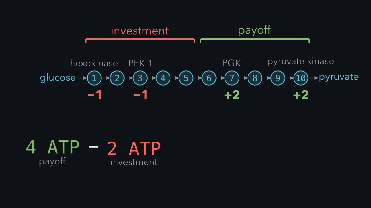 ▶ Watch Spend, Balance, Control · 2:37
Read the transcript of “Spend, Balance, Control”
Film transcript: Spend, Balance, Control
Glycolysis has two distinct phases. In the investment phase, two ATP are consumed to phosphorylate glucose. In the payoff phase, four ATP are produced. Two from phosphoglycerate kinase and two from pyruvate kinase. Subtract the investment from the payoff and the net yield is two ATP per glucose, plus two NADH. Aerobic respiration then extracts roughly 30 more, which is why oxygen-dependent metabolism is so much more efficient. The central problem fermentation solves is a redox bookkeeping problem. Glycolysis consumes NAD+, +, and produces NADH at the glyceraldehyde 3-phosphate step. But the cell has only a finite NAD+ +, pool. Fermentation regenerates NAD+ +, by passing electrons from NADH to pyruvate, making lactate, or to acetaldehyde, made from pyruvate, making ethanol.
The two cycles are tightly coupled, consume NAD+ +, here, regenerate it there, so that glycolysis can keep running. Here's a useful metaphor. Think of a metabolic pathway as a pipe. A reversible reaction is like a valve you can partially close. Pressure equilibrates on both sides. An irreversible reaction is a one-way valve. Close it and flow stops completely. Cells mainly regulate metabolism at irreversible steps, because those can be throttled. Control is still shared with other steps. Block any essential step and the whole line shuts down. Glycolysis is regulated at three enzymes, hexokinase, phosphofructokinase-1, and pyruvate kinase. Each responds to different energy signals.
Hexokinases 1 to 3 are inhibited by their product, glucose-6-phosphate. PFK-1 is the master throttle, activated by a MP in fructose 2, 6 -bisphosphate, inhibited by ATP and citrate. Liver pyruvate kinase is activated feed-forward by fructose-1,6-bisphosphate. Together, these three checkpoints match glycolytic flux to the cell's energy needs. Think of phosphofructokinase-1 as a turnstile. Glucose passes through it at step 3, which helps the cell regulate carbon flow into glycolysis. In the liver, fructose sneaks in downstream. It bypasses this gate and enters as DHAP and glyceraldehyde 3-phosphate, though other controls remain. A large fructose load bypasses this checkpoint.
When Usain Bolt runs 100 meters in under ten seconds, his leg muscles need ATP faster than oxygen-based metabolism can ramp up to make it. So his cells lean on fast routes that need no oxygen: stored phosphocreatine, and glycolysis, the breakdown of sugar from muscle glycogen. Most living things run glycolysis or a variant of it, from your neurons to brewer’s yeast to bacteria that have never met oxygen. It splits one glucose into two three-carbon pyruvate molecules, earns a little ATP, banks electrons as NADH, and hands carbon building blocks to the rest of the cell.
The puzzle: the cell’s whole point is to make ATP, yet glycolysis starts by spending two. Why pay a toll to enter a pathway whose job is to earn, and where does the profit come from?
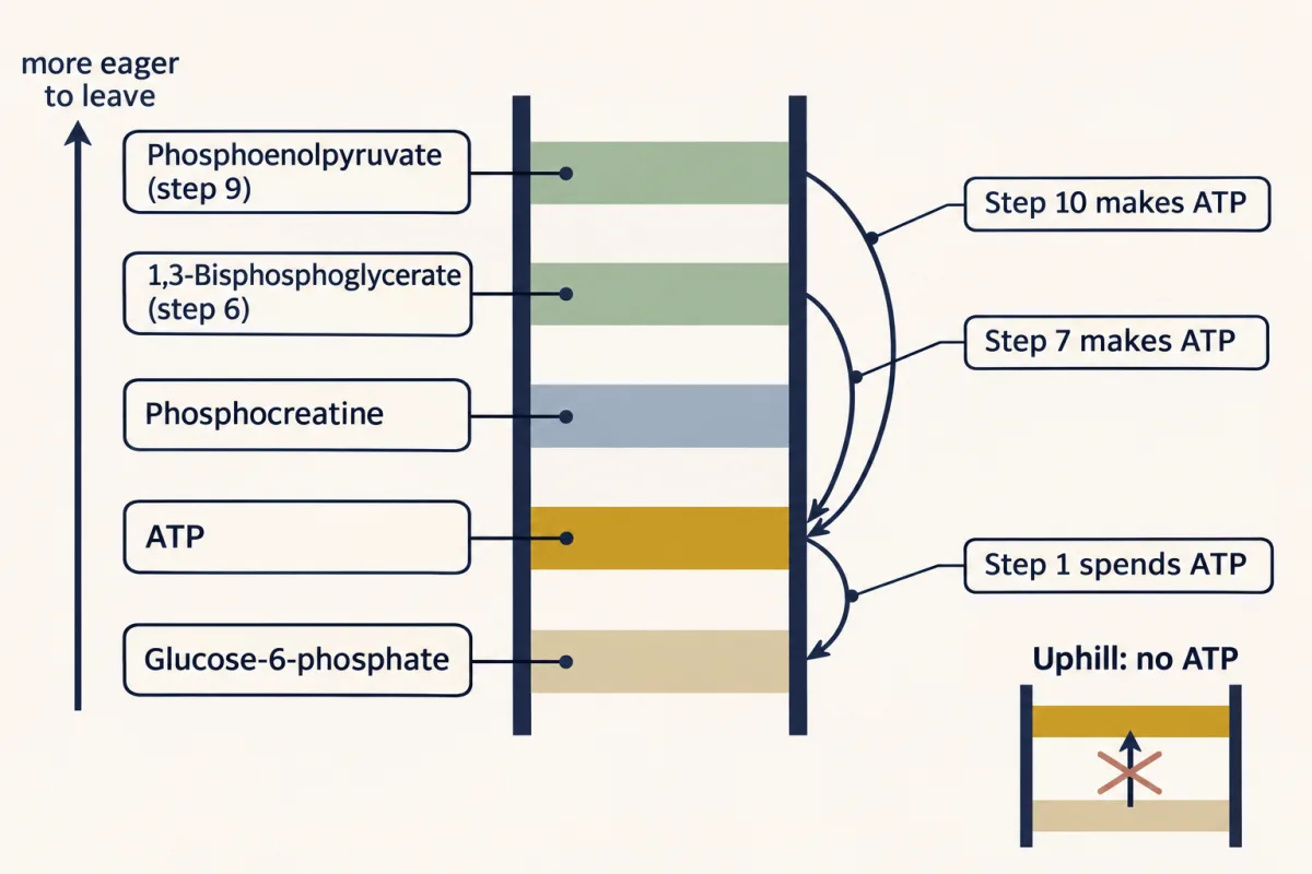
The one idea to carry through this lesson. Follow the phosphate. A kinase, an enzyme that moves a phosphate group, can load ADP favorably only from a phosphate more eager to leave than ATP’s own, on the standard ladder below. Glycolysis first turns glucose into two identical phosphorylated halves, then builds two such phosphates in each half, at steps 6 and 9, and a kinase cashes each one. Two consequences follow later: the step 6 oxidation needs NAD⁺, so the payoff runs only while NAD⁺ comes back; and the cell steers at the three steps that fall far downhill in the cell, which standard ΔG°′ values alone do not pick out.
The rule covers one glucose going to two pyruvate in the cytosol by the standard (Embden–Meyerhof–Parnas) route. It counts phosphate groups, not individual atoms (Going deeper), and the throttles’ details differ by tissue.
A ladder of phosphates
How eager a phosphate is to leave is measured by its standard free energy of hydrolysis, ΔG°′: the free-energy change when water takes it off, under standard conditions. The more negative the value, the higher the phosphate sits on the ladder (its phosphoryl-transfer potential). In the cell, concentrations shift each step’s ΔG (ΔG = ΔG°′ + RT ln Q, where Q is the ratio of products to reactants), enough to let a near-balanced step such as PGK’s run either way.
| Phosphate on | ΔG°′ of hydrolysis (kJ/mol) | |
|---|---|---|
| Phosphoenolpyruvate (PEP) | −61.9 | made at step 9 |
| 1,3-Bisphosphoglycerate (1,3-BPG) | −49.3 | made at step 6 |
| Phosphocreatine | −43.0 | the sprinter’s reserve |
| ATP (to ADP) | −30.5 | the cell’s currency |
| Glucose-6-phosphate | −13.8 | an ordinary phosphate ester |
In words The free-energy change for the move equals the ladder value where the phosphate starts, minus the ladder value where it ends up.
- ΔG°′(move)“delta G naught prime of the move”
- standard free-energy change for the transfer; negative means favorablekJ/mol
- ΔG°′(start)“delta G naught prime at the start”
- hydrolysis value of the phosphate on the donorkJ/mol
- ΔG°′(end)“delta G naught prime at the end”
- hydrolysis value of the phosphate on the acceptorkJ/mol
Sanity check Under standard conditions a phosphate moves favorably only down the ladder, so it can load ADP only from above ATP.
Try one number
PEP to ADP (step 10): −61.9 − (−30.5) = −31.4 kJ/mol, strongly favorable.
ATP to glucose (step 1): −30.5 − (−13.8) = −16.7 kJ/mol, favorable. Run backward, glucose-6-phosphate to ADP, it is +16.7: an ordinary phosphate ester cannot make ATP.
Need a refresher? units and prefixes
Phosphocreatine sits above ATP, which is why creatine kinase can refill ATP from it in a sprint’s first seconds. Glycolysis must build phosphates above ATP.
Investment, steps 1–5: trap, reshape, commit, split
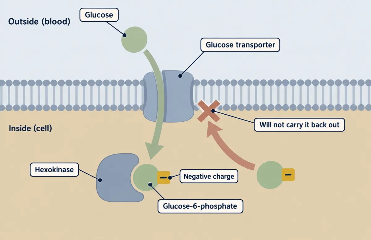
Step 1: trap. Hexokinase moves a phosphate from ATP onto carbon 6 of glucose. It closes around glucose by induced fit, which keeps water out, so ATP’s phosphate goes to glucose and not to water. Its negative charge traps it in the cell. PET scans exploit this: an ¹⁸F-labeled glucose analog is trapped after step 1, so tissues that take up glucose fast, including many tumors, light up.
Step 2: reshape. Phosphoglucose isomerase turns glucose-6-phosphate, an aldose (carbonyl on carbon 1), into fructose-6-phosphate, a ketose (carbonyl on carbon 2).
Predict. Step 4’s enzyme, aldolase, cuts the bond between the carbon next to the carbonyl and the one after it. Why move the carbonyl from carbon 1 to carbon 2 first?
Check your prediction
With the carbonyl at carbon 2, the cut falls between carbons 3 and 4, giving two three-carbon halves. With it at carbon 1, the cut would fall between carbons 2 and 3: a two-carbon and a four-carbon piece. The move also frees carbon 1 for the second phosphate.
Step 3: commit. Phosphofructokinase-1 (PFK-1) moves a second phosphate from ATP onto carbon 1, making fructose-1,6-bisphosphate. This is the committed step: its product is dedicated to glycolysis. The ledger reads −2 ATP, nothing earned.
Step 4: split. Aldolase cuts it into two three-carbon pieces, each with one phosphate: dihydroxyacetone phosphate (DHAP) and glyceraldehyde-3-phosphate (GAP).
Step 5: make the halves identical. Triose phosphate isomerase (TPI) turns DHAP into GAP. Only GAP has the aldehyde that step 6 oxidizes, so without TPI half the carbon would be stranded. Both halves now run the same steps, so every yield from step 6 onward counts double, a doubling students often forget.
Payoff, steps 6–10: make a phosphate worth more than ATP’s, then cash it
Each GAP’s phosphate, on carbon 3, is an ordinary phosphate ester like glucose-6-phosphate’s: below ATP on the ladder. The payoff phase does one job, twice in each half: build a phosphate above ATP, then let a kinase hand it to ADP.
Step 6: an oxidation pays for a new phosphate. GAPDH oxidizes GAP’s aldehyde carbon (carbon 1), and NAD⁺ takes the electrons to become NADH. Oxidizing an aldehyde to an acid is strongly downhill; done in one go, the energy would be lost as heat. GAPDH saves it: an active-site cysteine first bonds to the aldehyde, so the oxidation leaves a thioester, an activated bond, on the enzyme. Then inorganic phosphate (Pi, from the cell’s pool, not from ATP) attacks the thioester and carries the carbon away as an acyl phosphate: 1,3-BPG. Its new carbon-1 phosphate sits at −49.3 kJ/mol, well above ATP’s −30.5.
Step 7: cash the first one. Phosphoglycerate kinase (PGK) moves that phosphate onto ADP, making ATP and 3-phosphoglycerate. This is substrate-level phosphorylation: one enzyme passes a phosphate from a substrate to ADP, with no mitochondrion, oxygen or membrane gradient. Both halves do it: 2 ATP per glucose.
Steps 8 and 9: rearrange to make a better one. The remaining carbon-3 phosphate is still too low on the ladder to make ATP, so the cell raises it. Phosphoglycerate mutase shifts it from carbon 3 to carbon 2. Enolase then removes water, making a carbon–carbon double bond next to the phosphate: phosphoenolpyruvate (PEP). That step changes the free energy only slightly (ΔG°′ about +2 to +7.5 kJ/mol, by source), yet the phosphate it leaves behind sits at the top of the ladder, −61.9. A phosphate’s place on the ladder includes what happens to the rest of the molecule after it leaves: here the leftover enolpyruvate is an unstable enol that quickly turns into its much more stable form, pyruvate, so the phosphate leaves very readily.
Step 10: cash the second one. Pyruvate kinase hands PEP’s phosphate to ADP, making ATP and pyruvate: 2 more ATP per glucose.
The two ledgers
Steps 7 and 10 make 4 ATP per glucose; steps 1 and 3 spent 2. Counting phosphate groups shows where the profit comes from. Four phosphates leave to ADP: two repay the ATP lent at steps 1 and 3, and two are new, captured from inorganic phosphate at step 6 and paid for by the oxidation. So, for glucose, the net gain equals the new phosphate captured by oxidation. (Enter later, say as fructose-1,6-bisphosphate, and phosphates already on the substrate also reach ADP.) The oxidation also turned 2 NAD⁺ into 2 NADH, the second ledger.
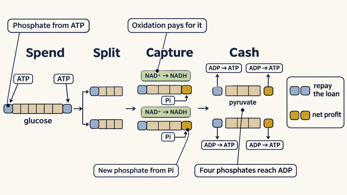
Net per glucose: +2 ATP, +2 NADH.
It is a thin margin, about a fifteenth of the 30 or so ATP that full respiration yields per glucose, about 28 more beyond glycolysis’s own 2. But glycolysis is fast, needs no oxygen, and for some cells is the only option: a red blood cell has no mitochondria, so glycolysis is its entire energy economy.
Check each step on the map against its reason, then run the ledger.
Net ATP (+2) = new phosphates captured at step 6 (2).
far downhill in the cell: a main control point×2 counts twice: two trioses per glucose
Investment steps 1–5 · trap, reshape, commit, split · spend 2 ATP
Spends one ATP to phosphorylate glucose. Induced fit closes the enzyme around the sugar, excluding water so ATP isn’t wasted on hydrolysis — and the charged product can no longer cross the membrane, trapping glucose inside the cell.
ΔG°′ −16.7 kJ/mol · ΔG in red cell −33.4 kJ/mol
Regulation. Inhibited by its own product, glucose-6-phosphate.
Aldose → ketose: a quiet repositioning of the carbonyl so the six-carbon sugar can later be split cleanly down the middle.
ΔG°′ +1.7 kJ/mol · ΔG in red cell −2.5 kJ/mol
The committed step. Once fructose-1,6-bisphosphate forms, the molecule is locked into glycolysis. The second and last ATP is spent here.
ΔG°′ −14.2 kJ/mol · ΔG in red cell −22.2 kJ/mol
Regulation. Inhibited by ATP and citrate; activated by AMP and fructose-2,6-bisphosphate. This is the master throttle of glycolytic flux.
Splits the six-carbon sugar into two three-carbon halves — the symmetry break the whole pathway is built around.
ΔG°′ +23.8 kJ/mol · ΔG in red cell −1.3 kJ/mol
Converts DHAP into a second glyceraldehyde-3-phosphate, so both three-carbon halves run the payoff phase. From here, everything counts twice.
ΔG°′ +7.5 kJ/mol · ΔG in red cell +2.5 kJ/mol
Clinical. TPI deficiency: when this enzyme fails, DHAP piles up and forms toxic methylglyoxal. It is the most severe glycolytic enzymopathy — hemolytic anemia plus progressive neurodegeneration.
Payoff steps 6–10 · build a phosphate above ATP, then cash it · ×2
The only oxidation. An active-site cysteine catches the aldehyde, so the oxidation leaves an activated thioester; inorganic phosphate (Pi, not ATP) then attacks it. Result: a new phosphate on carbon 1 at −49.3 kJ/mol, above ATP’s −30.5. The electrons go to NAD⁺ → NADH.
ΔG°′ +6.3 kJ/mol · ΔG in red cell −1.3 kJ/mol (steps 6 and 7 together)
The first ATP is made here by substrate-level phosphorylation: the high-energy phosphate goes straight from substrate to ADP. The ledger is now back to break-even. This cashes the phosphate built at step 6.
ΔG°′ −18.5 kJ/mol · ΔG in red cell counted with step 6
The carbon-3 phosphate is still below ATP on the ladder. Moving it to carbon 2 sets up the step that raises it.
ΔG°′ +4.4 kJ/mol · ΔG in red cell +0.8 kJ/mol
Removing water barely changes the free energy, but leaves PEP’s phosphate at the top of the ladder (−61.9): once it leaves, enolpyruvate snaps into stable pyruvate.
ΔG°′ +7.5 kJ/mol · ΔG in red cell −3.3 kJ/mol
Phosphoenolpyruvate hands its phosphate to ADP — the second substrate-level ATP — and the pathway ends at pyruvate.
ΔG°′ −31.4 kJ/mol · ΔG in red cell −16.7 kJ/mol
Regulation. Liver and red-cell forms: activated ahead of time (feed-forward) by fructose-1,6-bisphosphate from step 3 and inhibited by ATP and alanine; the main muscle form lacks the feed-forward control. In liver, glucagon switches it off via PKA phosphorylation.
For each card, is this token a real yield or cost of that step? Bank the real ones, toss the myths.
Step 1 · Hexokinase
ATP ×2 plus ATP
After the aldolase split: one card, counts twice.
Ledger closed
The investment phase spends 2 ATP; the payoff phase makes 4 ATP and 2 NADH, because everything after the aldolase split counts twice. Net: +2 ATP, +2 NADH. Where the profit comes from: of the 4 phosphates handed to ADP, 2 repay steps 1 and 3 and 2 are new, captured from inorganic phosphate at step 6.
Predict. Suppose arsenate, which looks like phosphate, replaces it completely at step 6: GAPDH uses it, but the product then breaks down on its own to 3-phosphoglycerate, passing nothing to ADP. What happens to the yields per glucose?
Check your prediction
NADH is unchanged at 2, because the oxidation still happens. But no new phosphate is captured, so step 7 makes nothing, and step 10’s 2 ATP only repay the 2 spent. Net ATP: 0. Glycolysis runs but earns nothing, as the rule predicts.
Why pyruvate’s fate matters: the NAD+ bottleneck
Glycolysis can stall with plenty of glucose, if the cell runs out of NAD⁺. Step 6 uses 2 NAD⁺ per glucose from a small, fixed pool, so NAD⁺ must be recycled. With plenty of oxygen, shuttles pass NADH’s electrons into the mitochondria, and NAD⁺ comes back. When oxygen runs short, or in a cell with no mitochondria, the electrons go to pyruvate or to a molecule made from it. A sprinting muscle hands them to pyruvate itself: lactate dehydrogenase (LDH) makes lactate and returns 2 NAD⁺ per glucose, so the electron ledger balances at zero. Yeast makes ethanol instead, which is convenient for brewers but incidental to the yeast, who care only about their NAD⁺.
Pick a route below and follow the electrons until NAD⁺ comes back to step 6.
- Product
- Acetyl-CoA → CO₂
- NAD⁺ regenerated?
- via shuttles + electron-transport chain
With oxygen, shuttles pass NADH’s electrons into the mitochondrion, where the electron-transport chain hands them to O₂, and NAD⁺ comes back to step 6. Pyruvate enters too and is oxidized to CO₂: about 30 ATP per glucose in all.
Fermentation makes no extra ATP. Its entire job is to regenerate NAD⁺ so glycolysis (and its 2 ATP per glucose) can keep running when oxygen is scarce.
Predict. Block LDH in a red blood cell and give it plenty of glucose. What happens to its ATP production?
Check your prediction
It stops. With no mitochondria, LDH is the red cell’s way to turn NADH back into NAD⁺. Without it the small NAD⁺ pool is used up at step 6, GAPDH stalls, and no payoff step runs.
Lactate is not a fatigue toxin. It regenerates NAD⁺ so you can keep running, it travels to the liver to be rebuilt into glucose (the Cori cycle), and it even acts as a signal. The burn in your legs is not lactate alone: it comes from several changes in hard-working muscle acting together, including rising acidity, and its exact causes are still debated.
Control: where the cell can turn the flow
Here is each step’s standard ΔG°′ beside its actual ΔG in a human red blood cell at 37 °C, worked out from measured concentrations.
| Step | Enzyme | ΔG°′ (kJ/mol) | ΔG in the cell (kJ/mol) |
|---|---|---|---|
| 1 | Hexokinase | −16.7 | −33.4 |
| 2 | Phosphoglucose isomerase | +1.7 | −2.5 |
| 3 | PFK-1 | −14.2 | −22.2 |
| 4 | Aldolase | +23.8 | −1.3 |
| 5 | Triose phosphate isomerase | +7.5 | +2.5 |
| 6 | GAPDH | +6.3 | −1.3 (steps 6 and 7 together) |
| 7 | Phosphoglycerate kinase | −18.5 | (in the row above) |
| 8 | Phosphoglycerate mutase | +4.4 | +0.8 |
| 9 | Enolase | +7.5 | −3.3 |
| 10 | Pyruvate kinase | −31.4 | −16.7 |
Values as tabulated in Lehninger; other texts give slightly different numbers.
In the cell, three steps fall far downhill. Hexokinase, for example, goes from −16.7 to −33.4 because the cell keeps its products, glucose-6-phosphate and ADP, scarce relative to glucose and ATP. Every other step sits near equilibrium (within a few kJ/mol of zero in this table, though for values that small the sign depends on the standard value and concentrations used).
Predict. PGK’s standard value, −18.5, is more negative than PFK-1’s, −14.2. Is PGK a control point?
Check your prediction
No. In the cell PGK, paired with GAPDH, sits near zero and runs freely in both directions. Aldolase is the mirror case: standard value +23.8, yet in the cell it sits near zero and carries the flow forward. Only the cellular values show which steps are one-way.
Why only the one-way steps? Near equilibrium, an enzyme runs forward and backward almost equally fast, usually with far more enzyme than the net flow needs, so halving it barely changes the flow. Far from equilibrium, almost nothing runs backward, so the net flow is the forward rate, and changing the enzyme’s activity changes it. These three are the main control points, not the only ones: glucose transport and some reversible enzymes are regulated too, and control of the overall flow is shared among several steps. (So of the explainer film’s two pictures, keep its later line, that cells regulate mainly at the one-way steps, not the valve you can partly close at a reversible step.)
Hexokinase is inhibited by its product, glucose-6-phosphate, so it slows when the trap is full. The liver’s form, glucokinase, is not.
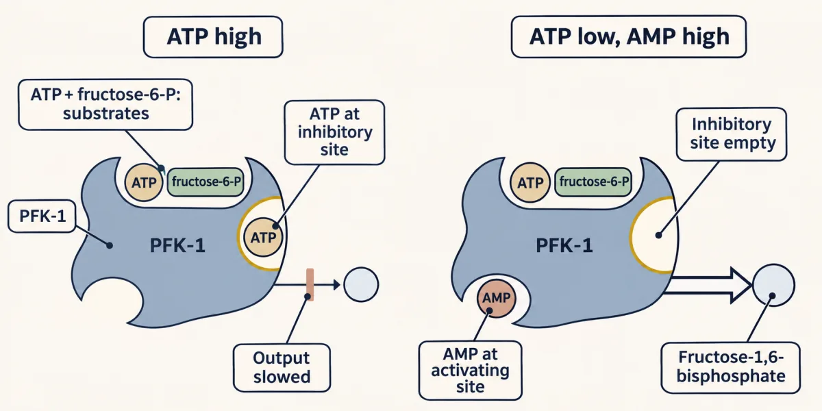
Phosphofructokinase-1 (PFK-1), the master throttle, carries a paradox: ATP is its substrate and also its inhibitor. When ATP is high, ATP binds a second site and shuts PFK-1 down. When ATP runs low, AMP rises and switches it back on (AMP is the alarm because adenylate kinase keeps 2 ADP ⇌ ATP + AMP close to balance and AMP is normally scarce, so a small fall in ATP raises AMP by a large fraction). Fructose-2,6-bisphosphate, below, is a second activator. The enzyme asks not “is there glucose?” but “does this cell need energy now?” Citrate, which builds up when the citric acid cycle is full, also inhibits it: carbon is backing up.
Pyruvate kinase in liver (and in red cells) is inhibited by ATP and alanine and activated ahead of time (feed-forward) by fructose-1,6-bisphosphate from step 3; the main muscle form lacks this feed-forward control. In the liver, glucagon also switches it off through phosphorylation by protein kinase A (PKA).
Tissue wiring and carbon supply
Tissues wire these steps differently, and make different forms of PFK-1. Muscle PFK-1 listens to the muscle’s own energy charge. Liver PFK-1 also hears pancreatic hormones through fructose-2,6-bisphosphate: insulin raises it and switches PFK-1 on; glucagon lowers it and slows PFK-1. Muscle burns glucose for itself. Liver keeps blood glucose up for everyone else, so when glucagon signals a shortage, liver turns glycolysis down and makes glucose instead (gluconeogenesis).
Glycolysis also supplies carbon building blocks: glucose-6-phosphate to the pentose phosphate pathway, dihydroxyacetone phosphate (DHAP) to glycerol-3-phosphate for lipids, 3-phosphoglycerate to serine, pyruvate to alanine.
Going deeper
Proving the pathway: isotope tracing and enzyme assays
The pathway was mapped, not assumed. One tool: label a single glucose carbon with a heavy or radioactive isotope and trace where it lands. Enzyme studies and the chemical identification of each intermediate built the map, and tracing confirmed that both three-carbon halves run the payoff phase. The step 1 PET scan scales the idea up: many cancers run glycolysis hard even when oxygen is plentiful. To find the throttles, you assay the regulated enzymes directly: add ATP, AMP or citrate to purified phosphofructokinase-1 (PFK-1) one at a time and watch the velocity climb or fall, the same initial-velocity discipline from enzyme kinetics.
About the free-energy table
Some standard values differ by source (enolase is given as anything from about +2 to +7.5 kJ/mol), which is also why the ladder arithmetic gives −18.8 for step 7 against the table’s −18.5. The enolase pair is the least self-consistent: with the same red-cell concentrations the +7.5 standard value would give a positive ΔG (about +7), so the −3.3 reflects a different standard value or different concentrations; enolase is better described as near equilibrium in the cell than assigned a sign. Recomputing from approximate red-cell concentrations gives about −34 kJ/mol for hexokinase, −18.8 for PFK-1, +0.5 for phosphoglycerate kinase (PGK) and −22.7 for pyruvate kinase: the same pattern. The standard and cellular columns rank hexokinase and pyruvate kinase in opposite orders, and the two cellular estimates rank PFK-1 and pyruvate kinase in opposite orders, so check which values a source quotes.
Counting phosphates, not atoms
The ledger counts phosphate groups, which is not quite the same as following atoms. In mammals, phosphoglycerate mutase passes the phosphate through a phosphorylated histidine on the enzyme and a 2,3-bisphosphoglycerate intermediate, so the phosphate on 2-phosphoglycerate is not the same atom that sat on carbon 3. The count still balances.
Fructose in the liver
Fructose takes its own route into liver glycolysis. Fructokinase makes fructose-1-phosphate, aldolase B cleaves it to dihydroxyacetone phosphate (DHAP) and glyceraldehyde, and triose kinase phosphorylates the glyceraldehyde to GAP. The carbon enters below PFK-1, bypassing that checkpoint, though other controls remain. This is what the explainer film means by fructose sneaking in downstream.
Your notes
Select any text in the lesson to highlight it or add a note to it. Your highlights and notes are saved in this browser and collected in your notebook.
This browser is not letting the site save anything (a private window or strict privacy settings), so highlights and notes will be lost when you leave the page.
How we measure it
ATP and NADH accounting (the ledger)
The net yield isn't asserted — it's counted. Tally ATP spent (steps 1, 3) against ATP made (steps 7, 10) and NADH made (step 6), doubling everything after the aldolase cleavage. The books balance at +2 ATP and +2 NADH per glucose, and the running ledger lets you check any yield question step by step.
Isotopic carbon tracing
Feed cells glucose labeled at a specific carbon — say ¹³C or ¹⁴C on C1 — and follow where that atom lands. Tracing confirms that the six-carbon sugar splits into two three-carbon halves, and shows which glucose carbons end up where in pyruvate. A related idea, scaled to a PET scanner, uses an ¹⁸F-labeled glucose analog that cells take up and trap after step 1, to map a tumor's glucose uptake.
Enzyme assays for the regulated steps
To find a throttle you measure the enzyme's velocity while changing one effector at a time. Adding ATP, citrate, AMP, or fructose-2,6-bisphosphate to purified PFK-1 and watching the rate rise or fall is how the regulators of the committed step were mapped — the same initial-velocity logic from enzyme kinetics, applied one molecule at a time.
Practice questions on this lesson (17) → MCAT-style, original, each with an explanation
Where this lesson fits
Before thisReview first
- Metabolism: Basic Concepts and DesignThis course · Lesson 15
It teaches ATP as the energy currency and how coupling an uphill step to a downhill one works, which the phosphate ladder here uses.
- Basic Concepts of Enzyme ActionThis course · Lesson 7
You need to know how enzymes speed up reactions and how free energy governs direction before reading steps far from equilibrium.
- Carbohydrates: One Bond Decides Fuel or FiberThis course · Lesson 10
Glucose's ring form and the aldose and ketose distinction explain the isomerase step that turns glucose-6-phosphate into fructose-6-phosphate.
After thisStudy next
- Gluconeogenesis: Pushing Glucose Back UphillThis course · Lesson 17
It runs the pathway backward, using bypasses for exactly the three far-downhill steps you identified here as the control points.
- The Pyruvate Dehydrogenase Complex: The One-Way Gate to the CycleThis course · Lesson 18
It takes the pyruvate made at step 10 and shows what the cell does with it when oxygen is available.
- Integration of Metabolism: How the Body Switches FuelsThis course · Lesson 30
It uses PFK-1 and pyruvate kinase control by hormones to show how the body switches fuels between fed and fasting states.
Elsewhere on the siteFilms, simulations and method films in other courses
- Spending ATP, Rebuilding ATPFilm · Biology
It shows how a cell spends ATP and rebuilds it from glucose, the same ATP economy glycolysis runs on.
Suggested by an AI model from the lessons themselves, then checked. How we use AI
Finish line
Test yourself
17 questions
Practice and MCAT-style, with explained answers.
Play it · about 3 min
Ledger Rush: Glycolysis
Bank the real ATP and NADH, toss the myths, catch the doubling after the split.
Play →Next · Lesson 17
Gluconeogenesis: Pushing Glucose Back Uphill
When food runs out, your liver (with help from the kidney) builds the glucose your brain and red blood cells still need, and it does it with four new enzymes, three compartments, and real ATP and GTP spent to make the climb.
Continue →Also in the arcade for this lesson: Metabolic Autopsy: Glycolysis