Organismal Biology · Unit 3: Supply and exchange
Circulation and the Heart
Rewritten on : new text, flashcards and film. Your notes and highlights stay, but the text around them may have moved.
An adult human holds about 5 litres of blood, yet at rest each side of the heart pumps about that much every minute. Where does it all go?
Pause when the reading reaches a film, and offer to play it. Saved on this device.
Watch the film now, or keep listening
Builds on:Size, Scale and SurfaceTissues, Organs and HomeostasisWhat an Organism Has to DoWhy these?
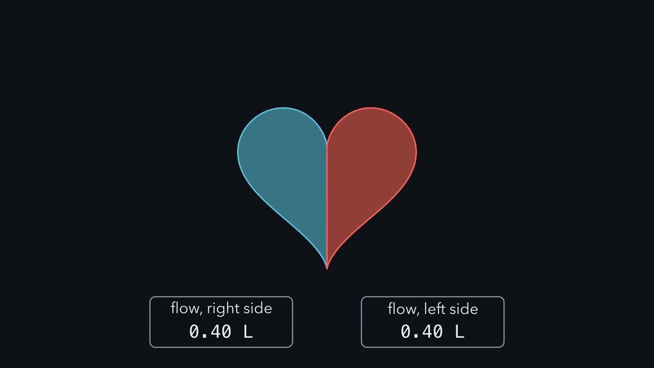 ▶ Watch One Flow, Two Pressures · 3:07
Read the transcript of “One Flow, Two Pressures”
Film transcript: One Flow, Two Pressures
Both sides of your heart pump the same flow of blood every minute. So why does the left side push so much harder than the right? Start with flow. Flow is the volume of blood moved each minute. One ventricle's flow is its heart rate times its stroke volume, the blood it ejects with each beat. Now follow the loop. The right ventricle sends oxygen-poor blood through the lungs. It comes back oxygen-rich to the left side, which sends it through the body. The two sides pump one after the other, in series.
Blood leaving one side must pass through the other before it comes back. So over time, their average flows must be equal. Now draw pressure as height. Each ventricle is a lift that raises blood up. Then blood rolls downhill through its circuit, from high pressure to low. Some circuits are harder to push through. Narrow vessels resist the flow, like a narrow pipe. Call that resistance. The pressure drop across a circuit equals the flow times its resistance. The lungs resist the flow only a little. So the right ventricle needs only a small lift to drive the whole flow through them. The body's circuit resists far more. So the left ventricle must lift the same flow much higher, to drive it down that long, narrow slope.
Same flow, different costs. At the same flow, the circuit with more resistance needs the bigger pressure drop. Two separate pumps let each circuit get its own pressure. One caution. This rule is bookkeeping on averages over many beats. In a living body, the heart and the vessels set the flow together. Now a heart with one pump. A teleost fish, such as a trout, has one ventricle. It drives blood through its narrow gill capillaries first, then on to the body. Here is the question. Blood leaves the gills and reaches the body. Is its pressure the same as the ventricle made, higher, or lower?
Lower. The gill capillaries resist the flow, so part of the pressure is spent there. The body is supplied with only what is left. Birds and mammals avoid that trade. They have two pumps. The lungs run at low pressure and the body at high pressure. Both carry the full flow. One flow, two costs. Both sides move the same blood. The harder circuit, the body's, takes the bigger pressure drop.
From the film
- At rest, both sides of a mammal's heart pump about the same flow, because blood goes round the two circuits in turn.
- Averaged over many beats, pressure drop = flow × resistance: at a given flow, the harder a circuit is to push through, the bigger the drop.
- The body's circuit resists far more than the lungs', so the left ventricle must make much higher pressure than the right.
- In a teleost fish, one ventricle pushes blood through the gills and then the body, so the body gets the lower pressure that is left.
- At the same flow, the harder circuit costs more pressure.
An adult human holds about 5 litres of blood, yet at rest each side of the heart pumps roughly that much every minute. The first film showed the left side paying far more pressure for that same flow. What follows adds the pressures, the valves, and animals that solve it another way.
The one idea. At a given flow, the pressure a circuit costs depends on how hard it is to push through, its resistance, just as water needs more push through a narrow pipe than a wide one: pressure drop = flow × resistance.
Scope: beat-averaged pressures in a steady state. The rule is bookkeeping, not a claim about control: in a living body the heart and vessels set the flow together, and changing a resistance can change the flow. Within one beat, each valve follows the pressures on either side of it at that moment.
Bulk flow, open and closed
Diffusion is quick over micrometres but far too slow over centimetres (size, scale and surface), so a large animal relies on bulk flow: a pump drives the whole fluid, and diffusion does only the last short step. A circulatory system is that pump, the fluid and the spaces it flows through.
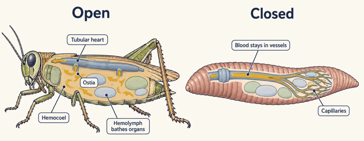
In an open circulatory system, the fluid, called hemolymph because it is blood and tissue fluid in one, leaves the vessels and flows through the hemocoel, a set of body spaces that bathe the organs directly. In a closed circulatory system, blood stays inside vessels, and exchange happens across the thin walls of capillaries, the smallest vessels.
Insects such as grasshoppers and Drosophila are open: a tubular heart along the back pumps hemolymph forward, and it drifts back over the organs into the heart through small valved openings, the ostia. It carries little oxygen, because air reaches the tissues mostly through air tubes, the tracheae (gas exchange). Most molluscs, such as snails and clams, are also open.
Open and closed do not split invertebrates from vertebrates. Earthworms are closed, with muscular vessels that pump. Squid and octopuses, which are molluscs, are closed too, with two branchial hearts that push blood through the gills and a systemic heart that sends it to the body. All vertebrates are closed. (See animal body plans.)
Through the mammalian heart, valve by valve
An atrium is a receiving chamber and a ventricle a pumping chamber. An artery carries blood away from the heart and a vein carries it toward the heart, whatever its oxygen content. “Right” and “left” mean the animal’s own right and left. In a mammal after birth, the route is:
- The superior and inferior venae cavae bring oxygen-poor blood from the body to the right atrium.
- It passes the tricuspid valve into the right ventricle.
- The right ventricle pushes it through the pulmonary valve into the pulmonary trunk and arteries, to the lungs.
- The pulmonary veins return oxygen-rich blood to the left atrium.
- It passes the mitral valve into the left ventricle.
- The left ventricle pushes it through the aortic valve into the aorta, and on to the body.
So the pulmonary artery carries oxygen-poor blood and the pulmonary veins oxygen-rich blood. Each valve is a one-way door: it opens when pressure behind it exceeds pressure ahead, and closes when that reverses.
Systole is a chamber’s contraction phase and diastole its relaxation phase. For the adult human left ventricle at rest, one beat has four phases (pressures in mmHg, millimetres of mercury):
- Filling (diastole). Atrium above ventricle, so the mitral valve is open and blood flows in; the atrium’s own contraction adds the last part.
- Isovolumic contraction (systole; same volume). Ventricle above atrium, so the mitral valve shuts; still below the aorta, so the aortic valve stays shut.
- Ejection (systole). Ventricle passes aortic pressure (about 80 mmHg), the aortic valve opens, and blood leaves; both peak near 120 mmHg.
- Isovolumic relaxation (diastole). Ventricle falls below the aorta, so the aortic valve shuts; both stay shut until ventricular pressure drops below atrial pressure and the mitral valve opens again.
So in “120 over 80 mmHg”, the 80 is the aorta’s lowest pressure, not the ventricle’s, which falls to a few mmHg as it fills. The right ventricle runs the same cycle against the lungs’ low resistance: its pulmonary valve opens once its pressure passes pulmonary-artery pressure, and it peaks near 25 mmHg, about a fifth of the left’s.
Predict. Same flow, peaks of 120 and 25 mmHg. Is the body’s circuit about five times as hard to push through as the lungs’?
Check your prediction
No, harder still. Resistance is the mean (beat-averaged) pressure drop divided by flow, not a ratio of peaks: about 90 / 5 = 18 for the body against 6 / 5 = 1.2 for the lungs (table), about 15 times as much.
| Resting adult human, illustrative | Lung (pulmonary) circuit | Body (systemic) circuit |
|---|---|---|
| Flow, one ventricle’s output | about 5 L per min | about 5 L per min |
| Mean pressure going in | about 14 mmHg (pulmonary artery) | about 93 mmHg (arteries) |
| Mean pressure coming out | about 8 mmHg (left atrium) | about 3 mmHg (right atrium) |
| Mean drop, flow × resistance | about 6 mmHg | about 90 mmHg |
| Peak ventricular pressure | about 25 mmHg | about 120 mmHg |
One circuit or two
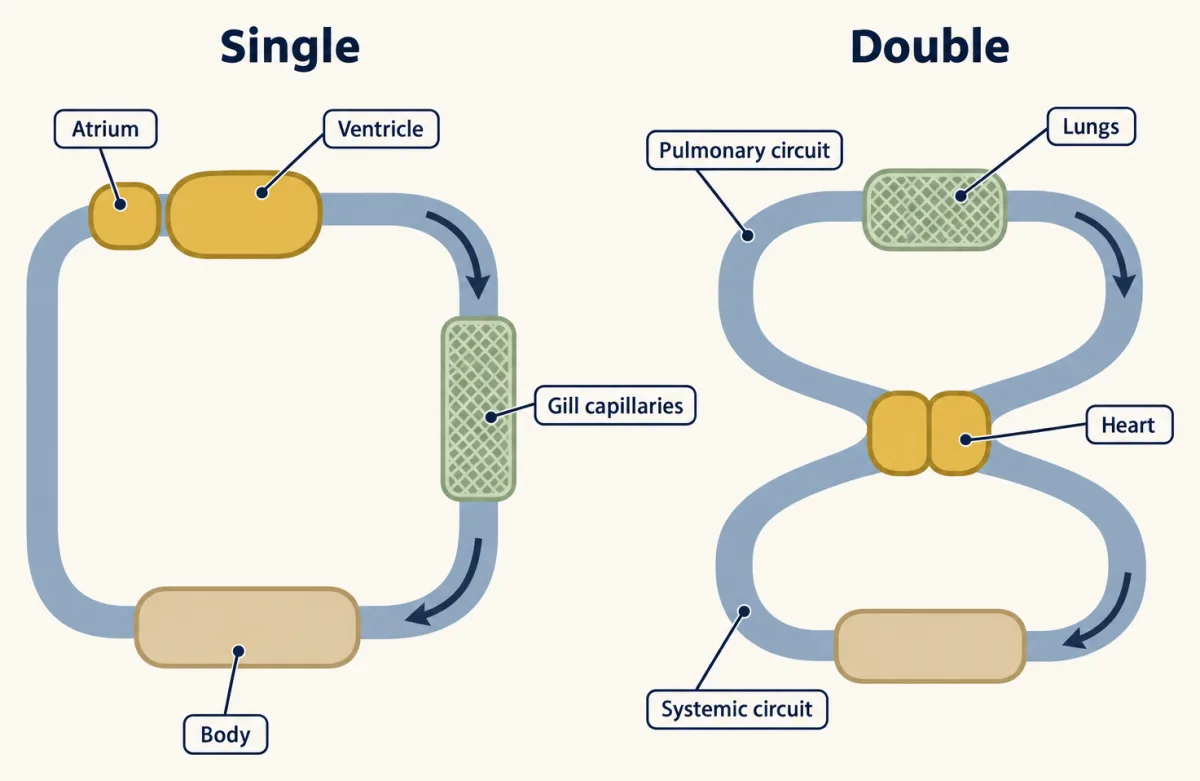
In single circulation, blood passes through the heart once per loop; in double circulation, twice: once on a pulmonary circuit to the gas-exchange organ and back, and once on a systemic circuit to the rest of the body and back.
Teleost fish (most bony fish, such as trout) have single circulation, the arrangement the first film priced, with one atrium filling the one ventricle.
Birds and mammals have four chambers that keep the circuits fully separate, each with its own pump.
Frogs have two atria and one ventricle. The right atrium receives body blood, including oxygen-rich blood from the skin; the left receives lung blood. The ventricle feeds a pulmocutaneous circuit (lungs and skin, since frog skin exchanges gas too) and a systemic circuit; the two streams are thought to stay partly separate in it, with some mixing.
Pythons have one ventricle, partly divided by a muscular ridge that lets it run at two different pressures during contraction. You will work the numbers in the design problem.
Crocodilians have four chambers, but the right ventricle opens into both the pulmonary artery and a left aorta. A small opening, the foramen of Panizza, joins the left aorta to the right aorta, which the left ventricle fills at body pressure. So the left aorta usually holds more pressure than the right ventricle makes, keeping the left aortic valve shut.
Predict. If resistance to flow into the lungs rises, where can right-ventricle blood go?
Check your prediction
Right ventricular pressure climbs until it passes left-aortic pressure; the left aortic valve then opens and part of the output leaves by the left aorta, bypassing the lungs: a shunt.
None of these hearts is a step toward the mammal’s: a frog heart that sends blood to the skin, or a crocodile heart that bypasses the lungs, works well in its own animal.
How much blood do these pumps move, and how did anyone first show it is the same blood going round? The second film follows Harvey’s arithmetic.
Read the transcript of “Harvey's Arithmetic”
Film transcript: Harvey's Arithmetic
The first film compared two circuits carrying the same flow. How big is that flow, and what did measuring it first prove? For centuries, the standard account came from Galen. Blood was made continually from food in the liver. The tissues it reached used it up. William Harvey, a London physician, tested this on the human arm. He tied a band round the upper arm. Tied tight, it stopped the pulse below it. Tied more loosely, it let the arteries drive blood in, but stopped its return. The veins below the band swelled. So blood enters a limb by arteries, and leaves by veins. Then Harvey asked a question of quantity. He made small guesses, on purpose, for how much blood leaves the heart at each beat.
He multiplied by the number of beats in half an hour. Even that low product was more blood than the whole body contains, and far more than food could replace. Now test the argument. Suppose his guess per beat was still too big, by a full half. Does his conclusion fall?
It stands. Even with half the scoop, half an hour still overflows the body many times. The heart moves far more than the body holds. So the same blood must go round. It leaves the heart by the arteries and comes back by the veins, again and again. Blood circulates. Harvey's product now has a modern name. Cardiac output is one ventricle's heart rate times its stroke volume, the blood it ejects per beat. So the heart has two dials on its flow. One dial sets the rate of beating. The other sets how much blood each beat ejects.
In a healthy mammal, the rate dial is the sinoatrial (SA) node. It is a patch of pacemaker muscle in the right atrium. Sympathetic nerves turn it up, and the vagus nerve turns it down. The stroke volume dial has three inputs of its own. They are how much the ventricle fills, how strongly it contracts, and the pressure it must push against. Over half an hour, rate times stroke volume adds up to far more blood than you hold. Food could never replace it. So the same few litres go round, again and again.
From the film
- Harvey's ligatures, bands tied on the arm, showed that blood enters a limb by arteries and leaves by veins.
- Even a deliberately small guess per beat, times the beats in half an hour, is more blood than the whole body contains.
- So the same blood must go round: it circulates rather than being made and used up.
- Cardiac output is heart rate times stroke volume, the blood one ventricle ejects per beat.
- The SA node and autonomic nerves set the rate; filling, contraction strength and the pressure the ventricle pushes against change the stroke volume.
Harvey’s numbers, redone
Beyond the second film’s ligatures, Harvey pressed along a vein to show that its valves pass blood only toward the heart. His argument did not show how arteries connect to veins: Marcello Malpighi saw capillaries in the frog lung in 1661.
Worked problem. Redo Harvey’s calculation for an illustrative adult human at rest in a steady state, with a heart rate of 75 beats per min, an end-diastolic volume (EDV, the ventricle’s volume when full) of 120 mL and an end-systolic volume (ESV, the volume left after ejection) of 50 mL. Ejection fraction is the share of the full volume each beat ejects.
stroke volume = EDV - ESV and cardiac output = heart rate x stroke volume and ejection fraction = stroke volume / EDV
- Stroke volume. 120 mL - 50 mL = 70 mL per beat.
- Cardiac output. 75 beats per min x 70 mL per beat = 5,250 mL per min, or 5.25 L per min, for one ventricle; the two pump in series, so their outputs are not added.
- Ejection fraction. 70 / 120 = 0.583, or 58.3 percent.
- Harvey’s comparison. Assume 5.0 L of blood in the body. A volume equal to all of it passes through one side of the heart in 5.0 / 5.25 = 0.95 min, about 57 seconds. That is an average turnover time; some blood takes short routes and some longer ones. In half an hour that side pumps 5.25 x 30 = 157.5 L, more than 30 times the body’s blood; in a day, 5.25 x 1,440 = 7,560 L. The second film’s halved stroke volume still gives about 79 L in half an hour (35 x 75 x 30 mL), nearly 16 times the body’s blood.
- Stroke volume = EDV - ESV
- Cardiac output = heart rate x stroke volume
- Ejection fraction = stroke volume / EDV
- Time for 5.0 L of blood to pass one side of the heart
- In half an hour (30 min)
- In a day (1,440 min)
In this sketch: adult human at rest, steady state, illustrative numbers, 5.0 L of blood in the body, one ventricle's output; slider ends go beyond the lesson's worked values and are not data; boxes and arrows are a schematic, not anatomy.
The rule to keep: stroke volume = EDV - ESV, cardiac output = heart rate x stroke volume, and because the left and right ventricles pump in series their outputs are equal over time, so one ventricle's output is the figure to use and the two are never added.
The rate dial: pacemaker and conduction
The second film named the rate dial of a healthy mammalian heart, the sinoatrial (SA) node, near the entry of the superior vena cava, but not how it keeps time. Its cells drift slowly upward in voltage to the threshold for an impulse, fire, and start again; that drift, the prepotential, sets the heart rate. Two mechanisms are thought to drive the drift: currents across the cell membrane, and rhythmic release of calcium inside the cells. How much each contributes is still debated (the action potential covers how impulses are made).
Excitation spreads across both atria, then reaches the atrioventricular (AV) node, where conduction slows.
Predict. Why would a heart slow its own signal here?
Check your prediction
The delay lets the atria finish contracting before the ventricles start. Without it the two would contract almost together, and the atria’s push, the last part of filling, would come too late.
Excitation then runs down the bundle of His, its right and left branches, and the Purkinje fibres in the ventricle walls.
Besides the second film’s nerves (sympathetic speeds the SA node, the parasympathetic vagus slows it), circulating adrenaline raises the rate. The control circuits, including the baroreflex, are in the hypothalamus and autonomic system, Otto Loewi’s frog hearts in transmitter systems, and how adrenaline speeds the pacemaker inside the cell in cAMP and PKA.
The stroke volume dial: filling, contractility and load
The second film named three inputs to stroke volume, but not which way filling turns the dial.
Predict. In Starling’s dog heart-lung preparation, the heart is cut off from its nerves and a reservoir sets how much blood returns to the right atrium. Raise the reservoir. What does output do?
Check your prediction
It rises to match. Patterson, Piper and Starling reported in 1914 that when more blood returned, the heart filled further and its output rose; Otto Frank had found in frog hearts in 1895 that, over a range, more filling gave a stronger contraction. With nerves removed, the change came from the heart itself.
Preload is how much the ventricle is filled and stretched before it contracts. By the Frank-Starling relation, over the normal range stroke volume rises along a curve as end-diastolic volume rises. Sympathetic stimulation raises contractility, the strength of contraction at a given filling, and so lifts the whole curve. The third input, afterload, is the pressure the ventricle ejects against; vessel resistance helps set it (blood and vessels).
Design problem
Wang and colleagues (Journal of Experimental Biology, 2003) reported mean arterial pressures of 75.7 cm H2O in the systemic circulation and 11.6 cm H2O in the pulmonary artery of conscious Python molurus. Does one ventricle mean one pressure? Convert the values to mmHg (1 mmHg = 1.36 cm H2O), say what they imply about how the ventricle works, and design a measurement that would test your answer.
Saved in this browser only.
One way to do it
Convert: 75.7 / 1.36 = 55.7 mmHg, and 11.6 / 1.36 = 8.5 mmHg. The systemic value is about 6.5 times the pulmonary one (75.7 / 11.6 = 6.5, the same ratio in either unit).
One open chamber would push the same peak pressure into both outflows while their valves are open. A gap in mean pressure alone could come from how the two sets of arteries drain between beats, but Wang’s systolic peaks were far apart too, so the ventricle behaves as two regions during contraction. Wang and colleagues’ simultaneous recordings inside the ventricle of anesthetized snakes placed the difference at the muscular ridge, and Jensen and colleagues (Journal of Experimental Biology, 2010), imaging python hearts by ultrasound and angioscopy, saw the ridge press against a structure called the bulbuslamelle during systole and divide the ventricle by function.
To test it, record pressure in both regions at once through the beat: separate systolic peaks support functional division. Compare a frog. Separate pressures do not rule out some mixing of blood, so measure that separately.
Going deeper
Python pressures
Among reptiles with an undivided ventricle, pressure separation like the python’s is known mainly in pythons and monitor lizards; in other snakes studied, such as vipers and garter snakes, it does not occur.
The crocodile shunt can be driven
Resistance to flow into the lungs can rise actively. Connective-tissue nodules in the right ventricle’s pulmonary outflow tract, the cog-tooth valve, move under active control and restrict flow to the lungs, raising right ventricular pressure toward the shunt threshold (Axelsson and colleagues, Journal of Experimental Biology, 1996; Franklin and Axelsson, Nature, 2000).
Fish chambers and the bird-mammal split
Blood reaching a teleost heart first collects in a sinus venosus before the atrium, and the ventricle ejects into the bulbus arteriosus, an elastic outflow rich in smooth muscle rather than heart muscle. Its low stiffness damps pressure swings before the delicate gill capillaries (Andersen and Østbye, Genes, 2025; Matsuki and colleagues, Proceedings of the National Academy of Sciences USA, 2026).
Birds and mammals reached four chambers independently; birds keep the right arch of the aorta, mammals the left (see reading phylogenetic trees).
Pacemaker and conduction detail
In rabbit sinoatrial (SA) node tissue, Brown and DiFrancesco (Journal of Physiology, 1980) recorded a current active over the pacemaker voltage range that adrenaline increased.
A textbook schematic for adult humans places the atrioventricular (AV) delay described above inside the PR interval on an electrocardiogram, an interval that also includes the spread across the atria.
Your notes
Select any text in the lesson to highlight it or add a note to it. Your highlights and notes are saved in this browser and collected in your notebook.
This browser is not letting the site save anything (a private window or strict privacy settings), so highlights and notes will be lost when you leave the page.
How we measure it
Ligature and venous compression
Tie a band around a limb tightly enough to stop arterial inflow, or loosely enough to stop only venous return, and see where blood collects; press along a vein and see which way it refills. Harvey used these on the human arm to show the direction of flow.
Isolated heart and heart-lung preparations
Keep a heart beating outside the body (Frank's frog hearts) or with only its lungs (Starling's dogs), and control its filling and the resistance it pumps against. Because nervous control is removed and the experimenter sets filling, load and the perfusing fluid, changes in output can be traced to the heart itself.
Electrocardiography
Record from electrodes on the skin the small voltages the heart produces as excitation spreads through it. Willem Einthoven's string galvanometer made reliable human recordings possible early in the twentieth century, work recognized with the 1924 Nobel Prize in Physiology or Medicine; the QRS complex marks the depolarization of the ventricles.
Intraventricular and vascular pressure recording
Place pressure catheters in a ventricle, an artery or both and record pressure through each beat. Comparing two regions of one ventricle at the same moment, as Wang and colleagues did in anesthetized pythons, shows whether a chamber without a full wall works as one pump or two.
Practice questions on this lesson (3) → MCAT-style, original, each with an explanation
Where this lesson fits
Before thisReview first
- Size, Scale and SurfaceThis course · Lesson 2
You need its point that diffusion fails over long distances to see why large animals need bulk flow.
- Tissues, Organs and HomeostasisThis course · Lesson 6
Heart rate and output are controlled by feedback loops, which that lesson introduces.
- What an Organism Has to DoThis course · Lesson 1
It sets up the problem of moving oxygen and nutrients to cells that this lesson's pump solves.
After thisStudy next
- Blood Vessels, Blood and LymphThis course · Lesson 11
It applies pressure drop = flow × resistance to the vessels themselves, showing why narrow vessels carry slow flow.
- Carrying Oxygen and Carbon DioxideThis course · Lesson 13
It follows the oxygen that the circulation delivers, using cardiac output in the Fick principle.
- Gas Exchange and BreathingThis course · Lesson 12
It covers the lung and gill circuits that the heart pumps blood through.
Suggested by an AI model from the lessons themselves, then checked. How we use AI
Finish line
Test yourself
9 questions
Practice and MCAT-style, with explained answers.
Next · Lesson 11
Blood Vessels, Blood and Lymph
Blood moves slowest in the narrowest vessels in your body. How can narrower mean slower?
Continue →