Neuroscience · Unit 4: Motor systems
Motor Units, Reflexes and Locomotion
Rewritten on : new text, figures, flashcards and film. Your notes and highlights stay, but the text around them may have moved.
A knee tap makes the quadriceps kick and inhibits the hamstrings, yet the sensory axon that starts both only ever excites. How?
Pause when the reading reaches a film, and offer to play it. Saved on this device.
Watch the film now, or keep listening
Builds on:The Resting Membrane and Passive SignalsIntegration, Inhibition and NeuromodulationSynaptic Transmission and Quantal ReleaseWhy these?
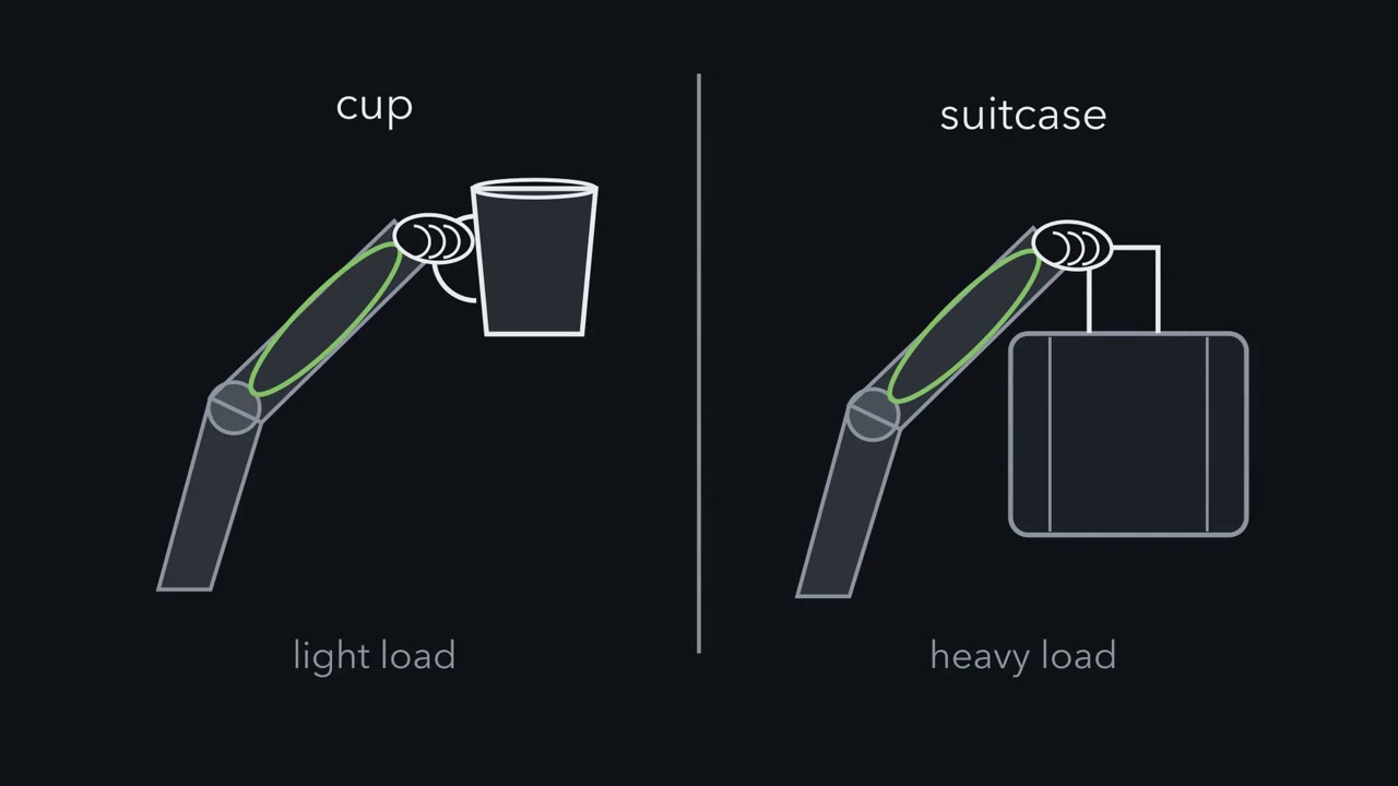 ▶ Watch Where Every Command Ends · 2:45
Read the transcript of “Where Every Command Ends”
Film transcript: Where Every Command Ends
Lifting a cup and lifting a suitcase can use the same muscle. Who decides how hard it pulls? The decision ends in the spinal cord, at cells called alpha motor neurons. Each motor neuron's axon branches to many muscle fibres. The neuron and its fibres make one motor unit. One spike normally makes every fibre of its unit twitch. All the motor neurons that supply one muscle make up its motor pool. Commands come from the brain, stretch sensors, pain and rhythm circuits. All of them end on the motor neurons. Sherrington called them the final common path. Picture one motor neuron's voltage as a bar, with a line at threshold. Excitation pushes the bar up, toward the line.
Inhibition holds it back. Usually it pulls the bar down. Its open channels also blunt excitation. When the net current lifts the bar to the line, the cell fires. So ask two questions of any spinal circuit. Which motor pools does it push up, and which down? And within a pool, which cells reach threshold first? Take a pool of small cells, which make weak units, and large cells, which make strong units. The input to the whole pool rises a little at a time. Which cells fire first?
The small ones. Many people guess the large ones, because big seems strong. Firing depends on how far the input moves the voltage. The same input moves a small cell further. A small cell has less membrane, so fewer ways for current to leave. Its input resistance is higher. Think of a bathtub with fewer drains. The same inflow raises the water higher. On screen is Ohm's law for a motor neuron. Higher input resistance means less current is needed. That order, small before large, is the size principle. Under shared input it is a strong tendency, not a law. There is a catch. A large cell has room for more synapses, so it can collect more input. In this simple model, small cells still go first if effective current grows less steeply with size than input resistance falls.
Real current distributions depend on the pathway. So, with equal thresholds, the small cells still go first. A pool has a second way to add force. Active units fire faster, so their twitches add up. Every command ends as current on motor neurons. Which pools go up or down, and who fires first?
From the film
- Every command to a muscle, from the brain, a stretch, pain or a rhythm circuit, ends as synaptic current on motor neurons.
- A motor neuron fires when the net current, times its input resistance, lifts it to threshold.
- Under shared input, small motor neurons usually fire before large ones, which is a strong tendency rather than a law.
Tap the tendon just below your kneecap and your leg kicks, through a path in the spinal cord with only one synapse between sensor and motor neuron.
The same tap also inhibits the motor neurons of the muscle on the back of your thigh. The same few segments of cord decide how hard a muscle pulls, keep a joint steady and can lay down the rhythm of a step. All of these commands funnel into one set of cells, and one idea about those cells ties the jobs together.
Motor units: where every command ends
The cells that command skeletal muscle are alpha motor neurons, whose cell bodies sit in the ventral horn of the cord and whose axons leave through the ventral roots. Each axon branches to many muscle fibres. A motor unit is one alpha motor neuron and all the fibres it innervates; a motor pool is all the motor neurons that supply one muscle. In mature muscle each fibre is contacted by one motor neuron, after an early period of multiple innervation that synapse elimination covers; how a motor neuron gets its identity is in the motor neuron code, and how its junction is built in the neuromuscular junction.
At the junction, acetylcholine released by the motor neuron depolarizes the fibre (synaptic transmission), and normally each motor-neuron spike gives every fibre of its unit an action potential and a twitch. So the unit is the smallest piece of force the cord can command. Units differ widely: small motor neurons supply few fibres and make weak units, large ones supply many and make strong units.
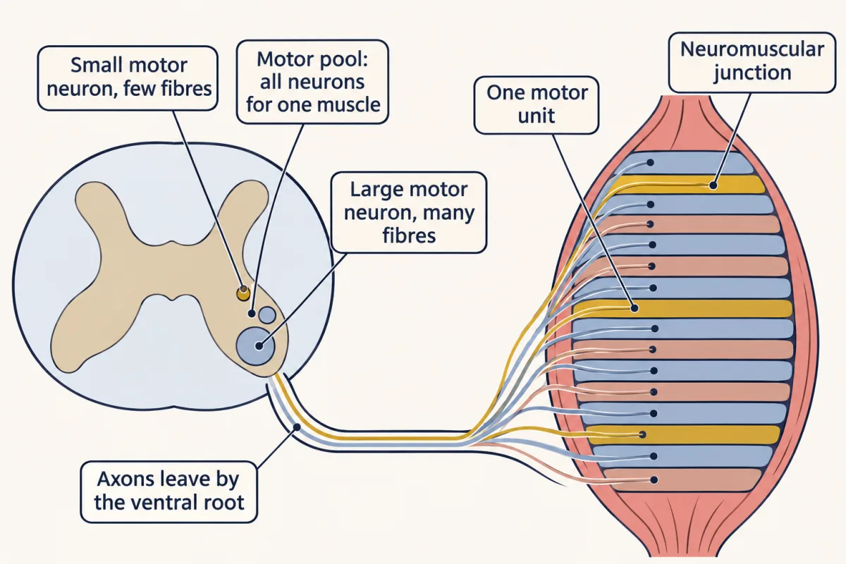
Every command that makes a limb muscle contract has to pass through these motor neurons. Charles Sherrington called them the final common path. Three puzzles show why that matters. Two words first: flexors are muscles that bend a joint, and extensors are muscles that straighten it.
- One axon, two opposite results. A tap below the kneecap stretches the quadriceps, on the front of the thigh. A sensory axon from the stretched muscle fires, and the quadriceps contracts. Yet the motor neurons of the hamstrings, on the back of the thigh, are inhibited, and that sensory axon only ever excites. How can it inhibit anything?
- Which units do the work? Lifting a coffee cup takes far less force than lifting a full suitcase, so fewer forearm motor units need to work. Which go first, and why those?
- Who keeps the beat? A cat’s spinal cord, cut off from the brain and from the sensory nerves of the hind legs, can still make the ankle flexors and extensors contract in turn. Where does the rhythm come from?
The idea that answers all three needs one more term. A cell’s input resistance, Rin, is how many millivolts its voltage changes for each nanoampere of steady current put into it. Picture a bathtub, where the water level is the voltage: with fewer drains, the same inflow raises the water higher.
The one idea to carry through this lesson. Every command the spinal cord sends to a muscle, whether it starts from a stretch, a pain, a rhythm or the brain, arrives as synaptic current onto motor neurons. Excitation adds current and inhibition, in effect, takes it away. A motor neuron fires when the net current, times its input resistance, lifts it to threshold, and then, normally, every fibre in its unit twitches. So ask two questions of any spinal circuit: which motor pools does it push up, and which down? And within a pool, which cells reach threshold first?
The rule is a simplification: its Ohm’s-law step describes a slow approach to threshold (Going deeper, “Where the rule is approximate”).
The size principle: small before large
Predict. A pool holds small motor neurons, which make weak units, and large ones, which make strong units. The drive to the whole pool rises a little at a time. Which start firing first, and why?
Check your prediction
The small ones. Many people guess the large ones, because big seems strong. But whether a cell fires depends on how far the input moves its voltage, not on how strong its unit is, and the same input moves a small cell further.
Picture three motor neurons side by side, small, medium and large, all receiving the same input. Under each one, draw a bar for its membrane voltage, with a line across the bar at threshold. Now turn the input up slowly. The small cell’s bar climbs fastest and crosses the line first. The medium cell crosses next, and the large one last. This order, small before large, is the size principle. Elwood Henneman described it in cats in 1957.
Why does the small cell’s bar climb fastest? Current that enters a cell has to leave through open channels in its membrane (the resting membrane and passive signals). A small cell has less membrane, so fewer channels in parallel and a higher input resistance: fewer drains.
Ohm’s law for a motor neuron. ΔV = I × Rin. To fire, a cell must depolarize by ΔVth, so the current it needs is Ith = ΔVth / Rin. Higher input resistance means less current is needed, so the cell is recruited earlier.
How synaptic currents sum on the way to the cell body is the subject of synaptic integration.
There is a catch. A large cell has more membrane, so it has room for more synapses and could collect more of the shared input. If its current grew as steeply as its resistance falls, the two would cancel, and cells needing the same depolarization would reach threshold together. For spindle input in cats, that does not happen. The same spindle input gives larger synaptic potentials in the smaller cells, and when the effective current reaching the cell body was measured in cat medial gastrocnemius motor neurons, it was on average larger, not smaller, in the high-resistance cells, reinforcing their head start (Heckman and Binder, 1988). Large motor neurons, which mostly supply fast units, also tend to have leakier membrane, which lowers their resistance further. In general, with equal required depolarizations, the small cells still go first whenever effective current grows less steeply with size than input resistance falls (Going deeper works through the scaling).
Why this order helps. Small units make small forces. Recruiting them first makes the first steps of force tiny, which is when an error of a fraction of a newton matters most: threading a needle, not lifting a suitcase.
Scope. The order is a tendency under shared input, first measured in cats, not a law. Differences in voltage threshold, persistent inward currents and neuromodulation can shift it, and an input that reaches only part of a pool can recruit cells out of size order.
Grading force: recruitment and rate
Worked problem. Three motor neurons, S, M and L, in one pool receive the same synaptic current, ramped up from 0 at 1 nA per second. These are illustrative round numbers, in the range reported for cat lumbar motor neurons, not readings from one recording. Input resistances are S 4.0 MΩ, M 2.0 MΩ and L 1.0 MΩ. Assume steady state (the ramp is slow compared with the membrane time constant) and that each cell must depolarize 10 mV from rest to fire. Both are simplifications: real thresholds differ, and identical effective current is only the simplest model of how an input is shared.
Step 1: current to threshold. A millivolt divided by a megohm is a nanoampere.
S: 10 mV / 4.0 MΩ = 2.5 nA; M: 10 mV / 2.0 MΩ = 5.0 nA; L: 10 mV / 1.0 MΩ = 10 nA
Step 2: time to threshold. At 1 nA per second, S fires at 2.5 s, M at 5.0 s and L at 10 s. The order is smallest first.
Step 3: force. Give the units forces of 0.05 N, 0.30 N and 1.50 N. As each joins, total force goes 0.05 N, then 0.35 N, then 1.85 N. The steps get bigger as force grows. In an illustrative model pool (Going deeper), each later recruit adds about 4% of the force already present, a fraction set by how unit forces are spread along the recruitment order, not by Ohm’s law.
- Current now
- —
- Units firing
- —
- Total force
- —
- Recruitment time = threshold current ÷ ramp rate
- —
—
In this sketch: steady state (the ramp is slow compared with the membrane time constant), every cell needs the same 10 mV depolarization to fire, each unit is simply on or off, and circle area is drawn in proportion to 1/Rin (relative sizes, not to scale). Round numbers, the lesson’s own.
I = ΔV / Rin: with the same current in every cell, the one with the highest input resistance reaches 10 mV first. In this sketch, with steady state and equal 10 mV thresholds, the input resistances are illustrative round numbers in the cat lumbar range; the forces are the lesson’s round numbers.
Recruitment is half the story.
Predict. A muscle’s force keeps rising after the last unit you are watching has joined. No new units are recruited. Where can the extra force come from?
Check your prediction
From the units already firing: they fire faster. Over the range where a motor neuron’s firing has not saturated, more excitatory drive generally raises its firing rate. That current-to-frequency relation is a property of the cell’s spiking, separate from the passive Ohm’s-law step, and it differs from cell to cell.
Picture a plot of one unit’s force against time. Each spike gives a twitch, a brief bump of force that rises and relaxes. With spikes far apart, the bumps stand separate. Bring the spikes closer, and each new twitch starts before the last has relaxed, so it rides on top of it and the line climbs higher, still rippling. Closer still, and the ripples merge into a smooth, high plateau, a tetanus (here a fused contraction; the word also names a disease). Fusion means the force has become smooth; it need not be the most force the unit can make, which keeps rising with rate until the force-frequency relation levels off. This is rate coding: an active unit makes more force by firing faster. So force rises both by adding units and by driving the active ones harder.
Full fusion is not how everyday force stays smooth: during ordinary voluntary contractions most units fire below it. The muscle’s force is smooth largely because its many units fire out of step with each other, so their ripples fill one another’s gaps.
In people, firing rate rises steeply just after a unit is recruited, then more slowly, and the shape differs from unit to unit (Going deeper).
One stretch, two commands
Read the transcript of “One Stretch, Two Opposite Commands”
Film transcript: One Stretch, Two Opposite Commands
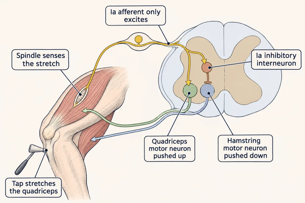
Tap the tendon below your kneecap, and your leg kicks. What happens to the muscle on the other side of the joint? The tap stretches the quadriceps. Inside it lie stretch sensors called muscle spindles. A spindle lies alongside the force-making fibres. It lengthens with the muscle, so it reports stretch. Its sensory axons, or afferents, carry signals into the cord. The main ones are called Ia fibres. The tap makes them fire. They enter the cord through the dorsal root, and they branch. One branch excites the quadriceps motor neurons directly. That is one synapse. The pool goes up, and the leg kicks.
But the motor neurons of the hamstrings, on the back of the thigh, are held back. A Ia fibre only ever excites. How can it hold anything back? Its second branch excites an inhibitory interneuron, on the same side of the cord. That interneuron inhibits the hamstring motor neurons. Their pool goes down. This path has two synapses. In this pathway, the excitatory Ia fibre inhibits the antagonist through an inhibitory interneuron. One stretch, two commands, to two pools. Sherrington called this pairing reciprocal innervation. A second sensor shows why the spindle needs help. The Golgi tendon organ sits where muscle meets tendon. Each one lies in line with a few fibres and senses their pull.
Together they signal force, not length. A unit pulling on one tendon organ twitches, so that organ fires. The shortened spindle can fall slack and go quiet. That slack spindle is a problem. Gamma motor neurons fix it. They supply only the spindle, and they contract its two ends. That pulls the centre of the spindle taut again, where the Ia endings wrap. Think of a tape measure that winds itself in, so it never goes loose. When alpha and gamma motor neurons fire together, the spindle can stay in range during a contraction. So ask the two questions of any reflex. A stretch pushes its own muscle's pool up and its antagonist's pool down. And gamma drive keeps the spindle signalling while the muscle shortens.
From the film
- A stretch excites the muscle's own motor neurons and, through an inhibitory interneuron on the same side, pushes its antagonist's motor neurons down.
- A Ia fibre only ever excites, yet it holds back the antagonist by way of a second synapse onto an inhibitory interneuron.
- Gamma motor neurons contract the spindle's ends so it stays in range and keeps signalling while the muscle shortens.
Directions here use the spinal-cord frame: dorsal means toward the back, ventral toward the belly. Ipsilateral means on the same side of the body, and contralateral on the opposite side. An afferent is a sensory axon carrying signals into the cord; an efferent carries signals out, as motor axons do.
A muscle spindle is a stretch sensor: a capsule of small, specialized intrafusal fibres lying parallel to the ordinary extrafusal fibres that make force, like a thread laid along a rope. When the muscle lengthens, the spindle lengthens with it, so it reports length. Its Ia afferents, said “one-A”, are its primary afferents. The name comes from an old numbering of nerve fibres by thickness, and Ia fibres are among the thickest and fastest. In relaxed human muscle, Ia afferents respond to both the speed and the amount of stretch, and the spindle’s group II (secondary) afferents mainly to the amount (reviewed by Dimitriou, 2022).
A tendon tap gives a brief stretch, and the Ia afferents fire. They enter the cord through the dorsal root and branch.
Predict. Ask the two questions of the tap. Which motor pools should the Ia afferents push up, which down, and through how many synapses each? Remember that a Ia afferent releases only an excitatory transmitter, glutamate.
Check your prediction
- One branch excites the motor neurons of the same muscle, the homonymous motor neurons, directly: one synapse, a monosynaptic path. That pool goes up, and the muscle contracts against the stretch. This is how the circuit helps hold a joint: a sudden stretch from an unexpected load is met by contraction of the stretched muscle.
- Another branch excites a Ia inhibitory interneuron on the same side of the cord, which inhibits the motor neurons of the antagonist, the muscle that moves the joint the other way. That pool goes down. The path has two synapses, so it is disynaptic, and it is ipsilateral.
In this fast pathway the Ia afferent itself makes only excitatory synapses; the inhibitory interneuron changes the sign.
That answers the first puzzle. The kick itself needs only one synapse. Inhibiting the hamstring motor neurons needs two, and that second command goes to a different pool, so the reflex as a whole is not purely monosynaptic. Sherrington called this pairing of excitation and inhibition reciprocal innervation; its inhibitory half is called reciprocal inhibition. Ia afferents also excite, directly, the motor neurons of synergists, muscles that help move the joint the same way.
A single central synapse keeps the delay inside the cord to a minimum; Going deeper compares that delay with the time spent in conduction.
The tendon organ: a force sensor
A second sensor shows why the spindle needs help. The Golgi tendon organ sits at the junction of muscle and tendon, in series with a small group of muscle fibres: in line with them, like a link in a chain. It senses the tension those fibres pass to the tendon, and together the tendon organs signal the force the muscle makes rather than its length.
Predict. A motor neuron fires, and its unit, whose fibres pull on one tendon organ, twitches. The muscle is free to shorten a little. Which sensor fires, and which may fall silent?
Check your prediction
That tendon organ fires, because the twitching fibres pull on it. The spindle lies alongside fibres that have just shortened, so it shortens too and can fall slack, and its firing drops or stops. Length and force are different signals, and the two sensors split them.
The tendon organ’s Ib afferents act through interneurons. The textbook pathway pushes the muscle’s own pool down, inhibiting its motor neurons. Real Ib effects change with the task, so the tendon organ is not simply a safety cutout against tearing.
The crossed-extensor reflex: four groups of pools at once
In the crossed-extensor reflex, the circuit crosses the midline. A painful stimulus to one foot acts through several interneurons, some of whose axons cross to the other side. Ask the two questions, and four groups of muscles answer, each with its own pools.
| Muscles | Same leg as the stimulus | Other leg |
|---|---|---|
| Flexors | pushed up: the leg withdraws | pushed down |
| Extensors | pushed down | pushed up: the leg takes the weight |
In the classic pattern two groups go up and two go down: reciprocal innervation again, now across both legs. The reflex is polysynaptic (many synapses in series) and bilateral, where the stretch reflex is local and one-sided.
Gamma: keeping the spindle in range
The twitch in the last check exposed a problem: a shortening muscle can leave its spindle slack and silent. Gamma motor neurons fix this. They supply muscle spindles only, ending on the contractile poles of the intrafusal fibres. When the poles contract, the centre of the spindle, where the Ia endings wrap, is pulled taut again.
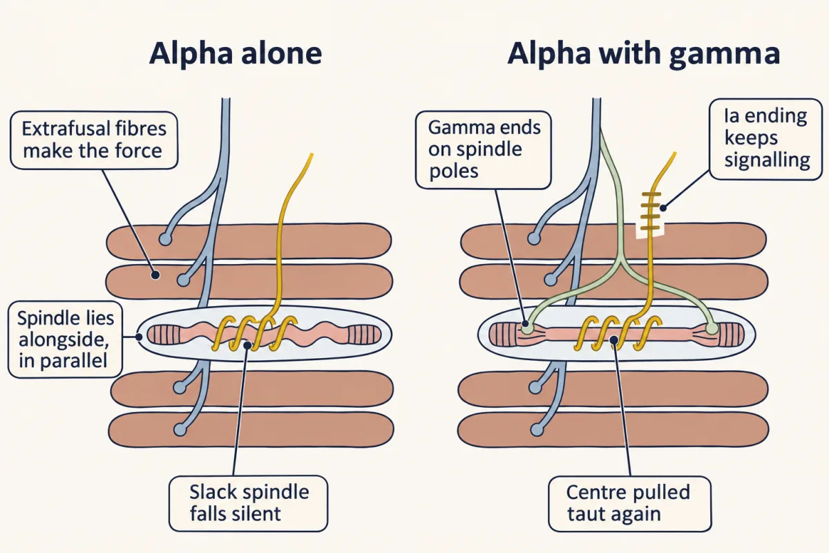
Think of a tape measure that winds itself in as the muscle shortens, so it never goes loose. When alpha and gamma motor neurons fire together, alpha-gamma coactivation, the spindle can be kept from falling slack during a contraction, so the stretch circuit keeps its length signal just as the muscle moves.
Gamma drive is not simply locked to alpha drive. One human study suggests it may be tuned to the goal of a coming movement, though gamma neurons themselves were not recorded (Going deeper).
Rhythm without a conductor
Read the transcript of “Rhythm Without a Conductor”
Film transcript: Rhythm Without a Conductor
When you walk, your legs bend and straighten in turn. Who keeps the beat? In the early twentieth century, a common view held that walking is a chain of reflexes, like falling dominoes. Graham Brown tested this in cats, in 1911. He cut the spinal cord to remove input from the brain. He also cut the dorsal roots carrying sensory signals from the hind legs. If walking is a chain of reflexes, what should be left?
Nothing rhythmic should remain. Instead, the ankle flexors and extensors still contracted in alternation. The rhythm arose within the cord. A network that can do this is called a central pattern generator. Modern work often uses a newborn rodent's cord in a dish. Drugs in the bath give a steady push, like holding down an accelerator. They carry no timing. Yet the flexor and extensor roots fire in alternating bursts. The beat comes from the cord. Graham Brown proposed two groups of neurons, one driving flexors and one driving extensors. Each group inhibits the other. Activity switches when the active group's inhibition fades, or the held-down group escapes.
It is reciprocal inhibition laid out in time. Whether real rhythm comes from such a simple pair is still debated. Sensory input still matters. In newborn mouse cords, ankle nerve pulses between flexor bursts could start a flexor burst early.
The cord makes the rhythm, and the senses adjust its timing. Which spinal neurons make the rhythm is still open. So is how much of this holds for human walking. The cord can keep the beat by itself. That beat reaches muscle as current, pushing pools up and down in turn.
From the film
- In cats, a spinal cord cut off from the brain and from sensory roots can still make alternating flexor and extensor contractions.
- A network in the cord that does this is a central pattern generator, and its rhythm arises within the cord.
- Sensory input adjusts the timing of the rhythm, and which spinal neurons make it, and how much holds for human walking, is still open.
In the early twentieth century a common view held that walking is a chain of reflexes: each step produces sensory signals that trigger the next.
Predict. In 1911 T. Graham Brown cut the spinal cord of cats, removing input from the brain, and also cut the dorsal roots that carry sensory signals from the hind limbs into the cord. If walking is a chain of reflexes, what should be left?
Check your prediction
If the chain idea were right, nothing rhythmic should remain. Instead, the ankle flexors and extensors still contracted in alternation. The rhythm arose within the cord. That answers the third puzzle.
A central pattern generator is a neural network capable of producing rhythmic output without patterned descending or sensory input.
Much modern evidence comes from fictive locomotion, locomotor-like motor output with no movement. It is also studied in paralysed adult cats, but a common preparation is an isolated spinal cord from a newborn rodent, bathed in drugs such as N-methyl-D-aspartate (NMDA) (a glutamate-receptor agonist) and serotonin, producing alternating bursts in the ventral roots that would drive flexors and extensors. The drugs are a steady push, like holding down an accelerator. They carry no timing, so the beat comes from the cord.
How could a network keep a beat? Graham Brown proposed the half-centre: two groups of neurons, one driving flexors and one driving extensors, that inhibit each other. Activity switches between them when the inhibition from the active group fades, or when the held-down group escapes, recovering and firing despite the inhibition. It is reciprocal inhibition laid out in time. Whether the rhythm comes from a simple pair of half-centres is still debated, and which spinal neurons make it is still open (Going deeper).
Whatever makes the rhythm, it reaches muscle the way every other command does: as synaptic current onto motor neurons, pushing the flexor pools up while the extensor pools go down, then the reverse. Within each pool, the second question still applies.
Sensory input still matters. In the work of Li, Garcia-Ramirez and Dougherty (2019) on cords of mice one to three days old, ankle nerve stimulation between flexor bursts could start a new flexor burst early. The network makes the rhythm and feedback adjusts its timing. For people, evidence for a spinal pattern generator is indirect; these results come from neonatal rodents and cats.
Design problem
Design an experiment to show that the mammalian spinal cord can generate a locomotor rhythm by itself, and that sensory input can reset it. Say what you record, what you predict, your controls and what your result cannot show.
Saved in this browser only.
One way to do it
Isolate the spinal cord of a newborn mouse (one to three days old) with ankle nerves attached, so there is no descending input and no movement feedback. Evoke activity with NMDA and serotonin in the bath; Li and colleagues (2019) used 7 µM and 8 µM. Record a flexor-related lumbar ventral root and an extensor-related one on both sides. Li and colleagues used L1 for flexor-related and L4 for extensor-related output; many laboratories use L2 and L5.
Predict that the flexor-related and extensor-related roots on one side alternate, and that the left and right roots of each pair alternate. Controls: record baseline activity in saline before adding the drugs, since neonatal cords can burst on their own, and check that the drug-evoked locomotor pattern fades on washout.
To test resetting, first transect the ventral roots proximal to the spinal nerve, so pulses cannot travel back into the cord along motor axons, then stimulate an ankle nerve with brief trains (for example 4 to 6 pulses at 20 Hz, onsets 50 ms apart) at several phases of the cycle. Plot the time to the next flexor burst against the phase of stimulation, using unstimulated cycles as the baseline. An early flexor burst after stimulation between flexor bursts, with the following cycles shifted in time compared with unstimulated cycles, shows resetting.
Limits: this is a neonatal cord driven by drugs, not adult or human walking. Rhythm with no patterned input shows the circuit is enough under these conditions; it does not identify a single pacemaker cell or prove that normal walking dispenses with the brain or with feedback.
Where this leads
What descending pathways add, and the signs that follow damage to them, are taken up in the basal ganglia and the cerebellum; diseases of motor neurons are in neurodegeneration.
Going deeper
Why one synapse?
A rough, illustrative delay budget for the human knee-jerk loop: the path between knee and cord is about 0.55 m each way, so at 60 to 70 m/s conduction alone takes about 16 to 18 ms. A further central synapse would add roughly 0.5 to 1 ms, about 2.5 to 5% of an illustrative 20-ms latency to the muscle’s electrical response; visible knee movement comes later still. Even that increment may matter for rapid feedback and timing precision. These are rough values, not a measurement, and the budget alone does not establish why the excitatory pathway is monosynaptic.
How the input scales with cell size
Suppose a cell’s effective share of a shared input grows with its membrane area A as Aα, and its input resistance falls as A−β. Then the depolarization scales as ΔV ∝ Aα−β. For cells of relative area 1, 2 and 4, these hypothetical cases illustrate the algebra; none is a measured scaling law:
| Case | α | β | Relative ΔV, small to large | Order |
|---|---|---|---|---|
| Identical current | 0 | 1 | 1, 0.5, 0.25 | small first |
| Current in proportion to area | 1 | 1 | 1, 1, 1 | none |
| Current grows more slowly than area (twice the area, about 1.4 times the current) | 0.5 | 1 | 1, 0.71, 0.5 | small first |
| Current in proportion to area, large cells leakier | 1 | 1.5 | 1, 0.71, 0.5 | small first |
Real current distributions depend on the input pathway and on dendritic filtering; the cat Ia measurement in the main text shows that effective current need not rise with area at all. Input resistance also differs between motor neurons not only because of how much membrane they have but because of how leaky that membrane is, which varies with motor-neuron type.
Where the rule is approximate
The Ohm’s-law part of the guiding idea describes a slow approach to threshold, with steady or slowly rising input. Inhibition works by opening channels, so it also lowers input resistance, and both effects raise the excitatory current a cell needs (next section). Persistent inward currents and neuromodulation amplify and prolong synaptic input, and they can make the current at which a unit stops firing differ from the current at which it started.
Inhibition opens channels
Inhibition does not literally subtract current; it opens channels. Take a cell with rest at −70 mV, threshold at −60 mV, input resistance 2 MΩ and an inhibitory reversal potential of −80 mV. With no inhibition it needs 5.0 nA of excitatory current to reach threshold. With inhibitory conductances of 0.05, 0.1 and 0.2 µS it needs 6.0, 7.0 and 9.0 nA. Even a pure shunt, with the inhibitory reversal potential at rest, raises the requirement, to 5.5, 6.0 and 7.0 nA. So in this passive model, with a steady excitatory current and an inhibitory reversal potential below threshold, “inhibition takes current away” is fair shorthand. Inhibition need not hyperpolarize a resting cell: a shunt leaves rest where it is, and an inhibitory reversal potential above rest can even depolarize it while still making firing harder. These are illustrative values.
Force steps across a large pool
In an illustrative model pool of 120 units, with recruitment thresholds spread tenfold and unit forces spread a hundredfold, both rising geometrically, recruitment order equals size order. With each active unit’s force held fixed, the force each new recruit adds, as a fraction of the force already present, falls from about 13% at the tenth unit to a nearly constant 4%. The first half of the units recruited carry only about 9% of the force that recruitment alone adds. The 4% holds only if unit force rises roughly geometrically along the recruitment order, and only after enough units have joined.
Smooth force from many units
As an explicit illustration, let one twitch be h(t) = P(t/τ)e1−t/τ, which peaks at t = τ, and sum identical twitches at regular intervals until the train reaches a periodic steady state. Define ripple as (maximum − minimum) / maximum within one steady-state interval. With τ = 80 ms (a slow unit), ripple is about 51% at 5 Hz and 4.7% at 20 Hz; with τ = 30 ms (a fast unit), it is about 28% at 20 Hz. These are outputs of the chosen linear model, not general physiological percentages: slow units tend to fuse at lower firing rates than fast ones, and the model does not capture force saturation at high rates.
Firing rate in human muscles
Avrillon and colleagues (2024) recorded the tibialis anterior and vastus lateralis of eight people per muscle with grids of 64 surface electrodes, during contractions up to 80% of maximal force, and sorted the signals into single motor units, about 129 units per participant in tibialis anterior. After an initial steep acceleration just above each unit’s recruitment, firing rates rose between force plateaus 10% of maximal force apart by 3.5 ± 1.7 pulses per second in tibialis anterior and 2.0 ± 1.1 in vastus lateralis. Most trajectories fitted a logarithm better than a straight line or an exponential. The input to each motor neuron was inferred from force, not measured inside the cell.
A spindle that changes before you move
Papaioannou and Dimitriou (2021) recorded single Ia afferents from wrist and finger extensors of volunteers with microneurography during an instructed delay before a reach. About 80 ms after the target was cued, Ia firing fell when the coming reach would stretch the spindle-bearing muscle, while kinematics and surface EMG showed no systematic change with the target. The principal analysis used eight afferents from six people, and separate experiments with a robot measured whole-arm stretch reflexes. The authors interpreted the results as compatible with goal-directed changes in gamma drive.
Who makes the rhythm?
Hägglund and colleagues (2013) used optogenetics in the isolated neonatal mouse cord to activate or inhibit selected populations, and found that locomotor-like bursting could be induced on one side alone, so rhythmic bursting does not need the other side of the cord. A 2021 review of mouse work reports that rhythm persisted after silencing or ablating each individual genetically defined ventral population it considered.
Your notes
Select any text in the lesson to highlight it or add a note to it. Your highlights and notes are saved in this browser and collected in your notebook.
This browser is not letting the site save anything (a private window or strict privacy settings), so highlights and notes will be lost when you leave the page.
How we measure it
Ventral-root recording of fictive locomotion
The spinal cord of a newborn rodent is removed into oxygenated saline, and drugs such as NMDA and serotonin evoke bursts of motor output. Electrodes on lumbar ventral roots record flexor-related and extensor-related bursts; their alternation shows a locomotor pattern with no limb moving and no brain attached.
High-density surface EMG with decomposition
Grids of many electrodes on the skin record muscle electrical activity, and a blind source separation algorithm sorts the mixture into the spike trains of individual motor units. It lets you follow dozens of units in a person, though the input to each motor neuron is inferred, not measured.
Microneurography
A fine tungsten needle inserted into a peripheral nerve in an awake person records the spikes of single sensory axons, such as a muscle spindle afferent. It shows what the spindle reports during a task, but not the gamma drive that shapes it.
Intracellular and patch recording from spinal neurons
An electrode inside or sealed onto a motor neuron or interneuron measures its membrane potential and synaptic responses while afferent nerves are stimulated. Latency and jitter of the responses suggest how many synapses lie between the afferent and the cell.
Practice questions on this lesson (3) → MCAT-style, original, each with an explanation
Where this lesson fits
Before thisReview first
- The Resting Membrane and Passive SignalsThis course · Lesson 3
Input resistance and how current becomes a voltage change in a cell come from the passive membrane there.
- Integration, Inhibition and NeuromodulationThis course · Lesson 7
You need how inhibition opens channels and shunts current there to see how pools are pushed up or down here.
- Synaptic Transmission and Quantal ReleaseThis course · Lesson 5
Acetylcholine release at the junction and quantal transmission from there underlie why each motor neuron spike twitches its unit.
After thisStudy next
- Descending Control and the Basal GangliaThis course · Lesson 16
Descending commands and the signs after damage to them act on the same motor neuron final common path.
- The CerebellumThis course · Lesson 17
The cerebellum tunes movement that ends on these motor pools, as shown in how it adapts reflexes and commands.
- When Mature Neurons Fail: NeurodegenerationThis course · Lesson 26
Diseases that kill motor neurons remove the final common path described here.
Elsewhere on the siteFilms, simulations and method films in other courses
- Every Response Has a Running TimeFilm · Organismal Biology
This film traces the knee-jerk route from receptor to effector, the simpler version of the stretch reflex here.
- Sliding, Not ShrinkingFilm · Organismal Biology
It shows how muscle fibres produce force, the output that motor units grade here.
- Length, load and powerSimulation · Physiology
It shows how muscle force depends on length, load and speed, which motor unit recruitment grades.
Suggested by an AI model from the lessons themselves, then checked. How we use AI
Finish line
Test yourself
8 questions
Practice and MCAT-style, with explained answers.
Next · Lesson 16
Descending Control and the Basal Ganglia
A stroke deep in the left hemisphere weakens the right side. A cut in the right half of the neck weakens the right side too. Why?
Continue →