Cell Biology · Unit 3: Traffic and movement
Molecular Motors and Cargo
Rewritten on : new text, flashcards and film. Your notes and highlights stay, but the text around them may have moved.
A motor protein turns ATP into steps along a track, and every track has a direction. Which motors walk which way, and how do we know?
Pause when the reading reaches a film, and offer to play it. Saved on this device.
Watch the film now, or keep listening
Builds on:Cytoskeletal Polymers and DynamicsVesicle Budding, Targeting and FusionMicroscopy and Quantitative ImagesWhy these?
 ▶ Watch Grip, Swing, Let Go · 3:09
Read the transcript of “Grip, Swing, Let Go”
Film transcript: Grip, Swing, Let Go
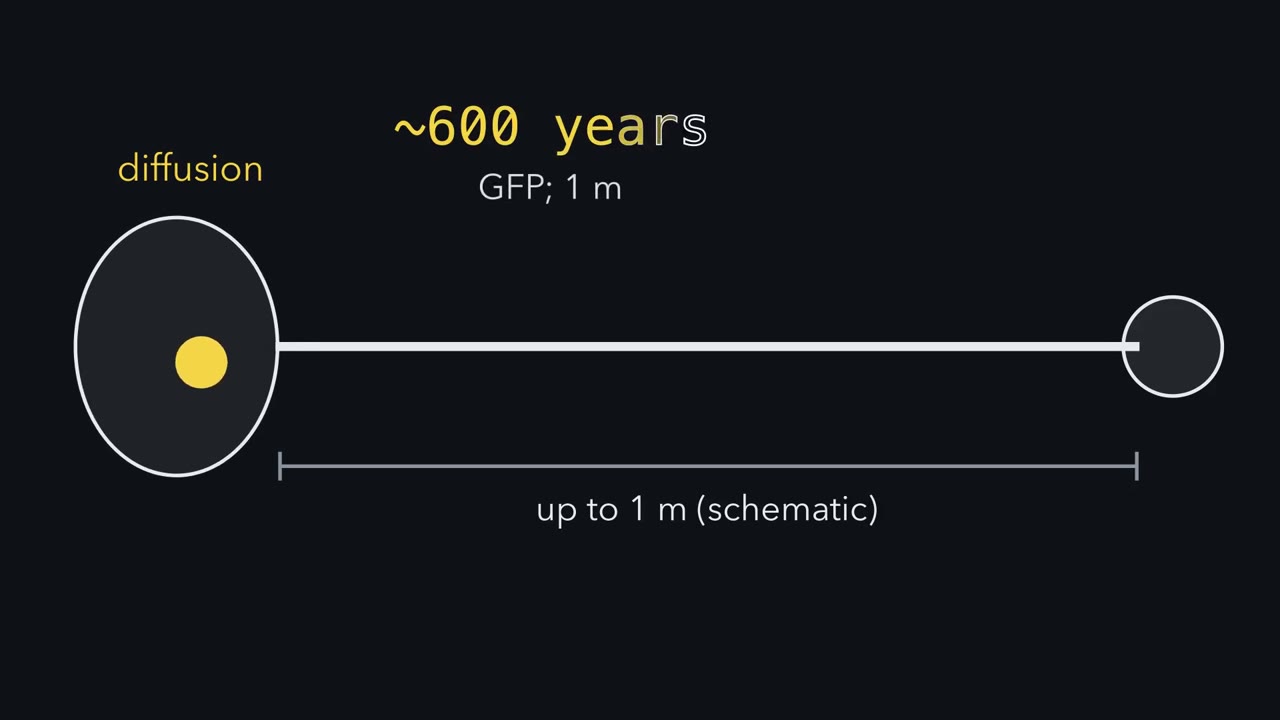
A protein made in a nerve cell's body may have to reach a synapse a metre away. Left to diffuse, it would take centuries, so how does it get there? The cell hands it to a motor, a protein that walks. Its road is a microtubule, and the road has a direction. In an axon, nearly every microtubule points its plus end out. Kinesin-1 walks toward the plus end. Dynein walks toward the minus end, back home. Watch one foot. It grips the track. It swings while gripping, which pulls the motor forward. Then it lets go and re-cocks. Each turn of this cycle is driven by one ATP. So a motor has feet on a polar track, each repeating grip, swing, let go, re-cock.
Which way the swing points helps set which way a motor goes, though dynein is less tidy. Kinesin-1 has two feet. The back foot lets go. The front foot, still gripping, swings and tips it past, onto the next site. Then the feet swap roles. Think of walking: your hips move one step, your back foot twice as far. The cargo rides at the hips, moving 8 nanometres per step. Each foot moves about 16. And at low load, each step costs one ATP. Here is the trick. The gripping foot nearly always stays on until the stepping foot lands. So one kinesin can take about 100 steps before it lets go. A motor that keeps walking like this is called processive. Now a test. Muscle myosin II also has two feet.
Could one molecule of it carry a vesicle all on its own? Pause and predict. No. Each myosin II foot spends most of its cycle off the track. The two feet do not take turns. One molecule soon falls away. So myosin II works in crowds, hundreds of heads per thick filament. That crowd shortens muscle. Heads grip the thin actin filaments, swing, and let go. The thin filaments slide inward between the thick ones. No filament gets shorter. They just overlap more. Back to the metre.
Cargo often carries several motors, so while one lets go, another still holds. How much this matters in cells is still being worked out. So a motor walks by repeating one small cycle. Keep a foot on the track, and one molecule can travel far. Let go too often, and only a crowd can pull.
From the film
- In an axon, microtubules have a direction: kinesin-1 walks to the plus end, outward, and dynein to the minus end, back toward the cell body.
- Each foot of a motor repeats one ATP-driven cycle: grip, swing, let go, re-cock.
- Kinesin-1 walks hand over hand: its cargo moves 8 nm per step while each foot moves about 16 nm.
- One kinesin foot is nearly always holding on, so one motor can take about 100 steps before letting go.
- Myosin II feet spend most of the cycle off the track, so it works in crowds, as in muscle.
The first film followed the walk itself; what it leaves open is how motor families, measurements and teams fit together.
The one idea to carry through this lesson. A motor is a set of feet on a polar track, and each foot runs one ATP-driven cycle: grip the track, swing while gripping, let go, re-cock. Three things follow. (1) The direction of the swing sets which way the motor goes; its reach, together with the spacing of binding sites on the track, sets the step size. (2) If one foot is always gripping, one molecule can walk alone (kinesin-1, myosin V). If its feet spend most of the cycle off the track, it has to work in a crowd (muscle myosin II). (3) One step’s work cannot exceed the energy of one ATP, so force × step is at most about 80–100 pN·nm (piconewton-nanometres, a unit of work).
Tracks with a direction, and three families of walkers
The first film used the simplest case; here is the fuller picture.
A motor needs a track, and the cell has two: actin filaments and microtubules, the polymers built in the lesson on cytoskeletal dynamics. Both are polar: their two ends differ, because every subunit in the filament faces the same way.
- An actin filament has a barbed end, also called the plus end, and a pointed end, the minus end. The names come from how the filament looks when it is decorated with myosin heads, which tilt like arrowheads.
- A microtubule has a plus end, which grows faster and in most animal cells points out toward the cell edge, and a minus end, which usually sits near the centrosome, the cell’s main microtubule-organizing centre. In an axon, nearly every microtubule has its plus end pointing away from the cell body.
| Family | Track | Usual direction | An exception you will meet | A job |
|---|---|---|---|---|
| Myosins | actin | toward the barbed (+) end | purified myosin VI moves to the pointed (−) end | myosin II contracts muscle; myosin V carries vesicles |
| Kinesins | microtubules | toward the plus end: outward | ncd, a fruit-fly kinesin, moves to the minus end in vitro | kinesin-1 (conventional kinesin) carries vesicles down axons |
| Dyneins | microtubules | toward the minus end: inward | none in this lesson | cytoplasmic dynein carries cargo back to the cell body |
In a nerve cell, kinesin-1 carries new cargo out to the axon tip, and dynein brings signals from the tip back to the cell body (the retrograde signal, long-distance signaling in neurons).
Direction belongs to the motor, and ncd breaks even the family pattern; a later section tests where direction comes from. The rule applies to single motors in vitro, outside the cell, at low load unless a load is stated, and its energy limit assumes one ATP per step, as shown for kinesin at low load. It says each foot lets go once per cycle, not what causes the release (ATP binding in myosin; in kinesin, a later step after the ATP is split). Dynein fits only loosely, and direction is still measured motor by motor.
One cycle, seen in one myosin head
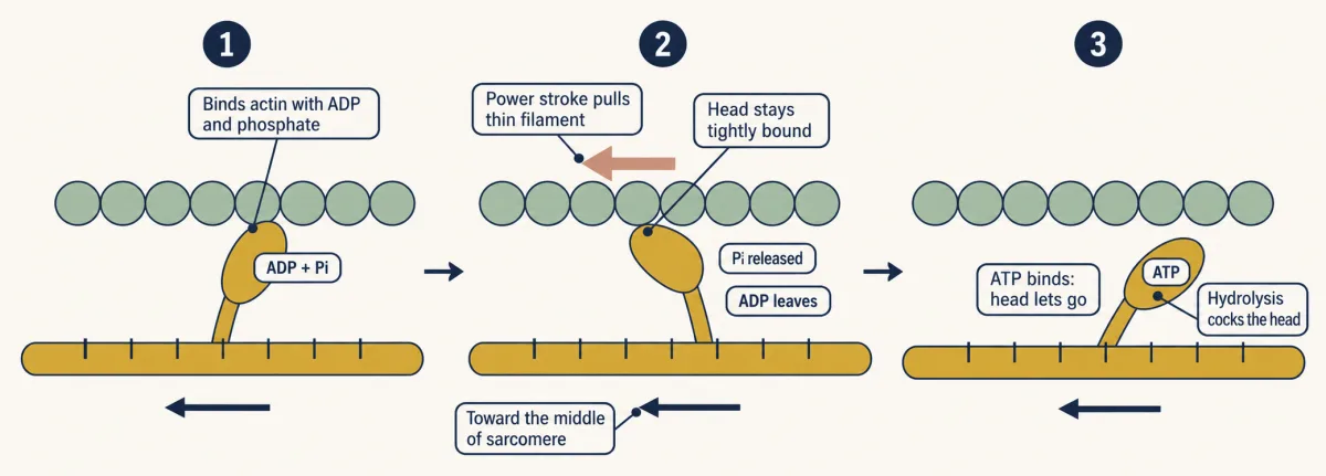
The four verbs hide chemical details. In muscle myosin II, one head reaches from a thick filament to a thin actin filament; its stiff neck is the swinging lever arm.
- Grip. Start with a cocked head that has just split an ATP and still carries the ADP and phosphate. It binds actin.
- Swing. The lever arm swings as phosphate leaves. This is the power stroke, and it pulls the thin filament toward the middle of the sarcomere, the repeating unit of muscle. ADP then leaves, and the head stays tightly bound.
- Let go. A new ATP binds, and the head releases actin.
- Re-cock. The head splits the ATP, which cocks the lever. ADP and phosphate stay bound, ready for step 1.
For exams, phosphate release is tied to the power stroke and is often taught as its trigger; researchers still debate whether phosphate leaves just before or just after the lever swings.
Predict. A muscle fibre is made permeable and all its ATP is washed out. Where are the myosin heads?
Check your prediction
Stuck to actin. ATP binding is the step that lets go, so with no ATP the heads finish their stroke and stay tightly bound. This is the rigor state, and it is why muscles stiffen after death (rigor mortis). So the pull itself needs no new ATP; letting go does.
Walking alone versus working in a crowd
Kinesin-1’s two identical heads join through a long coiled-coil stalk, a rope-like protein shaft whose tail and small partner proteins called light chains hold cargo. Each head binds one αβ-tubulin dimer, a paired tubulin building block, along a protofilament, one lengthwise row of the microtubule; binding sites sit 8 nm apart. The first film showed the walk, hand over hand: the front head, still gripping, swings its neck linker forward and tips the free head past it. The cargo, at the motor’s “hips” in the film’s walking picture, moves 8 nm per step; the stepping head moves about 16 nm (a dye on one head, tracked in a 2004 imaging study, moved about 17).
Because one head is nearly always holding on, a single kinesin can take about 100 steps in a row, under a micrometre in all, without falling off: it is processive. Its turn-taking is a coordination called gating (see Going deeper, “Why the two heads must take turns”). Unlike myosin, kinesin releases a head later in its cycle, after ATP is split. Myosin V also walks hand over hand along actin, with a much longer stride of about 36 nm.
Predict. Muscle myosin II also has two heads. Could one myosin II molecule carry a vesicle along actin?
Check your prediction
No. Each head spends most of its cycle off actin, and the two heads largely act independently, with no turn-taking. If, for example, each is bound about a twentieth of the time, both are off 0.95 × 0.95 ≈ 90% of the time, so one molecule falls off almost at once. Myosin II works only in crowds: a thick filament carries hundreds of heads, and a muscle millions, so at any moment some are gripping.
Back to the first film’s metre-long axon. Cargo in a cell often carries more than one motor, so while one kinesin has let go another can still be bound, and a motor that lets go can rebind. How much each contributes in cells is still being worked out.
The second film picks up here: it turns the invisible walk into measurements of direction, steps and force.
Read the transcript of “Turning Motion Into a Measurement”
Film transcript: Turning Motion Into a Measurement
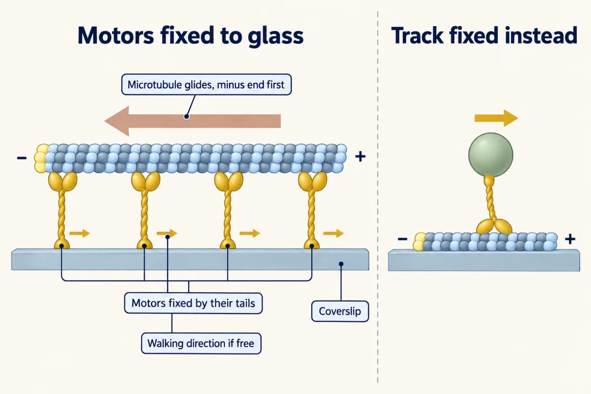
Chapter 1 showed a motor walking by gripping, swinging, releasing, and resetting. But how can experiments reveal the direction, steps, and pull of a machine too small to resolve? Begin with a gliding assay. Purified motors are fastened by their tails to a glass coverslip. Add fluorescent tracks and ATP. The motors cannot travel, but their feet can still walk. A fluorescence microscope follows the long glowing tracks; each motor is far too small to resolve. Imagine a free motor that walks right on a fixed track. Clamp that motor instead, and every stroke pushes the loose track left. The same interaction now moves the other partner. It resembles walking on a loose rug while bolted in place.
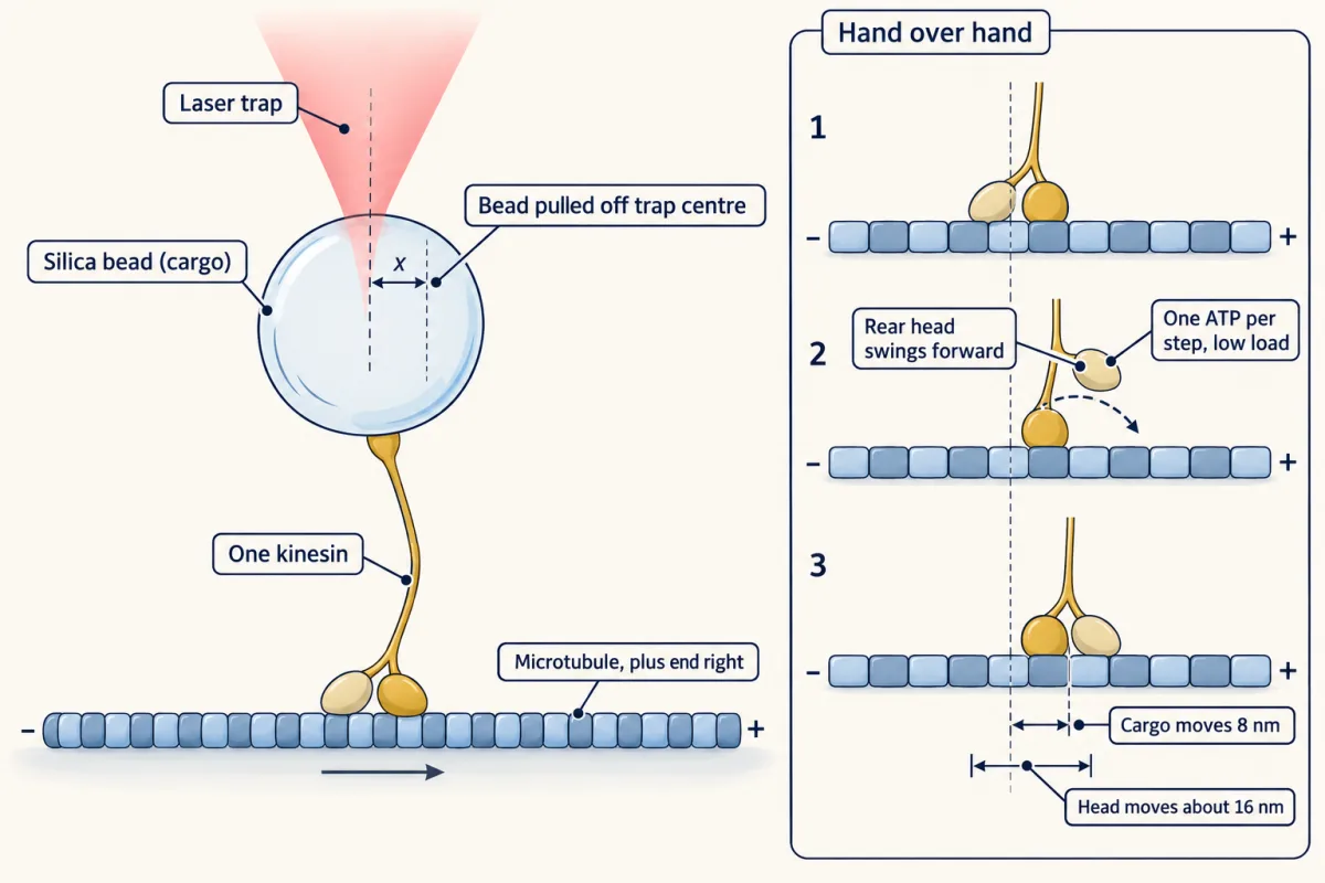
Mark the track's two ends, then watch which end leads. Because the track glides opposite the motor's walk, that movie reveals direction. It also reports speed across many motors at once. To see one motor, reverse the geometry again. Fix the blue track. Attach one motor to a yellow bead, then place that bead inside a focused laser trap. Sparse coating makes most beads carry at most one motor, separating one walker from the crowd. Suppose the motor advances in equal jumps, with waits between them. What shape should the bead's position record make? Pause and predict.
A staircase. Flat stretches are waits; sudden rises are steps. Kinesin bead records showed rises 8 nanometres high. Timing the flat stretches shows how long the motor waits before each jump. The invisible walk became a measured step size and a step rate. The trap also behaves like a spring. Pull the bead from the centre, and the restoring force grows. After calibration, displacement becomes force.
Trap stiffness says how much force each nanometre of displacement adds. Farther from centre means a larger opposing load. Eventually the motor may stall: it keeps trying, but makes no net progress. The bead's offset then reports the opposing force. Different loads reveal how the motor's speed changes as resistance grows. Position, time, and calibration turn motion into steps, speed, and pull. But a number belongs to its setup. In a fixed trap, load rises as the bead moves.
Feedback can hold load constant instead. Compare motors only under matched fuel and loading conditions. Otherwise, a neat calculation can combine measurements that never occurred together. One bench, two choices. Fix the motor, and the track reports direction. Hold the bead, and its motion reports steps and force. Change what is held still, and motion becomes a measurement.
From the film
- In a gliding assay, fixed motors drive a loose filament opposite to their walking direction.
- Marking the filament ends lets its leading end reveal motor direction.
- A bead carried by one motor can reveal discrete steps in its position record.
- In an optical trap, calibrated bead displacement reports force.
- Motor numbers should be compared only under matched ATP and loading conditions.
How we know: gliding assays and optical traps
Most numbers so far came from experiments on purified proteins, chiefly the two in the second film. The gliding assay turns the experiment upside down: stick motors by their tails to a coverslip, add fluorescent filaments and ATP, and watch with a fluorescence microscope (microscopy lesson). Movement needs ATP, a control the film did not show. The page now adds a polarity test, the trap numbers and the energy calculation.
Predict. Kinesin-1 walks toward the plus end. Fix many kinesins by their tails to glass and add a microtubule with ATP. Which end of the microtubule leads? (Think of walking on a loose rug: your feet push backward on it, so if you could not move, the rug would slide the opposite way to your walking.)
Check your prediction
The minus end leads. The motors are stuck, so each one, trying to walk toward the plus end, pushes the microtubule the other way, like the rug. Free on a fixed microtubule, the same motor walks toward the plus end.
Gliding usually measures many motors at once. To see one, use an optical trap: a focused laser that holds a small bead near its centre. A bead pulled off centre feels a restoring force, like a weight on a spring:
force = trap stiffness × the bead’s distance from the trap centre, or F = κx (Going deeper, “The sign in the spring law”)
The stiffness κ says how many piconewtons of pull the trap adds for each nanometre the bead is displaced. Coat beads so sparsely that most carry at most one motor, set one on a track, and record its position. Kinesin beads advance in 8-nm steps, and in a fixed trap kinesin kept moving against loads up to 5 to 6 pN. With load held constant by feedback, stall force depended on ATP; compare numbers only at the same ATP level and loading method (Going deeper, “How the load is applied”).
Worked problem: steps, force and the energy budget
These data are made up, though in the range of real measurements: one kinesin carries a bead along a microtubule in vitro, with plenty of ATP.
- Step rate. At low load the bead moves 400 nm in 0.5 s, in 8-nm steps. Velocity = 400 / 0.5 = 800 nm/s. Steps per second = 800 / 8 = 100. Near zero load kinesin uses one ATP per 8-nm step, so this motor burns about 100 ATP per second.
- Stall force. The trap stiffness is κ = 0.05 pN/nm. The motor walks until it stalls with the bead 120 nm from the trap centre. F = 0.05 × 120 = 6 pN, at the top of the fixed-trap range of 5 to 6 pN.
- The budget and its ceiling. Assume that hydrolysing one mole of ATP releases about 50 kJ, a round, conservative figure (the true value depends on the concentrations of ATP, ADP and phosphate; Going deeper, “How big the budget is”). Per molecule: 50,000 J / (6.022 × 10²³) = 8.3 × 10⁻²⁰ J, and since 1 pN·nm = 10−21 J, that is 83 pN·nm. Work is force × distance, so if each step costs one ATP, an 8-nm step can never push harder than 83 / 8 ≈ 10 pN (10 to 12.5 pN over the realistic range of 80 to 100 pN·nm).
- How much kinesin uses. At stall, one step would do 6 pN × 8 nm = 48 pN·nm. That is 48 / 83 ≈ 58% of the budget: about half, and nearer half with the larger ATP energy of assay conditions. Read it as an upper estimate, since it compares one forward step at the stall force with one ATP, and at true stall the motor makes no net progress.
- A longer stride. If myosin V also uses about one ATP per 36-nm step, the same budget caps its force at 83 / 36 ≈ 2.3 pN (2.2 to 2.8 pN over 80 to 100 pN·nm). Measured myosin V stall forces, about 2 to 3 pN, are of that size. A longer stride buys distance at the cost of maximum force, like a bicycle gear.
These readings assume a stiff link between bead and motor; a floppy tether stretches, and the bead under-reports the step.
Predict. Repeat step 2 in a trap twice as stiff, 0.1 pN/nm. Does the stall force change?
Check your prediction
No, in the ideal picture: the stall force is set by the motor (at a given ATP), not by the trap. The bead now stalls 60 nm from the centre instead of 120, and 0.1 × 60 = 6 pN. A stiffer trap only changes how far the motor gets.
The reader below uses the worked problem’s numbers and 50 kJ assumption. Its “energy share” row is step 4’s upper estimate (58% at stall), not a measured efficiency. Each load is held constant, as in a force clamp: the staircase is travel along the track, and x = F/κ is the offset the feedback keeps. Its straight speed-load line sketches the fixed-trap result.
Rule to keep: work per step is force times 8 nm, and the energy share is that work divided by the energy of one ATP, an upper estimate rather than a measured efficiency (83 pN·nm from the assumed 50 kJ/mol). The numbers are the lesson’s made-up numbers. The lesson ties one ATP per step to near-zero load and works out efficiency only near stall, so efficiency at other loads holds only if each step costs one ATP. The straight force-velocity line and the random timing of steps are a sketch of the logic, not data.
Keep one rule from this section: each number belongs to the preparation and loading condition that measured it, so do not combine numbers from different experiments (Going deeper, “Two myosin numbers that do not multiply”).
What sets direction and step: the swing
Short reminder: the swing helps set direction and, with track spacing, step size. Three motors test that rule.
Step size follows the lever. Myosin V’s lever arm is held straight by light chains. In a 2002 study, engineered myosin V with six light chains per lever stepped 36 nm, and a version with four stepped 24 nm: 6 nm per light chain both times. Over the range tested, shorten the lever and the stride shortens; the data do not show that ever-longer levers give ever-longer steps.
Direction follows the neck in ncd. The catalytic core of ncd resembles kinesin-1’s, but the neck joining core to stalk differs: a coiled coil in ncd, a different beta-strand structure in kinesin. Changing the neck residues reversed ncd’s direction, so the neck, more than the core, sets direction.
Direction follows the lever in myosin VI. An insert just before myosin VI’s lever arm redirects the lever, so the stroke points backward compared with other myosins and the motor moves toward the pointed end.
So the family gives you the track, and the design of the swing gives the direction. These results also show why direction is measured rather than assumed from family. Subfamily often predicts direction (ncd was the first of the kinesin-14s, a group of minus-end kinesins), but sequence alone does not predict it reliably enough to skip the experiment. A few yeast kinesin-5 motors even switch direction with conditions, and why some motors reverse in the cell is not fully resolved.
Dynein fits the rule only loosely. Its motor is a ring of six AAA+ domains, ATP-using enzyme modules, with a long stalk that reaches the microtubule. Its lever is a linker lying across the ring, which swings as ATP is used. The misfit is the step. Imaged without load at low ATP, human dynein-dynactin with an adaptor took forward steps anywhere from 8 to 32 nm and often stepped backward or sideways. In a 2026 optical-trap study, dynein-dynactin-BICD2 under load mostly took 8-nm steps, although earlier work had found variable steps at all the loads it tested.
Crowds I: muscle, where filaments slide and do not shrink
Muscle is point (2) of the rule on the largest scale. A skeletal muscle fibre is striped, and its repeating unit, the sarcomere, runs from one Z line (a protein disc) to the next. Thin filaments of actin anchor at each Z line and point inward. Thick filaments of myosin II sit in the middle. Three bands come from how the two overlap:
- the A band is the length of the thick filaments;
- the I band is thin filament only, on either side of a Z line;
- the H zone is the middle stretch of thick filament with no thin filament.
Predict. As the sarcomere shortens, which bands narrow and which keep their width?
Check your prediction
The I band and the H zone narrow, and the Z lines come together. The A band keeps its width, because it is the thick filaments’ length and they do not shorten. The thin filaments slide between them; no filament gets shorter.
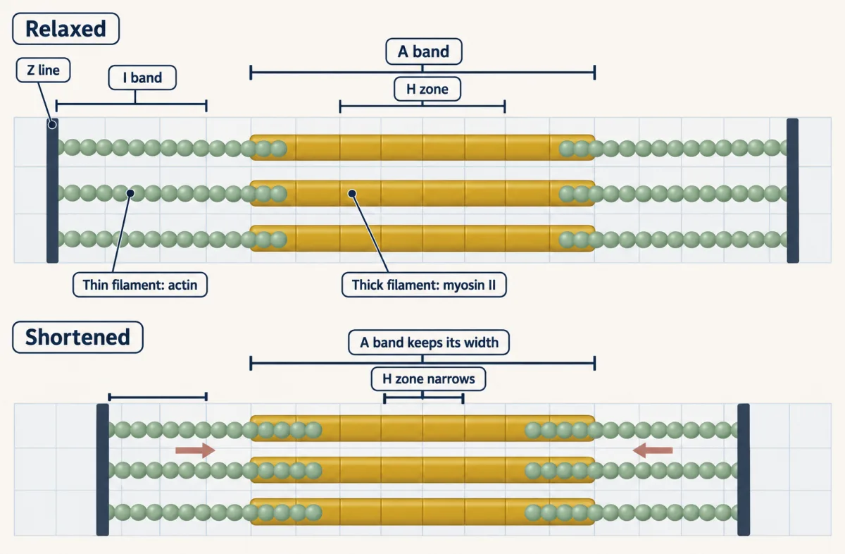
Two pairs of researchers saw exactly this in 1954, in back-to-back papers in Nature: Andrew Huxley and Rolf Niedergerke by interference microscopy of living frog fibres, and Hugh Huxley and Jean Hanson by phase-contrast microscopy of isolated myofibrils (the two Huxleys were not related).
Sliding predicts a length-tension curve: tension should track the number of heads within reach of a thin filament. In 1966 A. M. Gordon, Andrew Huxley and F. J. Julian held single frog fibres at fixed sarcomere lengths and measured tension during contraction. Tension peaked on a plateau between 2.05 and 2.2 µm. Stretched further, tension fell roughly in line with the lost overlap between thin filaments and myosin heads. At shorter lengths it fell too, first as the thin filaments crowd past one another, then more steeply as the thick filaments reach the Z lines and internal resistance to shortening grows.
In living muscle, calcium decides when heads may bind, through troponin and tropomyosin on the thin filament (calcium signaling covers that switch).
Crowds II: dynein teams
Dynein needs partners, and its partners help set how many dyneins pull together. In reconstitution, rebuilding a system from purified parts, human dynein becomes fast and processive when it binds dynactin, a large protein complex, and an activating adaptor, a cargo-binding protein (BICD2, BICDR1 and HOOK3 are examples). So the adaptor switches dynein on. It also biases how many dyneins ride together: a second dynein added pull, while speed depended on which adaptor was bound (Going deeper, “How many dyneins, and what they add”). More motors can give more pull and longer runs, but they need not mean faster cargo. Teams can also pull against each other, and many cargoes carry both kinesin and dynein; how such a cargo settles on a direction is still being worked out.
Design problem
You purify a new microtubule motor from a cell extract and must find which way it walks. Design the experiment, the controls, and the result that would tell you it is a plus-end motor.
Saved in this browser only.
One way to do it
Make polarity-marked microtubules: grow a short, brightly labelled seed, then extend it with dimly labelled tubulin. The minus end grows slowly, so the bright seed sits near the minus end and the long dim segment marks the plus end.
Run a gliding assay. Fix the motor to the coverslip, add marked microtubules and ATP, and film. The track moves opposite to the motor’s walking direction, so a plus-end motor glides microtubules with the bright minus end leading, and a minus-end motor glides them plus end first.
Confirm in the other frame. Fix marked microtubules to the surface and add beads coated with the motor, or the motor labelled with a single dye, and see which end it moves toward.
Controls: no ATP should stop movement, and so should the non-hydrolysable analogue AMP-PNP if the motor behaves like kinesin, which AMP-PNP locks onto the microtubule; a chamber with no motor should give no movement; conventional kinesin in a side-by-side chamber should glide microtubules minus end first, which checks your marking. Score many microtubules, not a few, and report the fraction moving each way.
Going deeper
The numbers behind the axon picture
Motor proteins are enzymes that turn ATP hydrolysis into steps along actin filaments or microtubules. A protein made in the cell body of a motor neuron may have to reach a synapse up to a metre away. Diffusion time grows with the square of distance, t ≈ x²/2D. Even a small protein like GFP (D ≈ 25 µm²/s in cytoplasm) would need about 600 years to diffuse one metre, and larger cargo diffuses more slowly still. A kinesin that never let go, walking at 800 nm/s (the worked problem’s speed), would cover that metre in about two weeks. The same machines pull chromosomes apart and contract muscle.
Who found kinesin
Kinesin was found by asking what moves organelles along nerve axons. In 1985 Ronald Vale, Thomas Reese and Michael Sheetz partly purified a protein from squid giant axons and optic lobes that moved microtubules, and named it kinesin. Scott Brady reported a candidate motor fraction from chick brain the same year.
The actin gliding assay, and gliding speeds
In 1986 Stephen Kron and James Spudich put myosin on glass with fluorescent actin on top, and the filaments moved only with ATP. Rabbit skeletal muscle myosin moved the filaments at 3 to 4 µm per second, and myosin from the amoeba Dictyostelium at 1 to 2 µm per second.
The single-kinesin trap records
The 8-nm steps were first seen in 1993 by Karel Svoboda, Christoph Schmidt, Bruce Schnapp and Steven Block. The 5 to 6 pN fixed-trap loads come from Svoboda and Block in 1994, and one ATP per step near zero load from Mark Schnitzer and Block in 1997.
The sign in the spring law
Written fully, the law is F = −κx, where κ is the trap stiffness and x is the bead’s displacement from the trap centre. The minus sign says the force points back toward the centre; the worked problem uses only its size, κx.
How the load is applied
In a fixed trap the load grows as the motor walks away from the centre, and kinesin’s speed fell roughly in proportion to the load (1994). In a force clamp, feedback holds the load constant. Coupling then stayed tight over a wide range of forces and, at high ATP, the speed-load curve was curved rather than straight; later estimates put the stall force near 7 pN.
How big the budget is
Under cell-like concentrations of ATP, ADP and phosphate, one ATP releases about 55 kJ/mol; under typical assay concentrations, about 59 kJ/mol (90 to 100 pN·nm per molecule). Against those, kinesin’s 48 pN·nm is 49 to 53% of the budget, below the 58% that the worked problem’s 50 kJ gives.
Why the two heads must take turns
A common shorthand says a motor is processive if each head spends most of its cycle bound (a high duty ratio). That is not enough. If two heads acted independently, a head bound half the time would give runs of about 1 step, and one bound 70% of the time about 2. A run of about 100 steps needs the partner head to be unbound at the moment of release only about 1% of the time. Independent heads would have to be bound over 99% of the time; kinesin’s heads are coordinated instead.
How many dyneins, and what they add
In a 2018 study of human dynein with pig brain dynactin, electron microscopy and single-molecule studies showed that the adaptors BICDR1 and HOOK3 mostly recruited two dynein dimers, and those complexes produced more force and moved faster. A 2019 study separated the two effects: adding a second dynein raised force but did not by itself set velocity, which depended on the adaptor and on how the closely packed dynein tails interact. Opposition within a team is measurable too: when many kinesins drove one microtubule in a 2019 in vitro assay, about a third were slowing it.
Two myosin numbers that do not multiply
In a 1994 trap study, one muscle myosin moved its actin filament about 11 nm at low load and pulled with 3 to 4 pN when held still. Those came from two loading conditions, so multiplying them does not give the work of one stroke. A 1995 trap study of a single myosin head put the working stroke nearer 4 nm, so 11 nm belongs to that preparation, not to the stroke itself.
Your notes
Select any text in the lesson to highlight it or add a note to it. Your highlights and notes are saved in this browser and collected in your notebook.
This browser is not letting the site save anything (a private window or strict privacy settings), so highlights and notes will be lost when you leave the page.
How we measure it
Gliding (in vitro motility) assay
Purified motors are fixed by their tails to a coverslip, and fluorescent filaments and ATP are added. The fixed motors push the filament, which moves opposite to the motors' walking direction. It gives filament speed and the fraction of filaments that move, with many motors at work.
Optical trap (optical tweezers)
A focused laser holds a bead carrying one motor. The bead's distance from the trap centre, times the calibrated trap stiffness, gives the force. Position records show single steps, the load at which the motor stalls, and how speed changes with load.
Single-molecule fluorescence localization
One dye on one motor head is imaged and the centre of its blurred spot is found to within a few nanometres. This follows each head rather than the cargo, and showed that a kinesin head moves about twice as far per step as the cargo does.
Interference and phase-contrast microscopy of muscle
These methods show the bands of living fibres or isolated myofibrils without stain. Measuring the A band, I band and H zone as the muscle shortens shows which filaments keep their length.
Practice questions on this lesson (3) → MCAT-style, original, each with an explanation
Where this lesson fits
Before thisReview first
- Cytoskeletal Polymers and DynamicsThis course · Lesson 14
You need actin and microtubule polarity, plus and minus ends, from there, since every motor's direction is defined against them.
- Vesicle Budding, Targeting and FusionThis course · Lesson 11
This lesson's motors are what carry the vesicles whose budding, targeting and fusion are taught there.
- Microscopy and Quantitative ImagesThis course · Lesson 2
Gliding assays and single-bead tracking rely on fluorescence imaging and resolution limits explained there.
After thisStudy next
- Cell Shape, Migration and CiliaThis course · Lesson 16
Actin and microtubule motors drive cell crawling and ciliary beating, including dynein arms that bend cilia.
- Mitosis and Chromosome SegregationThis course · Lesson 19
Kinesins and dyneins build the spindle and move chromosomes, using the direction rules learned here.
- Cytokinesis and Organelle InheritanceThis course · Lesson 20
Myosin II pulls on actin to contract the furrow, as it does in muscle.
Elsewhere on the siteFilms, simulations and method films in other courses
- Sliding, Not ShrinkingFilm · Organismal Biology
Muscle fibre shortening by sliding filaments is shown there with myosin and actin overlap.
- Power Peaks at a Middle LoadFilm · Physiology
Length, load and speed of muscle follow from the cross-bridge cycle and length-tension curve met here.
Suggested by an AI model from the lessons themselves, then checked. How we use AI
Finish line
Test yourself
8 questions
Practice and MCAT-style, with explained answers.
Next · Lesson 16
Cell Shape, Migration and Cilia
Four men had sperm that could not swim, and three could not clear mucus from their lungs. How could one missing part explain both?
Continue →