Organismal Biology · Unit 5: Defense, movement and reproduction
New to this? Review it at high school level: Germs, Vaccines and Antibiotics
Adaptive Immunity
Rewritten on : new text, flashcards and film. Your notes and highlights stay, but the text around them may have moved.
How can your body already hold cells that fit a virus you have never met?
Pause when the reading reaches a film, and offer to play it. Saved on this device.
Watch the film now, or keep listening
Builds on:Innate Immunity: Recognize and RespondGerms, Vaccines and AntibioticsWhy these?
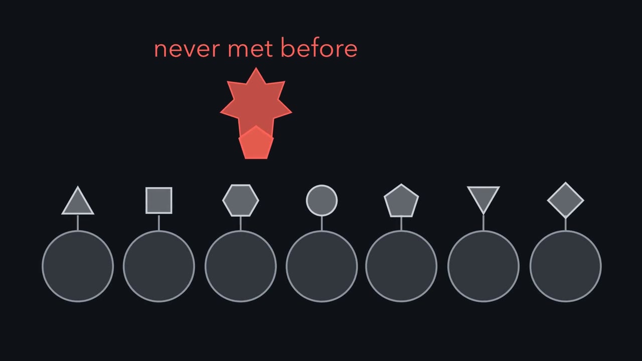 ▶ Watch Receptors First, Antigen Chooses · 3:21
Read the transcript of “Receptors First, Antigen Chooses”
Film transcript: Receptors First, Antigen Chooses
Your body already holds cells that can bind viruses you have never met. How could it prepare for something it has never seen? There are two possible answers. A soft, general receptor might fold itself around each new antigen, like clay. Or receptors could be made first, and the antigen picks one that fits. Adaptive immunity is carried by lymphocytes, a kind of white blood cell. The molecule a lymphocyte's receptor binds is called an antigen. Macfarlane Burnet proposed the second answer, called clonal selection. Nearly every lymphocyte makes receptors of one shape. It makes them before it meets the antigen they fit. But the human genome holds far fewer protein-coding genes than there are receptor shapes. So how can it make millions of them?
For a worked example, follow one developing B cell as it builds the heavy chain of its antibody. The tip of that chain is not encoded as one piece. It is built from short stretches of DNA, called gene segments. Here they sit in three rows: V segments, D segments and J segments. The cell picks one V, one D and one J. Enzymes cut the DNA, and the cell joins the three chosen pieces. That joined piece encodes the tip of one heavy chain. The screen shows illustrative human counts for each row. The cell takes one segment from each row. Do the choices add up, or multiply?
They multiply. Every V can go with every D, and every such pair with every J. So 69 segments give 5,520 V-D-J combinations for the heavy chain's variable region. And that is only one chain. Light chains use just V and J segments. T cells build their own receptor chains from segments of their own. Imprecise joins and chain pairing multiply the variety further. So the body holds a vast crowd of lymphocytes, each with its own receptor. It is like a shoe shop with millions of pairs already on the shelves, waiting for the right customer.
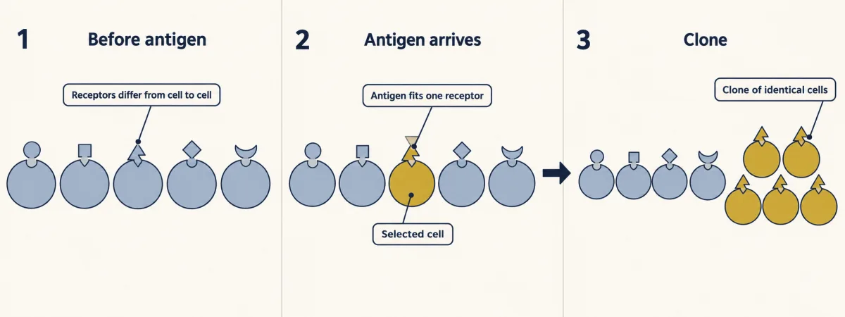
Now an antigen arrives. It drifts past B cell after B cell, and most receptors ignore it. It binds only the few whose receptors already fit. T cells need the antigen shown to them on another cell, as the text explains. Those cells divide. Their descendants form a clone, a population descended from one cell, all starting with the same receptor. The antigen did not mold that receptor. It existed before the antigen arrived. The antigen chooses; it does not shape.
From the film
- Nearly every lymphocyte makes receptors of one shape, and it makes them before it meets the antigen they fit.
- A developing B cell joins one V, one D and one J gene segment for its antibody heavy chain, so the choices multiply: 40 x 23 x 6 gives 5,520 V-D-J combinations for the heavy chain's variable region, from illustrative human counts.
- An antigen is bound only by the few cells whose receptors already fit (T cells need it shown on another cell), and those cells divide into a clone.
- The antigen chooses among receptors; it does not shape them.
You have never met most of the viruses that will ever infect you, yet your body already holds B cells whose receptors can bind them.
The one idea. Variety is multiplied before antigen; numbers are multiplied after it. Each developing lymphocyte assembles its receptor from gene segments chosen by chance (though not all equally often), so the possible choices multiply, and millions of receptor shapes exist before the antigen they fit arrives. That antigen picks the few cells that already fit, and their clone grows by doubling until there are enough. Memory leaves a larger starting clone, so the next response needs fewer doublings and arrives sooner.
Scope: conventional B and T cells of jawed vertebrates; the counts and the doubling are simplified models (Going deeper).
Receptors first: clonal selection
The first film’s rule is clonal selection (Macfarlane Burnet, 1957): receptors first, then antigen picks the few lymphocytes (B cells and T cells) that fit. Unlike innate immunity, whose receptors are encoded unchanged in the genome and recognize broad classes of microbes, adaptive immunity, the second layer in vertebrates, is specific for particular antigens and leaves memory, a faster and larger response the next time.
One shape does not mean one target. A receptor ignores almost every molecule, yet it can bind many that share the few features it contacts, even when they otherwise differ (Going deeper: one T cell clone responds to over a million peptides). That overlap is how a repertoire of millions covers microbes it never anticipated.
Making the variety: segments, joins and pairs
The first film assembled one heavy chain; it skipped the machinery and the other chains. An antibody is a Y-shaped protein of two identical heavy chains and two identical light chains; each arm’s tip, part heavy chain and part light chain, is the variable region that binds antigen. Each tip is encoded by gene segments, short stretches of DNA; RAG enzymes cut beside the chosen segments and the cell joins them: somatic recombination (in body cells, not eggs or sperm, so not inherited). The letters stand for variable, diversity and joining; a T cell pairs an alpha chain (V and J) with a beta chain (V, D and J). That is how about 20,000 protein-coding genes yield millions of receptors.
Two more steps multiply the variety: nucleotides added or removed at each joint (junctional variation), so cells that chose the same segments usually still differ, and each B cell’s pairing of one heavy chain with one light chain. Molecular detail: antigen receptors and checkpoints.
Worked problem: counting receptors. Continue from the film’s 5,520 heavy-chain V-D-J combinations. The segment counts are illustrative human values that differ between people and sources, and the count assumes every combination works.
Humans have two kinds of light chain, made from two separate sets of segments. Kappa: 40 V x 5 J = 200. Lambda: 30 V x 4 J = 120.
Predict. The film multiplied. To count light-chain combinations, do you add kappa and lambda, or multiply them?
Check your prediction
Add: 200 + 120 = 320, because a light chain is kappa or lambda. Pairing multiplies, because a B cell has a heavy chain and a light chain: 5,520 x 320 = 1,766,400 heavy-light combinations, about 1.8 million. Add for “or”, multiply for “and”.
receptor combinations = (V x D x J for heavy) x [(V x J for kappa) + (V x J for lambda)]
That is about 1.8 million combinations from fewer than 150 segments, before junctional variation multiplies the sequences many times over. Two later steps need antigen:
| Before antigen (during development, B and T cells) | After antigen (during a response) |
|---|---|
| segment choice, junctional variation, chain pairing | the fitting clone divides (B and T cells); B cells only: somatic hypermutation, point mutations in the variable region, with better binders favoured, so antibody affinity (how tightly it binds) rises |
Fixed in this sketch: humans have both light chains, kappa 40 V x 5 J = 200 and lambda 30 V x 4 J = 120.
- Heavy combinations (V x D x J)
- 5,520
- Light combinations
- 320
- Pairings (heavy x light)
- 1,766,400
- Segments used in total
- 148
receptors = 5,520 x 320 = 1,766,400 (about 1.8 million)
- Latest cell’s receptor
- No cell made yet
- Cells made
- 0
- Same receptor in two cells, in this sketch (junctional variation ignored): drawn
- 0
- Same receptor in two cells, in this sketch (junctional variation ignored): expected
- 0
In this sketch: every combination is functional and equally likely, any heavy chain pairs with any light chain, and junctional variation is ignored.
Each B cell assembles its antibody from gene segments before it meets any antigen.
Where B and T cells come from: a chicken’s bursa
Birds have a pouch of lymphoid tissue opening into the cloaca (the shared exit of gut, urinary and reproductive tracts), the bursa of Fabricius; in 1956 most chickens without it made little or no detectable antibody (Going deeper). J. F. A. P. Miller showed in 1961 that mice without a thymus from birth had impaired immune function. Neither result said whether the two organs fed one system or two.
Max Cooper, Raymond Peterson and Robert Good (1965, and in full with Mary Ann South in 1966) removed either the bursa or the thymus from newly hatched birds, then irradiated them to deplete lymphocytes that had already left the organ, and tested two jobs: making antibody and rejecting skin grafted from an unrelated chicken.
Predict. If one lineage of cells did both jobs, what would removing the bursa do to graft rejection? And if there were two lineages?
Check your prediction
One lineage: losing the bursa would cripple both jobs. Two lineages: each loss should hurt one job much more than the other. The birds showed the second pattern: without the bursa, antibody was largely lost while grafts were still rejected, only more slowly; without the thymus, graft rejection was impaired: two lineages, bursa-derived B cells and thymus-derived T cells.
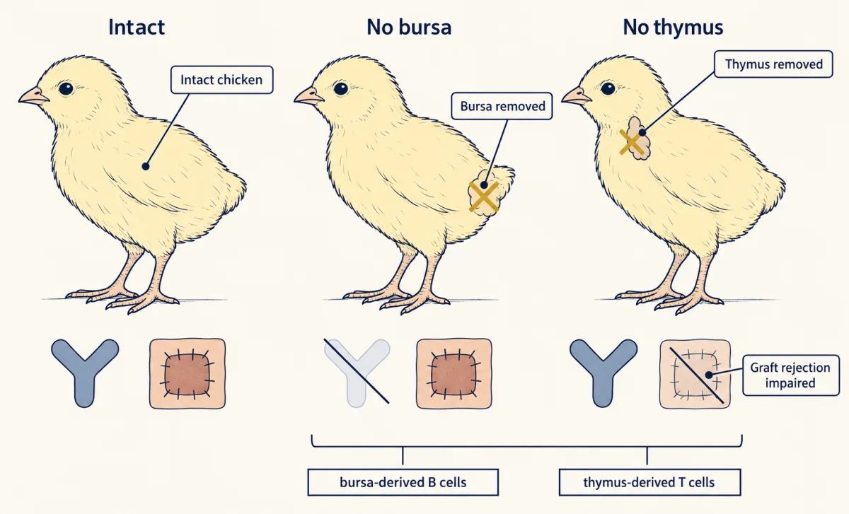
Mammals have no bursa: in humans and mice B cells develop from blood-forming stem cells in the fetal liver and then the bone marrow (stem cells), while T cells mature in the thymus in mammals and birds alike. The B honors the bird organ, not the bone.
Antibodies and killer T cells: class I and class II
B cells carry the humoral response, the part carried in body fluids; a B cell’s receptor binds intact antigen in lymph or blood, without processing. Activated, usually with signals from a helper T cell, it divides and some descendants become plasma cells, B cells that secrete large amounts of antibody. Most antibodies do not kill microbes on their own. They neutralize (coat a virus or toxin so it cannot attach to cells), opsonize (tag a microbe for phagocytes to engulf) and activate complement, the blood proteins you met in innate immunity.
T cells carry the cell-mediated response, and unlike the film’s B cells, a conventional (alpha-beta) T cell receptor does not bind free antigen. It needs something on another cell’s surface. A suspect is the major histocompatibility complex (MHC), a gene complex named for its role in graft rejection, whose classical cell-surface products come in many versions across a population.
Predict. Rolf Zinkernagel and Peter Doherty (1974) took cytotoxic T cells from virus-infected mice. Would those T cells kill infected cells from a mouse of a different MHC type?
Check your prediction
No. They killed infected cells only when the cells shared their MHC type. A T cell recognizes antigen only together with MHC.
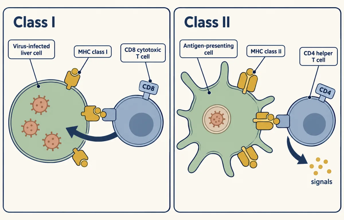
What the T cell reads is a short peptide displayed on an MHC molecule. There are two classes:
- MHC class I is on almost all nucleated cells. It displays peptides from proteins made inside the cell, so a virus-infected liver cell shows viral peptides. CD8 cytotoxic T cells read class I and kill the infected cell by triggering its self-destruction (apoptosis; the machinery is in death receptors, BCL-2 and caspases).
- MHC class II is normally found on antigen-presenting cells: dendritic cells, macrophages and B cells. It displays peptides from material the cell has taken in. CD4 helper T cells read class II and release signaling proteins (cytokines; see JAK-STAT signaling) that activate macrophages and help B cells.
A memory aid: CD8 x class I = 8, CD4 x class II = 8. The split by peptide source is not absolute: in cross-presentation, some antigen-presenting cells place peptides from engulfed material on class I.
Either way, a response starts from only the few cells that fit. How do they become enough, and why is the second time quicker?
Read the transcript of “Why Is the Second Time Faster?”
Film transcript: Why Is the Second Time Faster?
Suppose an infection leaves you a thousand times more cells that fit a virus. Is your second response a thousand times faster? The first film ended with an antigen choosing the few cells that fit. But a few cells cannot fight an infection. How do a few become enough? A chosen cell divides into two. Each of those divides again. With every round of division, the clone doubles.
So the number of cells is the starting number, doubled once for every round. Enough rounds turn a few cells into millions. Now draw it as a graph. Days run along the bottom. Cell number runs up the side. Each gridline is ten times the one below.
On this axis, steady doubling is a straight climb. A first response starts from a few cells, so it needs many doublings. It takes days to build. When the infection is cleared, most of the expanded cells die. But memory B cells and memory T cells remain. There are many more of them than the clone started with. Suppose the memory pool is a thousand times bigger than the starting clone. How many fewer doublings does a second response need?
About ten fewer. A thousandfold head start covers only about ten doublings. So it saves those rounds, not a thousandfold of the time. So the second curve starts high and reaches the line sooner. This second, secondary response needs fewer rounds of division.
Real secondary responses are usually larger too, for reasons this simple model leaves out. This doubling picture is a simplified model. It leaves out cell death during the climb. And many memory cells, though not all kinds, respond faster than first-time cells. Where does a head start come from?
Compare two mice. One gets a harmless viral protein, as a vaccine. Its own fitting cells expand, and memory cells stay behind. The other mouse gets antibody taken from an immunized mouse. It is protected at once. But the antibody is broken down, and no memory cells were made. Protection your own lymphocytes make is active immunity. Protection by antibody from another body is passive immunity. Six months later, both meet the virus. The vaccinated mouse starts high on the curve. The other starts from the bottom, like a mouse that never met it.
From the film
- A chosen clone grows by doubling, so a few cells need many rounds of division, and a first response takes days to build.
- When the response ends, most expanded cells die, but memory B and T cells remain, a larger population than the clone started with.
- A second response starts from that larger clone, so it needs fewer doublings and arrives sooner: in this simplified model a thousandfold head start saves about ten doublings, not a thousandfold of the time.
- A vaccine or an infection leaves your own memory cells (active immunity); injected antibody protects at once but fades and leaves no memory (passive immunity).
Memory, vaccines, mothers’ antibodies and blood groups
The film counted cells; it left out the antibody itself. After a first, primary response, antibody in plasma comes mainly from plasma cells, some long-lived, and falls as they and the antibody turn over. Memory B and T cells are a separate store that secretes no antibody while it waits, then drives the faster secondary response. Of the second film’s two kinds, only active immunity, from infection or a vaccine (antigen without the disease), leaves this store; passive immunity does not.
Mothers supply passive immunity in different ways in different animals. In humans the antibody class IgG crosses the placenta before birth. In cattle and horses, whose placentas let little antibody through, the newborn gets antibody mainly from colostrum, the first milk, absorbed by its gut in the first day or so. Birds deposit their antibody, IgY, in the egg yolk. More in animal reproduction.
Blood groups apply the same rules to red cells. In the ABO system, group A red cells carry the A antigen and group B red cells the B antigen; group O cells carry the unmodified H antigen, not no antigen. People normally have antibodies against the ABO antigens they lack: group O serum contains anti-A and anti-B, and group AB serum neither. That should puzzle you after clonal selection: how can serum hold antibody against cells it has never met? These antibodies are thought to arise in early life in response to similar sugars on gut bacteria and food, so they are already present before any transfusion, and they bind mismatched transfused cells at once. The genetics are in extending Mendel. The Rh antigen (in practice the RhD antigen) differs: an Rh-negative person normally makes anti-Rh only after exposure, and most such antibodies are IgG. An Rh-negative mother exposed to an Rh-positive fetus’s red cells can make anti-Rh IgG that crosses the placenta in a later pregnancy and can destroy fetal red cells.
The same machinery can misfire. In allergy of the immediate, IgE-mediated kind, a person makes IgE against a harmless antigen such as a pollen protein; IgE sits on mast cells, and when allergen binds and cross-links it the mast cells release histamine and other mediators within minutes. In autoimmunity, lymphocytes attack self. Normally self-reactive clones are deleted or silenced during development, and regulatory T cells restrain others; autoimmunity is a failure of this tolerance.
Adaptive immunity compared: lampreys and jawed vertebrates
The lymphocyte lineages are probably inherited from a common vertebrate ancestor, while the variable receptors evolved separately in jawless and jawed vertebrates (see reading phylogenetic trees for how to tell convergence from shared ancestry).
Jawed vertebrates, from sharks through bony fish, amphibians, reptiles, birds and mammals, broadly share the system above (RAG-assembled antibodies and T cell receptors, major histocompatibility complex (MHC) presenting peptides), with local variants: Atlantic cod, for example, have lost MHC class II and CD4. Invertebrates, such as insects, have no lymphocytes and rely on innate defenses, though some innate systems can be primed. Lampreys and hagfish, the jawless vertebrates, seemed to mount adaptive responses, yet no antibody, T cell receptor or RAG genes could be found. Then Pancer and colleagues (2004) found that sea lamprey (Petromyzon marinus) lymphocyte-like cells carry variable lymphocyte receptors, assembled in each cell from a library of leucine-rich-repeat modules. Each lamprey lymphocyte appears to make one receptor. They also have separate B-like and T-like lymphocytes, the T-like cells maturing in a thymus-like tissue in the gills.
So lampreys show the first half of the one idea with different molecules: variety made first, antigen choosing, the fitting clone expanding. How they keep memory is less well understood.
Design problem
Dan and colleagues (Science, 2021) sampled blood from 188 people up to eight months after SARS-CoV-2 infection. Spike antibody was fairly stable but drifted down, with an apparent half-life of about 140 days in a cross-sectional fit (different people sampled at different times; the 95% confidence interval, a range of values consistent with the data, ran from 89 to 325 days). Spike-specific memory B cells were more abundant at six months than at one month, while virus-specific CD4 and CD8 T cells declined with half-lives of about three to five months. A headline says “immunity fades in months.” Design a study that tests whether falling antibody means lost memory.
Saved in this browser only.
One way to do it
Measure each memory compartment separately in the same people: plasma antibody by ELISA (a plate test for antibody); antigen-binding memory B cells by antigen-probe flow cytometry (cells labelled with fluorescent antigen, counted one by one); and CD4 and CD8 T cells with a separate T cell assay, such as peptide stimulation followed by activation markers. Sample each person repeatedly (paired longitudinal samples), not different people at different times, and report each compartment’s trend. Memory B cells can persist or even rise while antibody falls, as they did here. State the limit plainly: none of these readouts is a measure of protection. Protection needs its own endpoint, such as reinfection rates, which this design lacks. It supports no advice about vaccines.
Going deeper
Going deeper: what the simple models leave out
The receptor count assumes every combination works and is equally used; real repertoires do not. The doubling picture leaves out the lag before the first division, cell death during expansion, and differences between memory and naive cells (cells that have not yet met their antigen). Those differences vary between memory subsets, but many memory cells respond faster or more efficiently on re-exposure.
Going deeper: how specific is a receptor?
Fit is not perfect one-to-one matching. Wooldridge and colleagues (2012) estimated, from tested peptides plus a mathematical model, that one human CD8 T cell clone from a person with type 1 diabetes responds to over a million different ten-amino-acid peptides presented on one major histocompatibility complex (MHC) class I molecule.
Going deeper: one receptor per cell, nearly
A minority of lymphocytes break the one-specificity rule: some T cells express two different alpha chains, and some B cells two light chains.
Going deeper: the RAG enzymes and the cut
In fibroblasts given a recombination test sequence, Oettinger and colleagues (1990) found that adding RAG2 to RAG1 raised recombination at least 1,000-fold. Depending on how the segments are oriented, the DNA between the joined segments is removed or inverted.
Going deeper: how the bursa experiment unfolded
It began by chance (Bruce Glick profile), then became a controlled test: Glick, Timothy Chang and R. George Jaap immunized bursectomized and intact chickens with a Salmonella antigen and compared agglutination titers (Poultry Science, 1956). Had the bursa no immune role, the titers would have matched; they did not. That showed the bursa is needed for normal antibody production in chickens. It did not identify the cell, or say anything about mammals.
Your notes
Select any text in the lesson to highlight it or add a note to it. Your highlights and notes are saved in this browser and collected in your notebook.
This browser is not letting the site save anything (a private window or strict privacy settings), so highlights and notes will be lost when you leave the page.
How we measure it
Ablation of a lymphoid organ (bursectomy, thymectomy)
Remove an organ early in life, wait, immunize, and compare the response with intact animals. Removing the chicken bursa reduced antibody, and neonatal thymectomy in mice impaired other immune functions, so each organ was needed for a different arm of the response in that species.
Agglutination titer
Mix serial dilutions of serum with bacteria or red cells and find the highest dilution that still clumps them. The titer is a relative measure of how much antibody the serum holds, and the same clumping principle underlies ABO blood typing.
Restriction digestion and hybridization
Cut genomic DNA with a restriction enzyme, separate the fragments by size, and probe them with labeled immunoglobulin sequence. A different fragment pattern in antibody-making cells than in embryo cells shows that the gene has been rearranged.
Antigen-probe flow cytometry
Label blood cells with fluorescent antigen and with antibodies against cell markers, then count, one cell at a time, the B cells that bind it (conventional T cells need antigen as peptide on MHC instead). This measures memory cells separately from the antibody in plasma, which is measured by ELISA.
Practice questions on this lesson (9) → MCAT-style, original, each with an explanation
Where this lesson fits
Before thisReview first
- Innate Immunity: Recognize and RespondThis course · Lesson 17
You need the innate layer, its broad receptors and complement, to see what adaptive immunity adds.
- Germs, Vaccines and AntibioticsBiology · High school
It introduces vaccines and how the body fights infection, which this lesson explains through memory and clonal selection.
After thisStudy next
- Animal ReproductionThis course · Lesson 20
It continues the mother-to-offspring antibody transfer and the Rh problem of pregnancy raised here.
- Animal DevelopmentThis course · Lesson 21
It shows how cells commit to one fate, which is a useful comparison with each lymphocyte committing to one receptor.
Suggested by an AI model from the lessons themselves, then checked. How we use AI
Finish line
Test yourself
14 questions
Practice and MCAT-style, with explained answers.
Next · Lesson 19
Muscle, Bone and Movement
In a skeletal muscle fibre, none of the filaments gets shorter, yet the whole fibre shortens. How can that be?
Continue →