Developmental Neuroscience · Unit 8: Building synapses
Building the Neuromuscular Junction: Agrin, Lrp4 and MuSK
Rewritten on : new text, flashcards and film. Your notes and highlights stay, but the text around them may have moved.
A mouse without the muscle kinase MuSK dies at birth: its motor axons reach the muscle, but no junction forms.
Pause when the reading reaches a film, and offer to play it. Saved on this device.
Watch the film now, or keep listening
Builds on:The Growth Cone: The Tip That Steers the AxonThe Neurotrophic Hypothesis TestedNeurotrophins, Trk Receptors and p75Why these?
 ▶ Watch The Muscle Proposes, the Nerve Disposes · 2:51
Read the transcript of “The Muscle Proposes, the Nerve Disposes”
Film transcript: The Muscle Proposes, the Nerve Disposes
A newborn mouse that lacks the muscle kinase MuSK cannot breathe. Its motor axons reach the diaphragm. So why does no junction form? A synapse is not built whenever an axon touches a target. At a mature junction, the acetylcholine receptors are packed under the nerve ending. Along the rest of the fibre, they are sparse. So building a junction is a sorting problem. Where do the receptors go, and who tells them?
The first clue came from frog muscle, in 1978. Fibres and nerve terminals were made to degenerate. Empty sheaths of basal lamina were left. Regenerating axons returned to the old synaptic sites. The basal lamina remembered where the synapse had been. McMahan's group searched the electric organ of the ray Torpedo. In 1987 they named the protein agrin. Motor neurons send their own form of agrin into the basal lamina. Mice lacking MuSK, a muscle receptor tyrosine kinase, make no junctions. Agrin acts through a complex that contains MuSK. Yet MuSK alone does not bind agrin. What kind of molecule would you look for?
A protein on the muscle surface that binds agrin and joins MuSK. That protein is Lrp4. Cells that are not muscle bind neural agrin once they are given Lrp4. Inside the muscle, Dok7 binds activated MuSK and boosts it. Rapsyn, a scaffold under the membrane, holds the receptors together. The chain runs: neural agrin, Lrp4, MuSK with Dok7, rapsyn, then clustered receptors.
Remove MuSK or Dok7, and no junctions form. Without rapsyn, receptors are made but never clustered. Remove agrin, and fewer, smaller clusters remain. Hold on to that odd result. Here is the idea for this lesson. The muscle proposes; the nerve disposes. The muscle builds clusters with its own machinery. The nerve decides which stay. Agrin, held under the terminal, raises MuSK activity there and protects those clusters. Acetylcholine disperses the rest. Takeaway: without MuSK, the muscle cannot build clusters, so no junction forms. When it can build, the nerve decides which clusters stay.
From the film
- A synapse is not built simply because an axon touches a target.
- The chain runs from neural agrin to Lrp4, MuSK with Dok7, rapsyn and then clustered acetylcholine receptors.
- The muscle proposes; the nerve disposes: the muscle builds clusters with its own machinery, and the nerve decides which stay.
A newborn mouse that lacks the muscle kinase MuSK cannot breathe. Its motor axons reach the diaphragm, but no neuromuscular junction forms.
The axon arrived and the muscle was there, yet no synapse formed. So a synapse does not simply happen when an axon touches a target. It is built, by signals passing in both directions, and MuSK sits at the centre of that exchange. This lesson asks how the junction between a motor neuron and a muscle fibre is assembled, and which partner decides where it goes.
The sorting problem
The neuromuscular junction (NMJ), where a motor axon meets a skeletal muscle fibre, shows the three signs of a chemical synapse: small vesicles of transmitter packed into the presynaptic terminal, a narrow cleft between the two cells, and a thickened postsynaptic membrane, where receptors and the proteins that hold them are concentrated.
Much of what we know about how synapses work and form was worked out at the NMJ. It is large, it lies outside the brain where you can reach it, and it is simple: one transmitter, acetylcholine, and one major receptor, the nicotinic acetylcholine receptor (AChR), a channel that opens when acetylcholine binds and lets cations into the muscle. (You met this receptor as a ligand-gated channel in that lesson.) The NMJ cleft also holds a sheet of basal lamina, the thin layer of extracellular matrix that wraps each muscle fibre. Keep that sheet in mind.
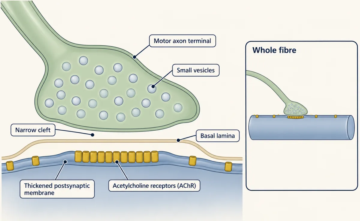
At a mature junction the receptors are packed under the nerve ending and sparse everywhere else on the fibre. Building the junction means solving that sorting problem: where do the receptors go, and who tells them?
The one idea to carry through this lesson. The muscle proposes; the nerve disposes. The muscle builds receptor clusters with its own machinery, and needs no nerve to do it. The nerve decides which clusters stay. Agrin, held in the basal lamina under the terminal, raises MuSK signaling there and protects those clusters. Acetylcholine disperses the clusters that are not protected.
What the rule covers.
- Which junction. The postsynaptic receptor clusters of developing mouse skeletal muscle, from the prepattern (about embryonic day 13) to birth. Most of the genetics is in the diaphragm, the sheet of muscle that drives breathing.
- Which effect. Where receptor clusters form and whether they survive. The rule does not cover how the nerve terminal differentiates, how the adult junction is maintained, or how an axon chooses where to land.
- A balance, not a switch. “Agrin protects” names agrin’s major job, not its only one; agrin can also start clusters (see Where the two forces meet).
How the nerve’s signal was found
The search for the nerve’s signal began with a strange experiment. In 1978 Sanes, Lawrence Marshall and U. J. McMahan damaged frog muscle so that the fibres and the nerve terminals degenerated, leaving the sheaths of basal lamina behind. Regenerating axons returned to the old synaptic sites on the empty sheaths. In later work from the same laboratory, regenerating muscle fibres clustered receptors at the old sites even with the nerve kept out. The basal lamina remembered where the synapse had been.
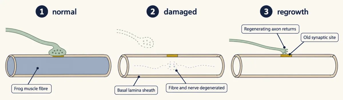
Think of a building demolished down to its foundation, with a mark on the slab showing where the front door stood. The new builders read the mark. McMahan’s group went looking for the ink. They needed tissue denser in cholinergic synapses than muscle, and found it in the electric organ of the ray Torpedo. Fractions of electric organ were tested on cultured chick muscle cells, counting receptor clusters stained with fluorescent alpha-bungarotoxin, a snake toxin that binds the acetylcholine receptor (AChR). In 1987 they named the active protein agrin.
Motor neurons make agrin and send it into the basal lamina, but muscle makes agrin too. The difference is splicing, in which one gene’s RNA is cut and joined in more than one way. Neural agrin carries a short insert of a few amino acids at one splice site. In culture assays on chick muscle cells, that insert makes it about a thousand times more potent at clustering receptors than the form without it, which muscle mainly makes.
Agrin had to be received. In 1996 the Regeneron group of David Glass and George Yancopoulos, with Burden, reported that mice lacking MuSK, a muscle-specific receptor tyrosine kinase, make no junctions, and that agrin acts through a complex containing MuSK. Like other receptor tyrosine kinases, MuSK signals when two kinase domains are brought together and phosphorylate each other (that lesson). Yet agrin did not bind MuSK on its own in binding assays. Glass and colleagues proposed that an unknown muscle component, which they called MASC, completed the receptor.
Predict. Agrin acts through a complex that contains MuSK, but MuSK alone does not bind agrin. What kind of molecule would you look for? How would you test a candidate?
Check your prediction
You want a protein on the muscle surface that binds neural agrin and also joins MuSK. One test: give the candidate to cells that are not muscle and ask whether they now bind agrin.
The missing link was Lrp4, a large membrane protein of the LDL receptor family, and it fits what Glass had called MASC. Mice lacking Lrp4 were reported in 2006 to have NMJ defects resembling those of MuSK mutants. In 2008 two laboratories showed that Lrp4 is the agrin receptor. Natalie Kim and colleagues in Burden’s laboratory found that nonmuscle cells given Lrp4 bound neural agrin and that Lrp4 forms a complex with MuSK. Bin Zhang and colleagues in Lin Mei’s laboratory showed that purified neural agrin binds purified Lrp4, and that Lrp4 also binds MuSK.
Two more proteins finish the chain. Dok7, an adaptor inside the muscle, binds phosphorylated MuSK and boosts its activity. Mice lacking Dok7 make no junctions, so that boost is not optional. Rapsyn, a scaffold on the inner face of the membrane, holds the receptors together (that lesson).
neural agrin → Lrp4 → MuSK (with Dok7) → rapsyn → clustered AChR
Remove one link at a time, and each mouse shows what that link does:
| Mouse lacks | Where the protein sits | What the junction looks like |
|---|---|---|
| agrin | neural form made by the nerve, held in the basal lamina | profoundly disrupted: markedly fewer and smaller clusters, abnormal nerve branching |
| Lrp4 | muscle surface | defects resembling those of MuSK mutants |
| MuSK | muscle membrane; the kinase | no junctions |
| Dok7 | inside the muscle | no junctions |
| rapsyn | inner face of the muscle membrane | receptors made but not clustered; nuclei beneath the synapse still transcribe receptor genes |
Every muscle link is essential. The nerve’s signal is the odd row: without agrin, fewer and smaller clusters remain. That row returns as the lesson’s puzzle.
The muscle starts without the nerve
Read the transcript of “A Band Without a Nerve”
Film transcript: A Band Without a Nerve
If agrin from the nerve switches on MuSK, then no nerve should mean no clusters. Is that true? Muscle cells grown in a dish, with no neurons at all, make small receptor clusters by themselves. But a dish is not an embryo. In an animal, a nerve might have marked the spot first. In 2001, two groups studied mouse embryos whose diaphragm receives no motor axons. The diaphragm is the muscle that drives breathing. They stained it with a toxin that labels the muscle's acetylcholine receptors. If the nerve chooses where receptors gather, what should the stain show? And if the muscle chooses?
A band of receptor clusters still formed across the middle of the muscle. That is where the nerve would normally arrive. Burden's group also found receptor RNA concentrated in the same band. The muscle had drawn a rough map of where junctions would go. It is called the prepattern. What does the prepattern need? Remove one part at a time. Embryos lacking agrin still build the early band. The nerve's signal is not needed to start. Embryos lacking MuSK make receptors, but no band. So the muscle's own kinase is needed for the band. Without rapsyn, there is no band of clusters either. So the muscle's own machinery is needed to start the band, and the nerve's signal is not. The muscle proposes.
The nerve still matters. Contact makes far more clustering and narrows the band. Without a nerve, the band was wider, and its clusters belonged to no terminal. Keep two statements apart. Clusters can form without a nerve. A synapse cannot. Takeaway: the muscle draws a rough map by itself. The nerve's work is to edit it.
From the film
- In mouse embryos whose diaphragm receives no motor axons, a band of receptor clusters still forms, so the muscle draws a rough map by itself.
- The muscle's own machinery, including MuSK and rapsyn, is needed to start the band; the nerve's signal is not.
- The nerve still matters: contact makes far more clustering and narrows the band.
If agrin from the nerve switches on MuSK, and MuSK gathers the receptors, a simple prediction follows: no nerve, no clusters. That was a strong form of the agrin hypothesis, and it was a reasonable one.
Culture gave the first hint that it was incomplete. Muscle cells grown with no neurons at all make small, spontaneous acetylcholine receptor (AChR) clusters. But a dish is not an embryo. In an animal, a nerve might have marked the spot before anyone looked.
Predict. In 2001 two groups, Xia Yang, Steven Burden and colleagues, and Weichun Lin, Joshua Sanes, Kuo-Fen Lee and colleagues, studied mouse embryos whose diaphragm receives no motor axons, either because the axons never reach it or because motor neurons were removed genetically. They stained each diaphragm with fluorescent alpha-bungarotoxin. If the nerve chooses where receptors gather, what should the stain show? If the muscle chooses, what should it show?
Check your prediction
The diaphragm still built a band of receptor clusters across its middle, the region where the nerve would normally have arrived. Burden’s group also found the RNA for receptor subunits concentrated in the same band. The muscle had laid down a prepattern: a rough map of where the junctions would go, drawn without the nerve.
Next, ask what the prepattern needs. Remove one part at a time:
| Embryo lacks | Central band of clusters? | What it tells you |
|---|---|---|
| motor axons | yes, wider than normal | the muscle can place clusters by itself |
| agrin | yes, in early embryos (most are lost later) | the nerve’s signal is not needed to start |
| MuSK | no; receptors are made but not concentrated in a band | the muscle’s own kinase is needed for the band |
| rapsyn | no | the scaffold is needed to hold receptors together at all |
The muscle’s own machinery is needed to start the band. The nerve’s signal is not.
The nerve still matters. Contact makes far more clustering and narrows the band, and without a nerve the clusters belong to no terminal. Keep two statements apart. “AChR clusters can form without innervation” is true. “The nerve is not needed for a synapse” is false.
A paradox, and a second force
Read the transcript of “Agrin Protects, Acetylcholine Disperses”
Film transcript: Agrin Protects, Acetylcholine Disperses
The muscle builds its band without agrin. So why do mice lacking agrin end up with so few clusters? Something must be removing the clusters the muscle made. What could the nerve bring that removes clusters? A second signal. Acetylcholine, released at every terminal, disperses clusters that agrin does not protect. To test that, use mice whose nerves release no acetylcholine. They lack the enzyme that makes it. Their receptors still cluster under the nerve. So acetylcholine is not what starts clusters. And they keep more clusters, spread over a wider band. Now lay out a grid. Agrin present or absent. Acetylcholine present or absent. Normal mice have a narrow band under the terminals.
Without agrin, early clusters form, then most are lost. Without acetylcholine, there are more clusters, over a wider band. Now predict the last cell. Remove both agrin and acetylcholine. What happens to the clusters? Clusters return, built by the muscle's own MuSK program. Two mouse studies reported this in 2005. Without acetylcholine, clusters persist even without agrin. So a major job of agrin in developing muscle is to protect clusters from acetylcholine. One puzzle remains. The cluster under the terminal gets the most acetylcholine. Why does it survive? In the working model, the two forces differ in reach. Agrin is anchored in the basal lamina, so its protection stays under the terminal.
The dispersing effect of acetylcholine reaches clusters along the rest of the fibre. Away from the terminal, they lack agrin's strong protection. How that effect reaches distant clusters is not fully settled. The two forces meet at one question. Does a cluster stay? Agrin, acting through MuSK, holds it. Acetylcholine's pathway pulls it apart. Without agrin, Lrp4 is thought to give MuSK some activity. That would explain why a few small clusters remain. Raise MuSK enough another way, and synapses can form even in mice lacking agrin. This rule is for receptor clusters in developing mouse muscle. There, the muscle proposes; the nerve disposes. Takeaway: agrin protects the clusters beneath it.
Acetylcholine disperses the rest.
From the film
- Acetylcholine, released at every terminal, disperses clusters that agrin does not protect.
- In mice lacking both agrin and acetylcholine, clusters return, so a major job of agrin in developing muscle is to protect clusters from acetylcholine.
- In the working model, agrin is anchored in the basal lamina, so its protection stays under the terminal.
Two results now pull against each other. The muscle builds its band of clusters without agrin. Yet mice lacking agrin end up with markedly fewer and smaller clusters.
Predict. Why do mice lacking agrin lose most of their clusters? Write down one explanation before reading on. (Hint: the nerve releases more than agrin.)
Check your prediction
The nerve brings a second signal. Acetylcholine, released at every terminal, disperses receptor clusters that agrin does not protect. Without agrin, little protects them, so most are lost.
To test that, you need a mouse whose nerves release no acetylcholine. Choline acetyltransferase (ChAT) is the enzyme that makes acetylcholine, so motor neurons in mice lacking ChAT release none. Their receptors still cluster under the nerve (Misgeld et al., Neuron 2002; Brandon et al., J. Neurosci. 2003), so acetylcholine is not the inducer. With the main dispersing force gone, the muscle keeps more clusters, spread over a wider band, and the nerve branches widely.
Predict. Here are the two inputs from the nerve, and what each mouse shows at birth. Fill in the last row, and say what it would tell you about agrin’s job.
| Mouse | Agrin from the nerve | Acetylcholine from the nerve | Receptor clusters at birth |
|---|---|---|---|
| normal | yes | yes | under the terminals, in a narrow band |
| lacking agrin | no | yes | early clusters, then markedly fewer and smaller |
| lacking ChAT | yes | no | more clusters, over a wider band |
| lacking both | no | no | ? |
Check your prediction
The tempting guess is no clusters, since both inputs from the nerve are gone. Instead, clusters return, built by the muscle’s own MuSK-dependent program. Two mouse studies published in 2005, one from Sanes and Lichtman’s laboratories and one from Kuo-Fen Lee’s laboratory with Weichun Lin, reported this result.
The double is the telling line. If agrin were required to make clusters, removing acetylcholine could not bring them back. The prepattern had shown that agrin is not needed to start clusters; the double shows what it is needed for. Acetylcholine is the force agrin opposes, and a major job of agrin in the animal is to protect clusters from dispersal by acetylcholine.
Other processes also affect cluster stability in developing mouse muscle, but these two forces, agrin-MuSK signaling and acetylcholine, explain the mutants above.
Why the cluster under the terminal survives
Predict. The cluster right under a nerve terminal receives the most acetylcholine. Yet it is the one that survives, while clusters farther along the fibre are lost. How can that be?
Check your prediction
The two forces do not have the same reach. In the working model, agrin is anchored in the synaptic basal lamina, so its protection stays under the terminal, while the dispersing effect of acetylcholine reaches clusters elsewhere on the fibre. Under the terminal, protection wins. Away from it, clusters lack agrin’s strong local protection, and in innervated muscle they usually disperse.
The mouse lacking ChAT fits this. Remove acetylcholine and the band widens, which fits acetylcholine removing clusters away from the terminals as well as beneath them, though these mice also have wider nerve branching. How the dispersing signal reaches distant clusters is not settled.
Where the two forces meet
Agrin and acetylcholine meet at one question: does a cluster stay? Agrin raises MuSK signaling through Lrp4, and that signaling stabilizes clusters. Acetylcholine drives a dispersal pathway through receptor activation and muscle activity, linked in mice to the kinase Cdk5. Whether acetylcholine works by turning MuSK signaling down, or by a separate route that strong MuSK signaling can resist, is not settled.
The balance explains a detail of the agrin-null mouse. Without agrin, MuSK is thought to keep a lower level of activity, driven by Lrp4, so a few small clusters are kept. A strictly on/off rule, “no agrin, no clusters”, would predict none.
The balance also allows that raising MuSK levels could stand in for agrin, and at high expression they do. Kim and Burden found that muscle expressing MuSK widely along the fibre, beyond its normal central band, formed clusters and synapses outside that band, and that synapses formed in agrin-mutant mice, rescuing their lethality. How much extra MuSK this takes is a dose question the abstract does not settle. In adult rodent muscle, neural agrin delivered to non-synaptic sites of innervated fibres induces new postsynaptic specializations there (Meier and colleagues, 1997). So MuSK signaling can both start new clusters and protect old ones. These are distinct outcomes, and “agrin protects” names a major job of agrin in the developing animal, not the only thing it can do.
In the terms of the neurotrophic lesson, agrin is instructive, not merely permissive: delivered somewhere new, it brings a postsynaptic specialization with it. That lesson’s rule was “make too many, then let the target keep what it can feed”. For the postsynaptic clusters, this one runs the other way: the target makes the surplus, and the neuron chooses what is kept. (On the nerve side, a fibre still begins with a surplus of inputs; see below.)
A second effect of activity: the nuclei
The nerve disposes in a second way, through activity and transcription. A muscle fibre is one long cell with many nuclei. When the nerve drives the fibre, receptor transcription is suppressed in nuclei away from the synapse, while the few nuclei beneath the terminal keep transcribing receptor genes, a pattern Burden’s group showed with transgenic mice in 1992. Cut the nerve and the suppression lifts: receptors appear along the whole fibre, and the denervated muscle becomes “supersensitive” to acetylcholine applied anywhere on it.
Keep the two effects apart. Dispersal removes clusters that already exist. Suppressed transcription means fewer receptors are made away from the synapse. The rapsyn-null mouse separates clustering from local transcription: no clusters, yet subsynaptic nuclei keep transcribing.
Signals in both directions
The muscle answers the nerve. In mice lacking MuSK or Lrp4, motor axons wander and nerve terminals fail to differentiate (DeChiara and colleagues, 1996; Weatherbee and colleagues, 2006). In mouse work, muscle Lrp4 also signals back to the nerve terminal, so the same complex helps organize the presynaptic side. The nerve’s own signals shape its growth too: mice lacking agrin show abnormal nerve branching in the muscle, and in mice lacking choline acetyltransferase (ChAT) the nerve branches widely.
NMJ vs. central synapses, and a junction that fails
The neuromuscular junction (NMJ) is a good guide to central synapses, but not a template. Many central neurons receive thousands of inputs that use different transmitters. Central synapses have no basal lamina in the cleft, and receptor clustering there uses other molecules, including neurexins, neuroligins and the scaffold PSD-95, taken up in that lesson. What carries over is the logic: each partner sends signals that make the other specialize, and stable synapses need both sides. How a muscle fibre that begins with several inputs ends with one is the subject of that lesson.
The NMJ also fails in a known way. In myasthenia gravis, the immune system makes antibodies against the junction, and muscles tire and weaken with use. Most patients have antibodies against the acetylcholine receptor (AChR). Some of the rest have antibodies against MuSK or Lrp4, two links from the chain above. Inherited mutations in several of the same genes, Dok7 among them, cause congenital myasthenic syndromes; other forms affect the nerve terminal or the basal lamina.
Putting it together
| Experiment | Animal and age | Observed result | What it supports | What it does not establish |
|---|---|---|---|---|
| Fibres and terminals degenerate, sheaths left | adult frog muscle | axons return to the old sites; regenerating fibres cluster receptors there with the nerve kept out (1978 and later) | the basal lamina keeps the synaptic address | which molecule |
| Torpedo fractions on muscle cells | cultured chick muscle cells | one protein, agrin, clusters receptors (1987) | agrin is enough to cluster receptors in a dish | that the nerve uses it in the animal |
| Delete MuSK, Lrp4, Dok7 or rapsyn | mouse | no junctions, or defects resembling MuSK mutants; rapsyn-null receptors not clustered | each muscle link is needed | which partner places the clusters |
| Lrp4 given to nonmuscle cells; purified proteins | cell line; in vitro | binds neural agrin; forms a complex with MuSK (2008) | Lrp4 is the agrin receptor | — |
| No motor axons reach the diaphragm | mouse embryo | a central band of clusters, wider than normal, with receptor RNA concentrated in it (2001) | the muscle proposes | that a synapse can form without a nerve |
| Delete agrin | mouse | early clusters, then markedly fewer and smaller | agrin is needed for a normal junction | that agrin starts clusters |
| Delete ChAT | mouse | clusters under the nerve; more clusters, wider band | acetylcholine disperses; it is not the inducer | how dispersal reaches distant clusters |
| Delete agrin and ChAT | embryonic mouse, 2005 | clusters persist without agrin when acetylcholine is absent | a major developmental job of agrin is to oppose acetylcholine-driven cluster loss | that agrin cannot induce clusters |
| Express MuSK beyond the central band (ectopic MuSK) | transgenic mouse | clusters and synapses form outside the normal band, and synapses form without agrin | MuSK level can be limiting, and sufficiently high MuSK expression can bypass the need for agrin | that acetylcholine acts by lowering MuSK activity |
| Receptor-gene reporter, with denervation | transgenic mouse, 1992 | expression restricted to nuclei beneath the synapse; after denervation, nuclei elsewhere express it too | innervation represses receptor-gene transcription away from the synapse | the molecular mechanism of that repression |
The guiding idea holds up across the table: the muscle proposes, the nerve disposes. Remove MuSK or rapsyn and no band is made. Remove the nerve, and the proposal is kept but belongs to no terminal. Remove agrin, and acetylcholine disposes of most of what the muscle proposed.
Design problem
Use the rule to predict receptor clusters in one new mouse, then revisit a row of the table with dose in mind: (1) a mouse lacking both MuSK and choline acetyltransferase (ChAT); (2) a mouse with normal innervation whose muscle makes extra MuSK along the whole fibre. Say what each would tell you.
Saved in this browser only.
One way to do it
- No MuSK, no ChAT. The rule predicts no clusters. Removing acetylcholine lets clusters survive only if the muscle can make them, and without MuSK it cannot. If clusters did appear, the muscle would have a second, MuSK-independent way to build them. This is a prediction of the rule, not a result reported in this lesson.
- Extra MuSK, normal nerve. The rule predicts clusters and synapses beyond the normal narrow band only if the extra MuSK signaling is strong enough to resist dispersal away from the terminals. A small increase might change nothing, so the prediction needs a dose. That is the Kim and Burden experiment above: MuSK expressed beyond the band formed clusters and synapses there, even without agrin. So MuSK level and location can be limiting, and enough MuSK signaling can bypass the requirement for agrin in forming synapses. Testing the dose would mean comparing lines that express different amounts.
Going deeper
The first contact
The first contact between axon and muscle is fast and physical. In culture, a growth cone from a frog spinal neuron adheres to a muscle cell rapidly after it touches. Adhesion holds the partners together. It does not by itself make a postsynaptic specialization.
What the axon does in a dish
When a motor axon is added to cultured muscle cells that already carry spontaneous clusters, it does not seek out those clusters, but where it touches, far more clustering follows.
What centres the band
An open question is what spatial cue centres the MuSK-dependent band in the first place. Candidates include intrinsic patterning of the muscle, Wnt signaling to MuSK and Lrp4, and the geometry of the fibres, and none is settled.
Your notes
Select any text in the lesson to highlight it or add a note to it. Your highlights and notes are saved in this browser and collected in your notebook.
This browser is not letting the site save anything (a private window or strict privacy settings), so highlights and notes will be lost when you leave the page.
How we measure it
Fluorescent alpha-bungarotoxin labeling
Alpha-bungarotoxin, a snake toxin, binds the muscle acetylcholine receptor tightly and nearly irreversibly. Coupled to a fluorescent dye, it shows where receptors sit. Labeling once in one colour and later in a second colour separates receptors that were already there from receptors added since.
Bioassay-guided purification
A tissue extract is split into fractions, and each fraction is tested on a living readout, here the number of receptor clusters on cultured muscle cells. Only fractions that keep the activity go forward. This is how agrin was found in Torpedo electric organ.
Knockout mice and mice without motor axons
A knockout mouse lacks one gene, usually in every cell. Mice that lack motor neurons, or whose motor axons never reach a muscle, show what the muscle can do alone. Comparing single and double knockouts places each gene in the pathway.
Whole-mount in situ hybridization
A labeled probe that pairs with one messenger RNA is applied to a whole piece of tissue, so you can see where the gene is expressed. In embryonic diaphragm it showed that receptor-subunit RNA is concentrated in a central band.
Method films · the methods library
Practice questions on this lesson (3) → MCAT-style, original, each with an explanation
Where this lesson fits
Before thisReview first
- The Growth Cone: The Tip That Steers the AxonThis course · Lesson 3
You need how the axon tip reaches and touches its target before asking why contact alone does not build a synapse.
- The Neurotrophic Hypothesis TestedThis course · Lesson 29
The lesson contrasts its rule of making too many and keeping what is supported with the muscle making surplus clusters.
- Neurotrophins, Trk Receptors and p75This course · Lesson 30
Receptor-kinase signaling from a target-derived factor is the model you reuse here for agrin and MuSK.
After thisStudy next
- Synapse Elimination: One Muscle Fiber, One AxonThis course · Lesson 37
A fibre that starts with several axons ends with one, building on the junction formed here.
- Synapses in the Brain: Adhesion, Scaffolds and AstrocytesThis course · Lesson 36
It asks which of this junction's signals carry over to brain synapses, which lack a basal lamina and use other molecules.
- Disorders of the Synapse: Built, Then KeptThis course · Lesson 49
It applies the idea that synapses must be built and then kept to disorders where that fails.
Elsewhere on the siteFilms, simulations and method films in other courses
- Counting failuresSimulation · Neuroscience
The acetylcholine released at the junction acts through the quantal release of transmitter that this simulation explores.
- Blips from a Silent NerveFilm · Neuroscience
It shows how acetylcholine release at the endplate works, which is the transmitter that disperses clusters here.
- Sliding, Not ShrinkingFilm · Organismal Biology
It shows the muscle fiber that the nerve contacts and drives through the junction.
Suggested by an AI model from the lessons themselves, then checked. How we use AI
Finish line
Test yourself
8 questions
Practice and MCAT-style, with explained answers.
Next · Lesson 36
Synapses in the Brain: Adhesion, Scaffolds and Astrocytes
A kidney cell that makes one neuronal protein, neuroligin, can persuade an axon to build a nerve terminal on it. So what happens to the synapses of a mouse that lacks neuroligins 1, 2 and 3?
Continue →← Lesson 34 · Pruning Without Dying: Axons That Cut Themselves Back